Özet
Akut pulmoner embolide BT pulmoner anjiyografide saptanan sağ ventrikül yüklenmesi veya disfonksiyonu bulguları kısa dönem prognozun tanınmış öngörücülerindendir. BTPA'da RV/LV oranı, interventriküler septumun sola bombeleşmesi ve IVC'ye kontrast reflüsü sağ kalp yüklenmesi değerlendirmesinde incelenebilir. Bu morfolojik bulgular, akut pulmoner embolide kısa süreli prognoz için tanınmış öngörücülerdir ve tedavi yaklaşımını yönlendirmede kullanılır. Akut pulmoner embolide sağ ventrikül yüklenmesi veya disfonksiyonu kısa dönem prognozla ilişkilidir; pıhtı morfolojisi uzun dönem komplikasyonları öngörebilir ve bronşiyal arter hipertrofisi kronik tromboembolik pulmoner hipertansiyonun tespitine yardımcı olabilir.
Faz ve pencere
- BTPA tanısal
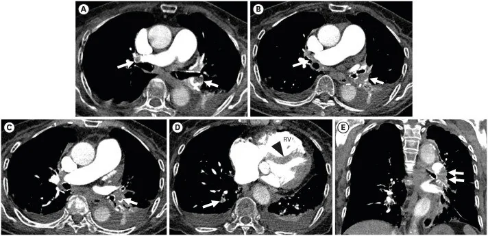
- BTPA'da RV/LV oranı ve sağ ventrikül genişlemesi değerlendirilir; interventriküler septumun sola bombeleşmesi ve IVC ya da hepatik venlere kontrast reflüsü de sağ kalp yüklenmesiyle ilişkili bulgulardır. Bu morfolojik bulgular, akut pulmoner embolide kısa süreli prognoz için tanınmış öngörücülerdir.
Önerilen pencereler: Mediasten (G 350 / M 50), Anjiyo (G 600 / M 150).
BT bulguları
- RV/LV iç çap oranında artış — ventriküller aynı aksiyel düzeyde veya dört boşluk düzleminde karşılaştırılır.
- İnterventriküler septum düzleşmesi — kısa eksende normal kavisli kontur kaybolur, septum düz veya sola bombeleşmiş görünür.
- Sağ ventrikül kavitesinde genişleme — sağ ventrikül boyutlarında artış gözlenir.
- Sağ atriyum genişlemesi — atriyal çap artışı sağ ventrikül disfonksiyonuyla ilişkili olabilir.
- IVC ve hepatik venlere kontrast reflüsü — sağ atriyumdan geriye doğru yoğun kontrast uzanımı görülür.
- Ana pulmoner arter genişlemesi — eşlik eden pulmoner hipertansiyon olasılığını düşündürür, akut yüklenmeye özgü değildir.
Ölçütler ve sınıflamalar
- Aksiyel veya dört boşluk düzleminde BTPA RV/LV iç çap oranı, sağ ventrikül büyümesi lehine
- >1.0
Normalde
Normal kalpte aynı aksiyel veya dört boşluk düzleminde sağ ventrikül iç çapı sol ventrikülden küçük, interventriküler septum sol ventriküle doğru hafif kavisli görünür. Karşılaştırmada ventrikül iç çaplarını aynı düzlemde ölçün; septumun kavisinin korunup korunmadığını ve IVC/hepatik venlerde geriye uzanan kontrast bulunup bulunmadığını değerlendirin.
Normal BT ile kıyasla
Aynı bölgenin, kaynak veri setinde patoloji içermediği belirtilen bir BT incelemesini kesit kesit kaydırın; organ sınırlarını açarak anatomiyi hasta görüntüsüyle karşılaştırın. Küçük rastlantısal bulgular tümüyle dışlanmamıştır.
Kaynak: TotalSegmentator v2.01 veri seti (Wasserthal ve ark., Radiology: Artificial Intelligence 2023), CC BY 4.0, vaka s1278; organ sınırları veri setinin otomatik segmentasyonundan, birleştirilerek sadeleştirildi. Görüntüler 2,5 mm kesit aralığına yeniden örneklendi.
Ayırıcı tanı
- Kronik tromboembolik pulmoner hipertansiyon
- bronşiyal arter hipertrofisi gibi bulgular kronik hastalığın tespitinde yardımcı olur.
- Sağ ventrikül infarktı
- sağ ventrikül genişlemesi olabilir; eşlik eden koroner dağılım bulguları ve klinik bağlam emboliden ayırır.
- Konstriktif perikard hastalığı
- kalp konturu/perikard değişikliği ve iki ventrikülde doluş etkilenimi ön plandadır; izole sağ ventrikül basınç yükü paterninden farklıdır.
- Konjenital kalp hastalığı
- kronik sağ boşluk genişlemesine eşlik eden septal veya büyük damar anatomik anomalileri aranır.
Tuzaklar
- RV/LV oranı ölçümü kalp eksenine ve kesit seçimine duyarlıdır; aynı aksiyel seviyede iç duvardan iç duvara ölçün veya dört boşluk düzleminde doğrulayın.
- IVC veya hepatik venlere kontrast reflüsü, ciddi pulmoner hipertansiyonla ilişkili olabilir ve akut pulmoner tromboemboli şiddetini değerlendirmede potansiyel bir görüntüleme belirtecidir.
- Septum düzleşmesi, sağ ventrikül basınç veya hacim yükünden kaynaklanır; akut pulmoner emboli ile kronik durumlar arasında ayrım yapmak için eşlik eden pıhtı bulguları ve klinik bağlam değerlendirilir.
Kendini dene
BTPA'da RV/LV iç çap oranı ≥1,0, septum düzleşmiş ve akut emboli görülüyor. En uygun BT yorumu nedir?
Cevabı göster
Sağ ventrikül yüklenmesi. Sağ ventrikül genişlemesi ile septum düzleşmesinin emboliyle birlikte bulunması BT'de sağ kalp yüklenmesini destekler. Diğer seçeneklerde farklı kalp veya aort morfolojisi beklenir. BT bulguları hemodinamik basıncı ölçmez.
BTPA'da IVC veya hepatik venlere kontrast reflüsü hangi durumla ilişkili bir bulgudur?
Cevabı göster
Ciddi pulmoner hipertansiyon. Bir CTPA çalışmasında inferior vena kava veya hepatik venlere kontrast reflüsü, ciddi pulmoner hipertansiyonun tek bağımsız öngörücüsü olarak saptanmıştır.
Bu konunun yer aldığı turlar
İlgili konular
Kaynaklar
Bu sayfadaki 34 cümle ve 2 quiz sorusu aşağıdaki kaynaklardan alıntılarla otomatik olarak karşılaştırıldı; 13 cümle bu karşılaştırmada düzeltildi (2026-10-09).
- Optimal approach to performing and reporting computed tomography angiography for suspected acute pulmonary embolism: a clinical consensus statement of the ESC Working Group on Pulmonary Circulation & Right Ventricular Function, the Fleischner Society, the Association for Acute CardioVascular Care (ACVC) and the European Association of Cardiovascular Imaging (EACVI) of the ESC, endorsed by European Respiratory Society (ERS), Asian Society of Thoracic Radiology (ASTR), European Society of Thoracic Imaging (ESTI), and Society of Thoracic Radiology (STR)pmc.ncbi.nlm.nih.gov
- Pulmonary tumour embolism and lymphangitis carcinomatosa: a case report and review of the literature.pubmed.ncbi.nlm.nih.gov
- Electrocardiogram Signal Analysis With a Machine Learning Model Predicts the Presence of Pulmonary Embolism With Accuracy Dependent on Embolism Burden.pubmed.ncbi.nlm.nih.gov
- Multimodality imaging of pericardial disease.pubmed.ncbi.nlm.nih.gov
- Placement of Simultaneous Inferior Vena Cava Filter During Emergent Open Pulmonary Thromboembolectomy.pubmed.ncbi.nlm.nih.gov
- Cardiopulmonary involvement in Puumala hantavirus infection.pubmed.ncbi.nlm.nih.gov
- Incidence and predictors of chronic thromboembolic pulmonary hypertension following first episode of acute pulmonary embolism.pubmed.ncbi.nlm.nih.gov
- [Diagnostics of acute pulmonary embolism : An update].pubmed.ncbi.nlm.nih.gov
- 36th International Symposium on Intensive Care and Emergency Medicine : Brussels, Belgium. 15-18 March 2016.pubmed.ncbi.nlm.nih.gov
- Pulmonary embolism critical care update: prognosis, treatment, and research gaps.pubmed.ncbi.nlm.nih.gov
- Recurrent massive pulmonary emboli in a critically ill patient with COVID-19.pubmed.ncbi.nlm.nih.gov
- To Thrombolyse or Not to Thrombolyse: Two Years Experience of Thrombolysis of Sub-Massive Pulmonary Embolism in a District General Hospital.pubmed.ncbi.nlm.nih.gov
- Clinical and echocardiographic predictors of mortality in acute pulmonary embolism.pubmed.ncbi.nlm.nih.gov
- Prognostic value of CT pulmonary angiography parameters in acute pulmonary embolism.pubmed.ncbi.nlm.nih.gov
- Clinical outcomes and factors associated with pulmonary infarction following acute pulmonary embolism: a retrospective observational study at a US academic centre.pubmed.ncbi.nlm.nih.gov
- The STEMI Mimic: Cocaine-Associated Pulmonary Embolism With ST-Segment Elevation.pubmed.ncbi.nlm.nih.gov
- Obese Adolescent with Factor V Leiden-Related Pulmonary Embolism: Case-Based Insight into Paediatric Thrombosis Management.pubmed.ncbi.nlm.nih.gov
- Recurrent Pulmonary Embolism After Pulmonary Endarterectomy During Rivaroxaban Therapy in a Resource-Limited Setting.pubmed.ncbi.nlm.nih.gov
- Review of D-Shape Left Ventricle Seen on Magnetic Resonance Imaging (MRI) or Computed Tomography (CT), Similar to the Movahed Sign Seen on Cardiac Gated Single-Photon Emission Computed Tomography (SPECT) as an Indicator for Right Ventricular Overload.pubmed.ncbi.nlm.nih.gov
- Advances in the imaging of pulmonary hypertension.pubmed.ncbi.nlm.nih.gov
- Pathophysiology and management of right ventricular failure in critically ill patients: A narrative review.pubmed.ncbi.nlm.nih.gov
- Artificial Intelligence in Pulmonary Hypertension: Current State and Future Prospects.pubmed.ncbi.nlm.nih.gov
- Single Ventricle Fontan: The Basics for General Cardiologists.pubmed.ncbi.nlm.nih.gov
- Perioperative Point-of-Care Ultrasound Utilization in Abdominal Organ Transplantation. Part II: Postoperative Care.pubmed.ncbi.nlm.nih.gov
- Indication for durable mechanical circulatory support: an expert opinion review.pubmed.ncbi.nlm.nih.gov
- Non-thrombotic pulmonary emboli: imaging findings and differential diagnoses.pubmed.ncbi.nlm.nih.gov
- External validation of a commercial AI system for pulmonary embolism detection on chest CTPA: a multicenter study.pubmed.ncbi.nlm.nih.gov
- Navigating the Complexity of PH-ILD: From Molecular Mechanisms to Integrated Clinical Evaluation.pubmed.ncbi.nlm.nih.gov
- The Role of Imaging in Pulmonary Vascular Disease: The Clinician's Perspective.pubmed.ncbi.nlm.nih.gov
- Diagnostic Evaluation of Pulmonary Hypertension: A Comprehensive Approach for Primary Care Physicians.pubmed.ncbi.nlm.nih.gov
- Hypertrophic cardiomyopathy: state of the art on diagnosis and current therapeutic approach.pubmed.ncbi.nlm.nih.gov
- An Intraoperative Fluoroscopic Predictor of Successful Micra Implantation on the Right Ventricular Mid-Septum.pubmed.ncbi.nlm.nih.gov
- Bicuspid Aortic Valve Disease-Associated Aortopathy in Pediatric Subjects-From Traditional Assessment to Current Advances and Future Perspectives: A Narrative Review.pubmed.ncbi.nlm.nih.gov
- High altitude pulmonary hypertension: overlap with other types of pulmonary hypertension and implications for treatment.pubmed.ncbi.nlm.nih.gov
- Agenesis of the left pulmonary artery and cardiac hydatid cysts: A dual rarity.pubmed.ncbi.nlm.nih.gov
- Atypically presented flank pain in a patient with pulmonary embolism; a case report and literature review of a diagnostic dilemma.pubmed.ncbi.nlm.nih.gov
- Epidemiology and Outcomes of Acute Pulmonary Embolism in Seychelles: A National Cohort Study With a Subgroup Analysis of Systemic Thrombolysis Efficacy and Safety in Intermediate-Risk Pulmonary Embolism.pubmed.ncbi.nlm.nih.gov
- When Pulmonary Embolism Opens the Door: Paradoxical Thalamic Stroke Through a Patent Foramen Ovale.pubmed.ncbi.nlm.nih.gov
- Primary pulmonary leiomyosarcoma in a patient with disseminated pulmonary tuberculosis and constrictive pericarditis.pubmed.ncbi.nlm.nih.gov
- Epicardial Lipomatosis as a Cause of Constrictive Pericarditis.pubmed.ncbi.nlm.nih.gov
- Imaging insights into systemic tuberculosis with cardiac involvement: A rare challenging case of tuberculous chronic constrictive pericarditis and myocarditis.pubmed.ncbi.nlm.nih.gov
- Cardiothoracic Imaging Findings in Obesity Hypoventilation Syndrome (Pickwickian Syndrome): A Case Series.pubmed.ncbi.nlm.nih.gov
- Multimodal imaging of infantile scimitar syndrome with Eisenmenger physiology: A case report of complex intracardiac and extracardiac shunts.pubmed.ncbi.nlm.nih.gov
- Prenatal diagnosis of a congenital pseudoaneurysm of the mitral-aortic intervalvular fibrosa: a multimodality imaging case report.pubmed.ncbi.nlm.nih.gov
- Tricuspid regurgitation: standardized stepwise work-up from referral to expert heart valve centre.pubmed.ncbi.nlm.nih.gov
- Relationship Between Routine Preoperative Chest CT-Based Cardiac Parameters and Recanalization After Mechanical Thrombectomy in Patients with Acute Ischemic Stroke.pubmed.ncbi.nlm.nih.gov
- Right Ventricular Compression From Pectus Excavatum: A Reversible Cause of Ventricular Dysfunction.pubmed.ncbi.nlm.nih.gov
- Imaging of inferior vena cava normal variants, anomalies and pathologies, Part 2: Acquired.pubmed.ncbi.nlm.nih.gov
- Free-breathing gradient recalled echo-based CMR in a swine heart failure model.pubmed.ncbi.nlm.nih.gov
- Contrast opacification on thoracic CT angiography: challenges and solutions.pubmed.ncbi.nlm.nih.gov
- Beyond the right ventricle: assessment of the left ventricular function in pulmonary hypertension.pubmed.ncbi.nlm.nih.gov
- Extensive bilateral pulmonary embolism revealing iron deficiency-masked JAK2-positive polycythemia vera: A case report.pubmed.ncbi.nlm.nih.gov
- The Initial Diagnostic Approach to Adult Congenital Heart Disease: A Practical Imaging Pathway for General Cardiologists and Cardiac Imagers.pubmed.ncbi.nlm.nih.gov
- How should we interpret excessive left ventricular trabeculation? Update on controversies from the cardiac imaging perspective.pubmed.ncbi.nlm.nih.gov
- Diagnostic Value and Operational Recommendations for Late Iodine Enhancement and ECV Quantification in Single-Energy Computed Tomography: A Narrative Review.pubmed.ncbi.nlm.nih.gov
- A nomogram integrating CTPA parameters and serum biomarkers to predict 30-day mortality and refine risk stratification in non-high-risk acute pulmonary embolism.pubmed.ncbi.nlm.nih.gov
- A Narrative Review of Right Heart Dysfunction in Pulmonary Embolism: Current Insights and Clinical Perspectives.pubmed.ncbi.nlm.nih.gov
- Prognostic Value of QT and QTc Interval Dispersion in Acute Pulmonary Embolism: A Retrospective Single-Center Study.pubmed.ncbi.nlm.nih.gov
- Transcatheter closure of patent ductus venosus in an adolescent using a KONAR-Multifunctional Occluder device.pubmed.ncbi.nlm.nih.gov
- Endovascular Management of Inferior Vena Cava-Right Atrial Luminal Narrowing Following Orthotopic Heart Transplantation: A Case Report.pubmed.ncbi.nlm.nih.gov
- Left Atrial Appendage: Mere Spectator or Protagonist? A Narrative Review of Its Multimodal Imaging Assessment and Its Closure Techniques.pubmed.ncbi.nlm.nih.gov
- Society for Cardiovascular Magnetic Resonance 2024 Cases of SCMR case series.pubmed.ncbi.nlm.nih.gov
- Atrial cardiomyopathy: From healthy atria to atrial failure. A clinical consensus statement of the Heart Failure Association of the ESC.pubmed.ncbi.nlm.nih.gov
- Saddle pulmonary embolism with right heart strain and incidental detection of aortic-origin splenic artery and dual renal arteries on CT angiography.pubmed.ncbi.nlm.nih.gov
- Unexpected Right Ventricular Uptake: A Sign of Pulmonary Hypertension from a Large Pulmonary Embolism.pubmed.ncbi.nlm.nih.gov
- Echocardiographic Assessment After Heart Transplant.pubmed.ncbi.nlm.nih.gov
- Quantitative CT Pulmonary Angiography and Echo Cardiography Analysis for Enhanced Cardiovascular Assessment of Right Ventricular Failure in Pulmonary Hypertension.pubmed.ncbi.nlm.nih.gov
- Evaluating inferior vena cava reflux and RV/LV ratio as predictors of acute pulmonary thromboembolism severity.pubmed.ncbi.nlm.nih.gov
- Correlative imaging integrates electrophysiology with three-dimensional murine heart reconstruction to reveal electrical coupling between cell types.pubmed.ncbi.nlm.nih.gov
- A workflow for the creation of photorealistic 3D cadaveric models using photogrammetry.pubmed.ncbi.nlm.nih.gov
- Contemporary Challenges and Strategies for Diagnosing and Managing in Type 2 Myocardial Infarction: A Narrative Review.pubmed.ncbi.nlm.nih.gov
- Machine learning and multi-omics technologies for precision cardiovascular medicine: advancing diagnosis, risk prediction, and therapeutic guidance.pubmed.ncbi.nlm.nih.gov
- Diagnostic Challenges in Takotsubo Syndrome: Bridging Mimics, Mechanisms, and Management.pubmed.ncbi.nlm.nih.gov
- Perioperative and Surgical Management of Constrictive Pericarditis.pubmed.ncbi.nlm.nih.gov
- Contemporary Review of Clinical Features, Multi-Modality Imaging, and Management of Pericardial Cysts.pubmed.ncbi.nlm.nih.gov
- Multimodal Cardiac Imaging in Systemic Lupus Erythematosus: From Clinical Suspicion to Diagnosis in Clinical Practice.pubmed.ncbi.nlm.nih.gov
- Prognostic Value of CT-Derived Indicators of Right-Heart Strain and Thrombus Burden for In-Hospital Adverse Events in Acute Pulmonary Embolism.pubmed.ncbi.nlm.nih.gov
- Suspected Acute Stanford Type A Aortic Dissection: A Case Report.pubmed.ncbi.nlm.nih.gov
- Cardiac Tamponade in Late Pregnancy Caused by <i>Corynebacterium amycolatum</i> Pericarditis and Managed by a Surgical Pleuro-Pericardial Window.pubmed.ncbi.nlm.nih.gov
- Case series: the right parasternal view and its prognostic value in detecting pathological abnormalities on transthoracic echocardiography.pubmed.ncbi.nlm.nih.gov
- Integrated diagnosis of submassive pulmonary embolism using computed tomography angiography, echocardiography, and artificial intelligence.pubmed.ncbi.nlm.nih.gov
- Imaging in chronic thromboembolic pulmonary hypertension: review of the current literature.pubmed.ncbi.nlm.nih.gov
- Multimodality imaging of acute and chronic pulmonary thromboembolic disease.pubmed.ncbi.nlm.nih.gov
- Admission Endothelial Activation and Stress Index and Echocardiographic RV-PA Coupling for Early Risk Stratification in Intermediate-Risk Acute Pulmonary Embolism.pubmed.ncbi.nlm.nih.gov
- Prognostic Role of Copeptin, H-FABP, and TTE in Pulmonary Embolism in the Emergency Department.pubmed.ncbi.nlm.nih.gov
- Impact of Left Ventricular Mass Index and Geometric Pattern on Right Ventricular Dimensions and Function in Hypertensive Patients Using Dual-Source Computed Tomography.pubmed.ncbi.nlm.nih.gov
- Phosphorescence lifetime microscopy and contrast-enhanced ultrasound imaging revealed strong left-to-right intraventricular shunt in anesthetized Xenopus laevis.pubmed.ncbi.nlm.nih.gov
- Comparison of pulmonary ventilation/perfusion tomography and conventional diagnostic techniques in the diagnosis of pulmonary thromboembolism.pubmed.ncbi.nlm.nih.gov
- Developing a novel deep learning-based model for automatic right ventricular parameters assessment on ctpa in pulmonary embolism.pubmed.ncbi.nlm.nih.gov
- The Role of Non-invasive Tests in Pulmonary Embolism.pubmed.ncbi.nlm.nih.gov
- Successful management of life-threatening acute pulmonary embolism during anesthesia induction for comminuted intertrochanteric fracture surgery: a case report.pubmed.ncbi.nlm.nih.gov
- Percutaneous Thrombectomy for Clot-in-Transit With Concomitant Left Ventricular Thrombus in a Patient With Acute Heart Failure Exacerbation: A Case Report.pubmed.ncbi.nlm.nih.gov
- Clinical-imaging-pathological correlation in pulmonary hypertension associated with left heart disease.pubmed.ncbi.nlm.nih.gov
- The right ventricle under pressure: Anatomy and imaging in sickness and health.pubmed.ncbi.nlm.nih.gov
- Ultra-low 70-kV cardiac CT angiography in neonates: a comparative dose-optimized approach with preserved image quality.pubmed.ncbi.nlm.nih.gov
- Electrocardiographic signs to differentiate between chronic thromboembolic pulmonary hypertension and intermediate-high risk acute pulmonary embolism.pubmed.ncbi.nlm.nih.gov
- From Dyspnea to Suspected Breast Cancer: A Post-traumatic Hematoma as a Diagnostic Pitfall.pubmed.ncbi.nlm.nih.gov
- A Case of Large Bilateral Pulmonary Embolism Presenting Only With Dyspnea in a Primary Care Clinic.pubmed.ncbi.nlm.nih.gov
- Extensive infiltrative cardiac lipomatosis with ventricular arrhythmias, superior vena cava compression, and biventricular dysfunction in a young woman: a case report.pubmed.ncbi.nlm.nih.gov
- Diagnostic Performance of Quantitative Computed Tomography Pulmonary Angiography Parameters in Patients With Pulmonary Embolism.pubmed.ncbi.nlm.nih.gov
- Clinical Characteristics, Treatment Response, and Prognosis Between Saddle and Non-Saddle Pulmonary Embolism in Intermediate- to High-Risk Acute Pulmonary Embolism.pubmed.ncbi.nlm.nih.gov
- Persistent Right Ventricular Dysfunction and Hemodynamic Evolution Predict In-Hospital Deterioration in Intermediate-High- and High-Risk Pulmonary Embolism.pubmed.ncbi.nlm.nih.gov
- Fulminant Pulmonary Hypertension Due to Pulmonary Lymphangitic Carcinomatosis and Tumor Embolism From Occult Pulmonary Adenocarcinoma.pubmed.ncbi.nlm.nih.gov
- Endovascular Treatment of Hemoptysis: Does Bronchial Artery Embolization Fit All?pubmed.ncbi.nlm.nih.gov
- Chronic thromboembolism phenotypes after acute pulmonary embolism: identification with the SEARCH algorithm.pubmed.ncbi.nlm.nih.gov
- Chronic Thromboembolic Pulmonary Hypertension: A Narrative Review.pubmed.ncbi.nlm.nih.gov
- Automated right ventricle-to-left ventricle diameter ratio predicts ICU stay for acute pulmonary embolism on CTPA examinations in the emergency department.pubmed.ncbi.nlm.nih.gov
- Quantitative assessment of risk stratification of acute pulmonary embolism based on three-dimensional computed tomography bronchography and angiography.pubmed.ncbi.nlm.nih.gov
- Unenhanced CT quantitative vascular morphometry for detecting central pulmonary embolism: a multicenter study.pubmed.ncbi.nlm.nih.gov
- Prediction of risk stratification in acute pulmonary embolism using a combined CTPA radiomics and machine learning model.pubmed.ncbi.nlm.nih.gov
- Multimodality cardiovascular imaging in pulmonary embolism.pubmed.ncbi.nlm.nih.gov
Eğitim amaçlıdır; hasta başında tanı ve tedavi kararının yerine geçmez, tıbbi tavsiye değildir. Metinler yapay zekâ desteğiyle yazıldı ve otomatik bir adımda kaynak alıntılarıyla karşılaştırıldı; bu bir tıbbi doğrulama değildir ve içerik uzman radyolog incelemesinden geçmedi. Klinik kararlarda güncel kılavuzları, kurum protokollerini ve radyoloji raporunu esas alın.