Özet
Paget–Schroetter sendromu, tekrarlayan kol ve omuz hareketleriyle kostoklaviküler aralıkta sıkışan subklavyen vende gelişen efor trombozudur; çoğunlukla genç ve aktif kişilerde kol şişliği, ağrı veya siyanozla ortaya çıkar. BT venografi, kostoklaviküler aralıkta subklavyen ven basısını veya trombozu ve venöz kollateralleri gösterebilir; görüntüleme, eşlik eden kitleleri ve anatomik daralma nedenlerini araştırmaya da yardımcı olur. Nötr pozisyon ve kol elevasyonundaki görüntülerin karşılaştırılması dinamik venöz darlığı gösterebilir; görüntüleme bulguları klinik belirtilerle birlikte değerlendirilmelidir. Tanı veya tedavide gecikme, pulmoner emboli, kronik venöz tıkanıklık ve posttrombotik sendrom gibi komplikasyonlarla ilişkili olabilir.
Faz ve pencere
- Venöz tanısal
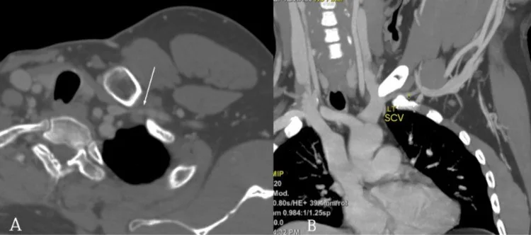
- Kontrastlı BT'de subklavyen ven trombozu veya oklüzyonu gösterilebilir; kronik venöz obstrüksiyonda geniş kollateral venöz kanallar eşlik edebilir. Nötr ve kol elevasyonlu seriler karşılaştırılarak istirahatte görülmeyen dinamik venöz darlık değerlendirilebilir; görüntüleme bulguları klinik belirtilerle birlikte yorumlanmalıdır.
Önerilen pencereler G genişlik, M merkez; değerler Hounsfield birimi (HU)
- MediastenG 350 / M +50
- KemikG 1800 / M +400
- Yumuşak dokuG 400 / M +40
BT bulguları
- Subklavyen ven kostoklaviküler aralıkta, klavikula ile birinci kosta arasında fokal yassılaşır veya incelir; daralma omuz elevasyonunda belirginleşebilir.
- Kontrastlı BT'de akut tromboz, subklavyen venlerde trombotik oklüzyon veya tıkanıklık olarak izlenebilir.
- Trombüs aksiller ve subklavyen venleri birlikte tutabilir.
- Kronik stenoz veya oklüzyonda segmental incelme, düzensiz duvar, kısmi rekanalizasyon ve omuz kuşağı çevresinde genişlemiş venöz kollateraller bulunabilir.
- Boyun, omuz veya göğüs duvarındaki belirgin venöz kollateraller venöz tıkanıklığın yerini ve kronikliğini değerlendirmeye yardımcı olabilir; kollateral yolların gelişmesi zaman alır.
- Birinci kosta, klavikula, subklavius kası ve kostoklaviküler bağın ilişkisi dar alanı açıklar; servikal kosta ya da eski travmaya bağlı kemik deformitesi ek anatomik daralma oluşturabilir.
- Kitleye bağlı sekonder venöz bası varsa akciğer apeksi, supraklaviküler yumuşak dokular ve mediastinal lenf düğümleri ayrıca taranır.
- Üst ekstremite derin ven trombozu şüphesinde ilk görüntüleme genellikle dupleks ultrasonografidir; ultrasonografi tanısal olmadığında BT veya MR venografi ek görüntüleme seçenekleri olarak kullanılabilir.
Ölçütler ve sınıflamalar
- Kolu abduksiyonda venöz çap azalması
- Kolu abduksiyonda dinamik inceleme, subklavyen vende istirahatte belirgin olmayan hemodinamik olarak anlamlı darlığı gösterebilir; görüntüleme bulguları tanıda klinik belirtilerle birlikte değerlendirilmelidir.
Normalde
Normal nötr pozisyonda subklavyen ven klavikula ve birinci kosta arasında açık lümenli, kesintisiz bir damar olarak brakiyosefalik vene uzanır; belirgin omuz kuşağı kollateralleri beklenmez. Dinamik görüntüleme, istirahatte belirgin olmayan venöz darlığı gösterebilir; bulgular klinik belirtilerle birlikte yorumlanmalıdır.
Normal BT ile kıyasla
Aynı bölgenin, kaynak veri setinde patoloji içermediği belirtilen bir BT incelemesini kesit kesit kaydırın; organ sınırlarını açarak anatomiyi hasta görüntüsüyle karşılaştırın. Küçük rastlantısal bulgular tümüyle dışlanmamıştır.
Kaynak: TotalSegmentator v2.01 veri seti (Wasserthal ve ark., Radiology: Artificial Intelligence 2023), CC BY 4.0, vaka s0975; organ sınırları veri setinin otomatik segmentasyonundan, birleştirilerek sadeleştirildi. Görüntüler 2,5 mm kesit aralığına yeniden örneklendi.
Ayırıcı tanı
- Kateter ilişkili üst ekstremite derin ven trombozu, üst ekstremite derin ven trombozunun sekonder nedenlerindendir; Paget–Schroetter sendromu ise torasik çıkıştaki anatomik basıyla ilişkili primer trombozdur.
- Santral venöz kateter veya kalp pili elektrotlarına bağlı venöz tıkanıklık subklavyen, brakiyosefalik ya da üst vena kava düzeyinde görülebilir.
- Pancoast tümörü
- akciğer apeksinde yumuşak doku kitlesi birinci kosta/pleksus komşuluğunda invazyon veya venöz bası yapar.
- Mediastinal lenfoma
- çoklu büyümüş lenf düğümleri ve mediastinal yumuşak doku kitlesi santral venleri dıştan sarabilir.
- Üst ekstremite derin ven trombozu kateter, malignite veya torasik çıkış basısı gibi farklı nedenlerle gelişebilir; Paget–Schroetter sendromu eforla ilişkili primer aksiller-subklavyen ven trombozudur.
Tuzaklar
- Kol elevasyonunda görülen venöz darlığı tek başına Paget–Schroetter sendromu olarak adlandırma; görüntüleme bulgularını klinik belirtilerle birlikte değerlendir.
- Subklavyen venin klavikula ile birinci kosta arasındaki kostoklaviküler aralıktan geçtiği segment, bu anatomik sınırlar dikkate alınarak değerlendirilmelidir.
- Üst ekstremite BT anjiyografisinde artefaktlar damar bulgularını taklit edebilir veya maskeleyebilir; değerlendirme sırasında bu olasılık göz önünde bulundurulmalıdır.
- Venöz kollateral yolların gelişmesi genellikle zaman alır.
Kendini dene
Yoğun üst ekstremite egzersizi sonrası tek kolda şişlik gelişen hastada kontrastlı BT'de aksiller-subklavyen ven trombozu görülüyor. En olası tanı nedir?
Cevabı göster
Paget–Schroetter sendromu. Egzersiz sonrası üst ekstremitede gelişen aksiller-subklavyen ven trombozu, efor trombozu olarak da bilinen Paget–Schroetter sendromuyla uyumludur. Kateter ilişkili tromboz santral intravasküler kateterlerle ilişkilidir; Pancoast tümörü ise akciğer apeksinden subklavyen veni invaze ederek oklüde edebilir.
Venöz torasik çıkış sendromunda kostoklaviküler aralıktan geçen ve basıya uğrayabilen damar hangisidir?
Cevabı göster
Subklavyen ven. Venöz torasik çıkış sendromu, kostoklaviküler aralıktan geçen subklavyen venin dıştan basısıyla ilişkilidir.
Kaynaklar
Bu sayfadaki 37 cümle ve 2 quiz sorusu aşağıdaki kaynaklardan alıntılarla otomatik olarak karşılaştırıldı; 24 cümle bu karşılaştırmada düzeltildi (2026-10-09).
- Paget–Schrotter Syndrome in the Primary Care Setting: A Case Report and Scoping Review of Clinical Presentation and Diagnostic Delaypmc.ncbi.nlm.nih.gov
- An Unexpected Complication of Training: A Rare Case of Paget-Schroetter Syndrome in an Adolescent Athletepmc.ncbi.nlm.nih.gov
- A Case of Paget-Schroetter Syndrome Successfully Treated With Endovascular Treatmentpmc.ncbi.nlm.nih.gov
- Spontaneous upper extremity deep vein thrombosis as initial manifestation of a malignant mediastinal teratomapmc.ncbi.nlm.nih.gov
- Upper extremity deep vein thrombosis associated with systemic lupus erythematosus and antiphospholipid syndromepmc.ncbi.nlm.nih.gov
- Upper Extremity Deep Vein Thrombosis: Symptoms, Diagnosis, and Treatment.pubmed.ncbi.nlm.nih.gov
- Paget-Schroetter Syndrome Resulting from Thoracic Outlet Syndrome and KAATSU Training.pubmed.ncbi.nlm.nih.gov
- Point-of-Care Ultrasound in the Diagnosis of Venous Thoracic Outlet Syndrome.pubmed.ncbi.nlm.nih.gov
- Venous Thoracic Outlet Syndrome with an Upper Extremity Deep Vein Thrombosis Caused by a Dislocated Clavicle Fracture: A Case Report.pubmed.ncbi.nlm.nih.gov
- Chronic non-bacterial osteitis presenting with upper limb deep vein thrombosis: diagnostic clues and therapeutic considerations.pubmed.ncbi.nlm.nih.gov
- Endovascular treatment of acute upper extremity deep vein thrombosis in Paget-Schroetter syndrome: A longitudinal analysis of three endovascular techniques.pubmed.ncbi.nlm.nih.gov
- Subacute Paget-Schroetter with rapid recurrence and concurrent pulmonary emboli: a case report.pubmed.ncbi.nlm.nih.gov
- An Unexpected Complication of Training: A Rare Case of Paget-Schroetter Syndrome in an Adolescent Athlete.pubmed.ncbi.nlm.nih.gov
- Anatomical Reasons for an Impaired Internal Jugular Flow.pubmed.ncbi.nlm.nih.gov
- Central vein stenosis in hemodialysis vascular access: clinical manifestations and contemporary management strategies.pubmed.ncbi.nlm.nih.gov
- Venous thoracic outlet syndrome.pubmed.ncbi.nlm.nih.gov
- Thoracic Outlet Syndrome in the Overhead Athlete: Diagnosis and Treatment Recommendations.pubmed.ncbi.nlm.nih.gov
- Imaging of venous compression syndromes.pubmed.ncbi.nlm.nih.gov
- Upper Extremity Compressive Neuropathies in the Pediatric and Adolescent Populations.pubmed.ncbi.nlm.nih.gov
- Paget-Schrotter Syndrome in the Primary Care Setting: A Case Report and Scoping Review of Clinical Presentation and Diagnostic Delay.pubmed.ncbi.nlm.nih.gov
- Delayed Diagnosis of Congenital Heart Diseases in Adults: Opportunities for Improving Diagnostic Strategies.pubmed.ncbi.nlm.nih.gov
- Venous Thromboembolism in Neonates, Children, and Adolescents: A Comprehensive Narrative Review of Risk Factors, Diagnosis, Treatment, and Prevention.pubmed.ncbi.nlm.nih.gov
- Acute Innominate Vein Dissection During Pacemaker Revision Procedure: A Case Report and Conservative Management.pubmed.ncbi.nlm.nih.gov
- When Cancer Clots: An Extensive Radiologic Analysis of Cancer-Associated Thromboembolism.pubmed.ncbi.nlm.nih.gov
- Systemic Venous Recanalization: Iliocaval and Superior Vena Cava Reconstruction in Children.pubmed.ncbi.nlm.nih.gov
- UK Kidney Association Clinical Practice Guideline on vascular access for haemodialysis.pubmed.ncbi.nlm.nih.gov
- Long-term safety and effectiveness of the "OptEase" vena cava filter.pubmed.ncbi.nlm.nih.gov
- Multimodality Imaging of Thoracic Outlet Syndrome: Etiological and Anatomical Correlates.pubmed.ncbi.nlm.nih.gov
- Case Report: HHS-spectrum distal palmar arterial thrombosis in a volleyball athlete with provocative-position thoracic outlet narrowing.pubmed.ncbi.nlm.nih.gov
- Rare Presentation of Thoracic Outlet Syndrome with First Rib Fracture - Case Report.pubmed.ncbi.nlm.nih.gov
- Thoracic Complications in Behçet's Disease: Imaging Findings.pubmed.ncbi.nlm.nih.gov
- 2025 Heart Disease and Stroke Statistics: A Report of US and Global Data From the American Heart Association.pubmed.ncbi.nlm.nih.gov
- Sharp recanalization of extensive multilevel central venous occlusions in a young patient with familial Mediterranean fever: A case report.pubmed.ncbi.nlm.nih.gov
- Ultrasound-guided hydrodissection of the subclavius muscle region in neurogenic thoracic outlet syndrome: a case report.pubmed.ncbi.nlm.nih.gov
- The American Venous Forum clinical practice guideline on the care of patients with upper extremity deep venous thrombosis.pubmed.ncbi.nlm.nih.gov
- Single-center experience in hybrid approach for subclavian vein effort thrombosis.pubmed.ncbi.nlm.nih.gov
- Operative Procedures for Ultrasound Assessment of Extracranial Artery Disease: A Narrative Review by the Italian Society for Vascular Investigation (SIDV).pubmed.ncbi.nlm.nih.gov
- Understanding Local Hemodynamic Changes After Liver Transplant: Different Entities or Simply Different Sides to the Same Coin?pubmed.ncbi.nlm.nih.gov
- Embolization in Pediatric Patients: A Comprehensive Review of Indications, Procedures, and Clinical Outcomes.pubmed.ncbi.nlm.nih.gov
- Subclavian Vein Thrombosispubmed.ncbi.nlm.nih.gov
- Anatomy, Thorax, Subclavian Veinspubmed.ncbi.nlm.nih.gov
- Asymptomatic central venous occlusion secondary to central venous catheter-use complicating pacemaker implantation: a leadless solution.pubmed.ncbi.nlm.nih.gov
- Pacemaker Lead-Associated Innominate Vein Thrombosis Despite Non-vitamin K Oral Anticoagulant (NOAC) Therapy: An Incidental CT Finding.pubmed.ncbi.nlm.nih.gov
- Angioplasty in Patients with Central Venous Occlusion Prior to Device Lead Implantation.pubmed.ncbi.nlm.nih.gov
- Marked Leukocytosis and Subclavian Vein Occlusion from Pancoast Tumor Mimicking Cellulitis: A Case Report.pubmed.ncbi.nlm.nih.gov
- Pancoast tumors: characteristics and preoperative assessment.pubmed.ncbi.nlm.nih.gov
- Therapeutic modalities for Pancoast tumors.pubmed.ncbi.nlm.nih.gov
- Refractory chylothorax caused by massive thymic hyperplasia in an infant: thoracoscopic thymectomy.pubmed.ncbi.nlm.nih.gov
- A Massive Cavernous Mediastinal Haemangioma Causing Superior Vena Cava Obstruction and Extending to the Supraclavicular Space: A Case Report.pubmed.ncbi.nlm.nih.gov
- Thoracic Outlet Syndrome Part I: Systematic Review of the Literature and Consensus on Anatomy, Diagnosis, and Classification of Thoracic Outlet Syndrome by the European Association of Neurosurgical Societies' Section of Peripheral Nerve Surgery.pubmed.ncbi.nlm.nih.gov
- CT angiography: current technology and clinical use.pubmed.ncbi.nlm.nih.gov
- Patterns of injury and violence in Yaoundé Cameroon: an analysis of hospital data.pubmed.ncbi.nlm.nih.gov
- Mitigating limb ischemia in veno-arterial extracorporeal membrane oxygenation: A narrative review of evolving role of distal perfusion catheters.pubmed.ncbi.nlm.nih.gov
- Systematic Echocardiographic Assessment of the Univentricular Heart Across the Stages of Fontan Palliation: A Practical Guide with Focus on Emerging 3D and 4D Imaging Modalities and Thromboembolic Complications from AEPC Imaging Working Group.pubmed.ncbi.nlm.nih.gov
- Giant cervicothoracic malignant peripheral nerve sheath tumor in neurofibromatosis type 1: a case report of multidisciplinary management and literature review.pubmed.ncbi.nlm.nih.gov
- Venous compression syndromes: clinical features, imaging findings and management.pubmed.ncbi.nlm.nih.gov
- International Society of Paediatric Surgical Oncology (IPSO) Surgical Practice Guidelines.pubmed.ncbi.nlm.nih.gov
- Cardiothoracic Applications of 3-dimensional Printing.pubmed.ncbi.nlm.nih.gov
- A Case of Paget-Schroetter Syndrome Successfully Treated With Endovascular Treatment.pubmed.ncbi.nlm.nih.gov
- Operative management of vascular thoracic outlet syndrome (VTOS) over two decades: patency, complications, and functional outcome.pubmed.ncbi.nlm.nih.gov
- Lead-Related Venous Obstruction in Patients With Implanted Cardiac Devices: JACC Review Topic of the Week.pubmed.ncbi.nlm.nih.gov
- Blood finds a way: pictorial review of thoracic collateral vessels.pubmed.ncbi.nlm.nih.gov
- 2.5-mm articulated endoluminal forceps using a compliant mechanism.pubmed.ncbi.nlm.nih.gov
- CT Angiography of the Upper Extremities: Review of Acute Arterial Entities.pubmed.ncbi.nlm.nih.gov
- Diagnostic and Therapeutic Approach to Thoracic Outlet Syndrome.pubmed.ncbi.nlm.nih.gov
- Improved image quality and micronodule detection in thyroid spectral computed tomography using modified swimmer's position.pubmed.ncbi.nlm.nih.gov
- The spectrum of computed tomography findings in blunt trauma of the subclavian/axillary artery: a pictorial essay.pubmed.ncbi.nlm.nih.gov
- Iatrogenic percutaneous vascular injuries: clinical presentation, imaging, and management.pubmed.ncbi.nlm.nih.gov
- Imaging Spectrum of Thoracic Outlet Syndrome: A Case Series Highlighting Arterial, Venous, and Neurogenic Subtypes.pubmed.ncbi.nlm.nih.gov
- Intermittent upper limb cyanosis revealing Paget-Schroetter syndrome: the value of patient-provided photographs.pubmed.ncbi.nlm.nih.gov
- An Overview of Vascular Compression Syndromes and Associations with Autonomic Dysfunction: A Review.pubmed.ncbi.nlm.nih.gov
- Paget-Schroetter Syndrome (PSS) and Adjunctive Treatment With Mechanical Aspiration Thrombectomy and Catheter Directed Thrombolysis: A Case Report and Review of Literature.pubmed.ncbi.nlm.nih.gov
- Paget-Schroetter syndrome: An unfamiliar cause of upper-limb deep venous thrombosis.pubmed.ncbi.nlm.nih.gov
- Subclavian Effort Thrombosis: Pathophysiology, Diagnosis, and Management.pubmed.ncbi.nlm.nih.gov
- An Unusual Presentation of Paget-Schroetter Syndrome: A Reminder on the Importance of Clinical Judgment.pubmed.ncbi.nlm.nih.gov
- Thoracic Outlet Syndrome Case Report: Appropriate Diagnosis Can Expedite Patient Treatment and Prevent Negative Outcomes.pubmed.ncbi.nlm.nih.gov
- Robotic-assisted thoracoscopic surgery first rib resection-surgical technique.pubmed.ncbi.nlm.nih.gov
- Breaking the blockage by advancing treatment strategies for upper extremity deep vein thrombosis.pubmed.ncbi.nlm.nih.gov
- Current Clinical Concepts: Rehabilitation of Thoracic Outlet Syndrome.pubmed.ncbi.nlm.nih.gov
- Patient-reported outcomes after upper-extremity deep vein thrombosis due to compression syndromes: a multinational longitudinal retrospective study.pubmed.ncbi.nlm.nih.gov
- A 12-Year Review of Upper Extremity Deep Vein Thrombosis-Are They the Same as Lower Extremity Deep Vein Thrombosis?pubmed.ncbi.nlm.nih.gov
- Pediatric Venous Thromboembolism: Considerations for the Interventional Radiologist.pubmed.ncbi.nlm.nih.gov
- Utilizing Provocative Maneuvers Intraoperatively in Conjunction With IVUS to Diagnose vTOS: The New Gold Standard.pubmed.ncbi.nlm.nih.gov
- Participation of Arterial Ischemia in Positional-Related Symptoms among Patients Referred for Thoracic Outlet Syndrome.pubmed.ncbi.nlm.nih.gov
- Thoracic Outlet Syndrome: Single Center Experience on Robotic Assisted First Rib Resection and Literature Review.pubmed.ncbi.nlm.nih.gov
- Pulmonary Artery Thromboembolism (PATE) in Paediatrics: Three Clinical Case Presentations.pubmed.ncbi.nlm.nih.gov
- Chronic Venous Intervention Using a Large-Bore, Vacuum-Assisted Aspiration System: A New Era?pubmed.ncbi.nlm.nih.gov
- Contemporary management of superior vena cava syndrome.pubmed.ncbi.nlm.nih.gov
- Non-invasive Vascular Laboratory Evaluation of the Upper Extremity.pubmed.ncbi.nlm.nih.gov
- High-resolution three‑dimensional contrast‑enhanced magnetic resonance venography in children: comparison of gadofosveset trisodium with ferumoxytol.pubmed.ncbi.nlm.nih.gov
- Intracranial venous thrombosis imaging by anatomical location: an educational review with mimics and diagnostic pitfalls.pubmed.ncbi.nlm.nih.gov
- Thrombosis and Thrombotic Risk in Athletes.pubmed.ncbi.nlm.nih.gov
- SCREENING FOR REFERRAL BY A SPORTS PHYSICAL THERAPIST REVEALS AN EFFORT THROMBOSIS IN A COLLEGIATE PITCHER: A CASE REPORT.pubmed.ncbi.nlm.nih.gov
- MR Lymphangiography in Pediatrics: Indications, Technique, Findings, and Management.pubmed.ncbi.nlm.nih.gov
- Idiopathic upper extremity deep vein thrombosis: A systematic review.pubmed.ncbi.nlm.nih.gov
- Blood pool contrast agents for venous magnetic resonance imaging.pubmed.ncbi.nlm.nih.gov
- Clinical Applications of Dual-Energy CT.pubmed.ncbi.nlm.nih.gov
- Split-Bolus Single-Pass Multidetector-Row CT Protocol for Diagnosis of Acute Pulmonary Embolism.pubmed.ncbi.nlm.nih.gov
- CT and MR imaging findings of systemic complications occurring during pregnancy and puerperal period, adversely affected by natural changes.pubmed.ncbi.nlm.nih.gov
Eğitim amaçlıdır; hasta başında tanı ve tedavi kararının yerine geçmez, tıbbi tavsiye değildir. Metinler yapay zekâ desteğiyle yazıldı ve otomatik bir adımda kaynak alıntılarıyla karşılaştırıldı; bu bir tıbbi doğrulama değildir ve içerik uzman radyolog incelemesinden geçmedi. Klinik kararlarda güncel kılavuzları, kurum protokollerini ve radyoloji raporunu esas alın.