Özet
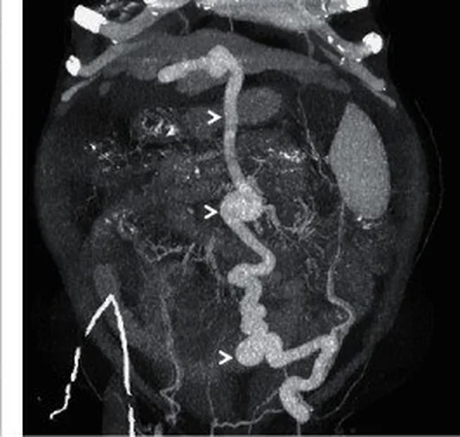
Spontan portosistemik şantlar, portal hipertansiyon sırasında portal venöz sistemi sistemik venöz dolaşıma bağlayan genişlemiş kollateral kanallardır; özellikle sirozlu hastalarda değerlendirilir. Portal venöz fazlı BT, şantın portal girişini ve sistemik çıkışını tüm güzergâhı boyunca göstererek doğrudan splenorenal bağlantıyı gastrik varisler üzerinden sol renal vene ulaşan gastrorenal yoldan ayırmaya yardımcı olur. Büyük şantlar hepatik ensefalopatiyle ilişkili olabilir; varis terimi tek başına portal ve sistemik dolaşımı birleştiren şant yolunu tanımlamaz. Şantın portal girişini, damar güzergâhını ve sistemik çıkışını birlikte tanımlayın; bu anatomik haritalama eşlik eden portal ven trombozu veya başka kollaterallerin değerlendirilmesine yardımcı olur.
Faz ve pencere
- Portal tanısal
- Portal venöz kontrastlanmada portal/splenik ven veya bu sistemden beslenen gastrik kollateraller ile renal ven, vena kava ya da epigastrik venler arasında kontrastla dolu, kıvrımlı venöz kanal izlenir; kanalın iki ucunu ve çevre varislerle ilişkisini aksiyel görüntüler ile koronal/sagittal MPR'de takip et.
Önerilen pencereler: Anjiyo (G 600 / M 150), Batın (G 400 / M 50), Karaciğer (G 150 / M 30).
BT bulguları
- Splenorenal şant — splenik ven veya dalak hilusu venöz kanallarından sol renal vene doğrudan uzanan geniş, kıvrımlı bağlantı.
- Gastrorenal şant — fundik gastrik varislerden sol renal vene uzanan kıvrımlı retroperitoneal venöz bağlantı.
- Paraumbilikal şant — sol portal ven dalından çıkarak falciform ligament ön kenarı boyunca umbilikusa uzanan ve epigastrik ya da internal thoracic venlerle anastomoz yapan rekanalize damar.
- Mezokaval veya mezorenal yol — mezenterik venöz dallardan retroperitoneal kollateraller aracılığıyla vena kava ya da renal vene ulaşan bağlantı.
- Portal venöz fazlı BT'de şant boyunca kontrastlanma ve damar devamlılığı değerlendirilir; portal giriş, gövde ve sistemik çıkış ayrı ayrı gösterilebilir.
- Şantla birlikte portal veya splenik ven lümeninde trombüs/darlık ve tıkanıklık çevresinde gelişmiş alternatif venöz yollar bulunabilir.
- Sirotik karaciğer morfolojisi, splenomegali, asit ve gastroözofageal ya da mezenterik varisler portal hipertansiyona eşlik edebilir.
Ölçütler ve sınıflamalar
- Büyük spontan portosistemik şant (derlemede kullanılan tanım)
- çap ≥8 mm
- Drenaj yollarına göre örnekler
- Anatomik örnekler arasında sol renal vene bağlanan splenorenal ve gastrorenal yollar ile retroperitoneal mezokaval, mezorenal ve mezogonadal yollar bulunur. Paraumbilikal yol sol portal daldan ön karın duvarı venlerine uzanır; bu adlandırma drenaj anatomisini tarif eder, klinik şiddeti tek başına ölçmez.
Normalde
Normal portal venöz anatomide splenik ven ile SMV pankreas boynu arkasında birleşerek portal veni oluşturur ve portal ven karaciğer hilusuna yönelir; hilus, sol renal ven, vena kava ya da epigastrik venler arasında geniş, kıvrımlı bir kollateral kanal beklenmez.
Normal BT ile kıyasla
Aynı bölgenin, kaynak veri setinde patoloji içermediği belirtilen bir BT incelemesini kesit kesit kaydırın; organ sınırlarını açarak anatomiyi hasta görüntüsüyle karşılaştırın. Küçük rastlantısal bulgular tümüyle dışlanmamıştır.
Kaynak: TotalSegmentator v2.01 veri seti (Wasserthal ve ark., Radiology: Artificial Intelligence 2023), CC BY 4.0, vaka s0119; organ sınırları veri setinin otomatik segmentasyonundan, birleştirilerek sadeleştirildi. Görüntüler 2,5 mm kesit aralığına yeniden örneklendi.
Ayırıcı tanı
- Portal kavernom
- kronik ana portal ven tıkanıklığı sonrası hilusta çok sayıda kıvrımlı portoportal kollateral gelişir; tek bir portal-sistemik çıkış kanalı değildir.
- İzole sol taraflı portal hipertansiyon
- kısa gastrik ve fundik varisler ön plandadır; splenik ven trombozu gösterilebilir ve karaciğer sirozu bulunmayabilir.
- Cerrahi splenorenal şant, spontan kollateral olarak adlandırılmamalıdır; bir çalışmada cerrahi şant varlığı spontan şant analizinin dışında tutulmuştur.
- Gonadal veya lumbar venöz kollateral
- retroperitoneal kollateral portal/splenik ya da mezenterik bir damardan sistemik venlere uzanabilir; portal besleyiciyle devamlılık gösterilmeden yalnız geniş bir sistemik ven portosistemik şant olarak adlandırılmaz.
- Portal ven anevrizması
- portal ven içinde fokal genişleme vardır; renal ven veya başka sistemik vene uzanan devamlı bir drenaj kanalı yoktur.
Tuzaklar
- Spontan splenorenal şant BT'de dalak ve sol renal hilus bölgesindeki geniş, kıvrımlı venlerin genişlemiş sol renal vene drenajıyla tanımlanır; damarlar çok kıvrımlı olduğunda bağlantının splenik ven üzerindeki kesin başlangıcı seçilemeyebilir.
- Gastrorenal şantı splenorenal şant diye adlandırmak drenaj yolunu değiştirir; gastrorenal tipte fundik varisler yolun parçasıyken splenorenal tipte splenik ven ile sol renal ven doğrudan birleşir.
- Şant kanalı tek kesitte izlenmiyorsa komşu aksiyel görüntülerde ve çok düzlemli rekonstrüksiyonlarda anatomik devamlılığı kontrol edin; görüntüdeki kesintiyi tek başına trombüs olarak yorumlamayın.
Kendini dene
Portal fazlı BT'de splenik ven ile sol renal ven arasında doğrudan geniş damar görülüyor; fundik varisler bu yola katılmıyor. En uygun ad nedir?
Cevabı göster
Splenorenal şant. Splenik ven ile sol renal venin doğrudan bağlantısı splenorenal şanttır. Gastrorenal yol mide fundusundaki varisleri içerir; portal kavernom hilus çevresinde portoportal kanallardan, paraumbilikal ven ise ön karın duvarına uzanır.
Sol portal dalından çıkan damar ligamentum teres boyunca umbilikusa ve epigastrik venlere ulaşıyor. Bu bağlantı hangi kollateraldir?
Cevabı göster
Paraumbilikal şant. Sol portal dalından umbilikus üzerinden epigastrik venlere uzanan yol paraumbilikal şanttır. Gastrorenal ve mezokaval yolların çıkışları farklıdır; portal kavernom ise portal hilusta portoportal damar ağı biçimindedir.
İlgili konular
Kaynaklar
Bu sayfadaki 38 cümle ve 2 quiz sorusu aşağıdaki kaynaklardan alıntılarla tek tek karşılaştırıldı; 3 cümle bu karşılaştırmada düzeltildi (2026-10-09).
- Portal hypertension: Imaging of portosystemic collateral pathways and associated image-guided therapypmc.ncbi.nlm.nih.gov
- Clinical Implications and Management of Spontaneous Portosystemic Shunts in Liver Cirrhosispmc.ncbi.nlm.nih.gov
- Closing spontaneous portosystemic shunts in cirrhosis: Does it make sense? Does it work? What does it take?pubmed.ncbi.nlm.nih.gov
- Clinical Implications and Management of Spontaneous Portosystemic Shunts in Liver Cirrhosis.pubmed.ncbi.nlm.nih.gov
- Pathophysiological management of gastric varices: From hemodynamics to targeted therapies.pubmed.ncbi.nlm.nih.gov
- Management of Large, Spontaneous Portosystemic Shunts in Liver Transplantation: Case Report and Review of Literature.pubmed.ncbi.nlm.nih.gov
- Imaging of spontaneous portosystemic shunts in portal hypertension: clinical significance and structured reporting.pubmed.ncbi.nlm.nih.gov
- Management of Portal vein Thrombosis in Cirrhosis.pubmed.ncbi.nlm.nih.gov
- Clip-Assisted Endoscopic Cyanoacrylate Injection: A Novel Technique for Acute Gastroesophageal Variceal Bleeding.pubmed.ncbi.nlm.nih.gov
- Clinical Outcomes of Portosystemic Shunts on the Outcome of Liver Transplantation.pubmed.ncbi.nlm.nih.gov
- Spontaneous portosystemic shunts in cirrhosis: Detection, implications, and clinical associations.pubmed.ncbi.nlm.nih.gov
- Association Between Portosystemic Shunts and Increased Complications and Mortality in Patients With Cirrhosis.pubmed.ncbi.nlm.nih.gov
- Simultaneous embolization of a spontaneous porto-systemic shunt and intrahepatic arterioportal fistula: A case report.pubmed.ncbi.nlm.nih.gov
- Development and validation of machine learning models to predict esophagogastric variceal rebleeding risk in HBV-related cirrhosis after endoscopic treatment: a prospective multicenter study.pubmed.ncbi.nlm.nih.gov
- Friend or Foe? Spontaneous Portosystemic Shunts in Cirrhosis-Current Understanding and Future Prospects.pubmed.ncbi.nlm.nih.gov
- Can Preoperative Multidetector Computed Tomography Identify Predictive Features of Difficult Native Hepatectomy at Liver Transplantation?pubmed.ncbi.nlm.nih.gov
- Reversal of hepatic encephalopathy following a trans-scrotal approach to mesogonadal shunt embolization: a case report.pubmed.ncbi.nlm.nih.gov
- Clinical applications of 4D flow magnetic resonance imaging in hepatic vascular disorders: practical biomarkers, protocol design, and translational implementation.pubmed.ncbi.nlm.nih.gov
- Congenital Extrahepatic Portosystemic Shunts: Spectrum of Findings on Ultrasound, Computed Tomography, and Magnetic Resonance Imaging.pubmed.ncbi.nlm.nih.gov
- A 7-mm Covered TIPS Reduces Hepatic Encephalopathy Without Increasing Rebleeding in Cirrhotic Patients With Small Liver: A Randomized Study.pubmed.ncbi.nlm.nih.gov
- Radiologic Interventions for Gastrointestinal Bleeding.pubmed.ncbi.nlm.nih.gov
- Portal vein recanalization and transjugular intrahepatic portosystemic shunt in chronic portal vein thrombosis: a narrative review.pubmed.ncbi.nlm.nih.gov
- Right portal branch to ovarian vein anastomosis: An unusual portosystemic shunt secondary to pelvic venous congestion.pubmed.ncbi.nlm.nih.gov
- Contemporary sequential segmental approach to congenital heart disease using four-dimensional magnetic resonance imaging with ferumoxytol: an illustrated editorial.pubmed.ncbi.nlm.nih.gov
- The Role of Imaging in Portal Vein Thrombosis: From the Diagnosis to the Interventional Radiological Management.pubmed.ncbi.nlm.nih.gov
- Preoperative vascular parameters and proximal splenorenal shunt patency in pediatric extrahepatic portal vein thrombosis.pubmed.ncbi.nlm.nih.gov
- Percutaneous retroperitoneal splenorenal shunt creation after failed PVR-TIPS attempt.pubmed.ncbi.nlm.nih.gov
- Efficacy and safety of EUS-guided coil embolization in combination with cyanoacrylate injection versus conventional endoscopic cyanoacrylate injection in the treatment of gastric varices with spontaneous portosystemic shunts.pubmed.ncbi.nlm.nih.gov
- Endoscopic ultrasound in portal hypertension: navigating venous hemodynamics and treatment efficacy.pubmed.ncbi.nlm.nih.gov
- A Comprehensive Review of Portosystemic Collaterals in Cirrhosis: Historical Aspects, Anatomy, and Classifications.pubmed.ncbi.nlm.nih.gov
- Navigating Portal Hypertension and Splenomegaly: A Clinical Encounter With an Unusual Variant of Cruveilhier-Baumgarten Syndrome.pubmed.ncbi.nlm.nih.gov
- All about portal vein: a pictorial display to anatomy, variants and physiopathology.pubmed.ncbi.nlm.nih.gov
- Non-variceal Extrahepatic Portosystemic Shunts: A Review of Pathogenesis, Diagnosis, and Treatment.pubmed.ncbi.nlm.nih.gov
- Contrast-Enhanced CT Finding of Hot Spot Sign Secondary to Supra Hilar Mass Causing Superior Vena Cava Syndrome: A Case Report.pubmed.ncbi.nlm.nih.gov
- Umbilical vein recanalization with a diameter greater than 6.48mm may promote deterioration of liver reserve function: A retrospective cohort study.pubmed.ncbi.nlm.nih.gov
- Gastric varices: a contemporary review of vascular anatomy and therapeutic strategies.pubmed.ncbi.nlm.nih.gov
- Use of a recanalized umbilical vein to embolize bleeding varices: A case report.pubmed.ncbi.nlm.nih.gov
- Budd-Chiari Syndrome Manifesting in Pregnancy: Case Report and Review of Management and Outcomes.pubmed.ncbi.nlm.nih.gov
- Ultra-Large Spontaneous Portosystemic Shunt is Correlated With a Higher Hepatic Venous Pressure Gradient and Increased Risk of Hepatic Encephalopathy.pubmed.ncbi.nlm.nih.gov
- Risk factors for hepatic encephalopathy in patients following transjugular intrahepatic portosystemic shunt: a meta-analysis and systematic review.pubmed.ncbi.nlm.nih.gov
- Preoperative CT-based 3D analysis of middle colic vein anatomy for complete mesocolic excision in right-sided colon cancer: a retrospective cohort study from a prospective clinical trial.pubmed.ncbi.nlm.nih.gov
- Oral food challenge unmasks porto-systemic shunting: a case report of a novel provocation test for congenital porto-systemic shunts.pubmed.ncbi.nlm.nih.gov
- Type Ib Abernethy Malformation Complicated by Hepatopulmonary Syndrome: A Case Report of a Rare Entity in a Pediatric Patient.pubmed.ncbi.nlm.nih.gov
- Splenic infarction after endoscopic glue injection for gastric varices: proportion, exploratory correlates, and clinical outcomes from a propensity score-matched analysis.pubmed.ncbi.nlm.nih.gov
- Long-term outcome of percutaneous transhepatic venous embolization for ectopic peristomal variceal bleeding: A case report.pubmed.ncbi.nlm.nih.gov
- Understanding Portosystemic Collateral Anatomy.pubmed.ncbi.nlm.nih.gov
- Septic Pylephlebitis and Cavernous Transformation Presenting as Pyrexia of Unknown Origin: A Diagnostic Challenge.pubmed.ncbi.nlm.nih.gov
- Portal Vein Invasion Secondary to Hepatic Hydatid Disease: A Rare Case Report.pubmed.ncbi.nlm.nih.gov
- Pseudotumoral portal cavernoma mimicking hilar cholangiocarcinoma: A diagnostic pitfall.pubmed.ncbi.nlm.nih.gov
- A Rare Pediatric Intraductal Papillary Mucinous Neoplasm Presenting as a Pancreatic Pseudocyst With Splenic Vein Thrombosis and Gastric Variceal Hemorrhage.pubmed.ncbi.nlm.nih.gov
- Massive Upper Gastrointestinal Bleeding From Ruptured Gastric Varices: A Rare Presentation of Type 1 Autoimmune Pancreatitis.pubmed.ncbi.nlm.nih.gov
- Long-Term Renal Outcomes Following Left Renal Vein Ligation Versus Direct Splenorenal Shunt Ligation in Living Donor Liver Transplantation: A 10-Year Single-Center Study.pubmed.ncbi.nlm.nih.gov
- Portal Vein Thrombosis, Management and Approaches in Liver Transplantation: A Narrative Review.pubmed.ncbi.nlm.nih.gov
- Computed Tomography Images of Spontaneous Portosystemic Shunt in Liver Cirrhosis.pubmed.ncbi.nlm.nih.gov
- Chylothorax after patent ductus arteriosus ligation in a dog with persistent left cranial vena cava.pubmed.ncbi.nlm.nih.gov
- Hemobilia: A Narrative Review of Current Diagnostic Techniques and Emerging Management Strategies.pubmed.ncbi.nlm.nih.gov
- Portal vein aneurysm-etiology, multimodal imaging and current management.pubmed.ncbi.nlm.nih.gov
- Society for Cardiovascular Magnetic Resonance/European Society of Cardiovascular Imaging/American Society of Echocardiography/Society for Pediatric Radiology/North American Society for Cardiovascular Imaging Guidelines for the Use of Cardiac Magnetic Resonance in Pediatric Congenital and Acquired Heart Disease: Endorsed by The American Heart Association.pubmed.ncbi.nlm.nih.gov
- Society for Cardiovascular Magnetic Resonance/European Society of Cardiovascular Imaging/American Society of Echocardiography/Society for Pediatric Radiology/North American Society for Cardiovascular Imaging Guidelines for the use of cardiovascular magnetic resonance in pediatric congenital and acquired heart disease : Endorsed by The American Heart Association.pubmed.ncbi.nlm.nih.gov
- Case report: A rare case of IgA nephropathy associated with type II Abernethy malformation.pubmed.ncbi.nlm.nih.gov
- Congenital Portosystemic Shunts: A Review.pubmed.ncbi.nlm.nih.gov
- Management Strategies for Refractory Esophageal Varices.pubmed.ncbi.nlm.nih.gov
- Collaterals in portal hypertension: anatomy and clinical relevance.pubmed.ncbi.nlm.nih.gov
- Portosystemic shunts and refractory hepatic encephalopathy: patient selection and current options.pubmed.ncbi.nlm.nih.gov
- Case Report: Varyingand Unique Symptomatic Presentations of Congenital Intrahepatic Portosystemic Venous Shunts.pubmed.ncbi.nlm.nih.gov
- Clip-assisted endoscopic cyanoacrylate injection <i>vs</i> endoscopic ultrasound-guided coil and cyanoacrylate injection for gastric varices: A propensity score-matched study.pubmed.ncbi.nlm.nih.gov
- Non-tumoral portal vein thrombosis in liver transplantation: Surgical perspectives and institutional protocol.pubmed.ncbi.nlm.nih.gov
- Portosystemic Shunt Surgery for Extrahepatic Portal Venous Obstruction Beyond Endoscopic Variceal Eradication: Two Decades of Pediatric Surgical Experience.pubmed.ncbi.nlm.nih.gov
- EUS-Guided Vascular Interventions: Recent Advances.pubmed.ncbi.nlm.nih.gov
- Partial Splenic Artery Embolization for the Management of Symptomatic Hypersplenism in Portal Hypertension: Clinical Insights from a Case Series.pubmed.ncbi.nlm.nih.gov
- Management of Preoperative Recipient Portal Vein Thrombosis in Living-donor Liver Transplantation.pubmed.ncbi.nlm.nih.gov
- Infarcts and ischemia in the abdomen: an imaging perspective with an emphasis on cross-sectional imaging findings.pubmed.ncbi.nlm.nih.gov
- Developmental Anomalies of the Inferior Vena Cava and its Tributaries: What the Radiologist Needs to Know?pubmed.ncbi.nlm.nih.gov
- Portosystemic shunt surgery in the era of TIPS: imaging-based planning of the surgical approach.pubmed.ncbi.nlm.nih.gov
- Hepatic encephalopathy due to non-cirrhotic portal hypertension associated with non-alcoholic steatohepatitis and chronic hepatitis B: a Case Report.pubmed.ncbi.nlm.nih.gov
- Porto-sinusoidal Vascular Disease: Classification and Clinical Relevance.pubmed.ncbi.nlm.nih.gov
- Transjugular intrahepatic portosystemic stent-shunt in the management of portal hypertension.pubmed.ncbi.nlm.nih.gov
- Patterns of injury and violence in Yaoundé Cameroon: an analysis of hospital data.pubmed.ncbi.nlm.nih.gov
Eğitim amaçlıdır; hasta başında tanı ve tedavi kararının yerine geçmez. İçerik yapay zekâ destekli bir süreçle güncel literatüre göre cümle cümle kaynaklarla karşılaştırıldı, ancak uzman radyolog incelemesinden geçmedi. Klinik kararlarda güncel kılavuzları, kurum protokollerini ve radyoloji raporunu esas alın.