
Özet
VCI veya iliak ven içinde görülen kontrastlanmayan alanlar her zaman trombüs değildir; bu bulgular sıklıkla yalancı lezyonlardır ve doğru tanı için ayırıcı tanıda dikkate alınmalıdır. Gerçek tromboz ile yalancı lezyonların ayrımı kritiktir; yanlış tanı yönetim stratejisini doğrudan değiştirir ve klinik sonuçları etkiler. Şüpheli VCI bulguları; damar anatomisi, ardışık kesitler ve çok fazlı kontrastlı BT değerlendirmesiyle yalancı lezyonlardan ayırt edilmelidir. Karışım artefaktını trombüs diye raporlamak gereksiz ek incelemeye, gerçek kalıcı trombüsün artefakt sanılması ise tanıda gecikmeye yol açabilir.
Faz ve pencere
- Portal tanısal
- VCI'de görülen her dolum kusuru trombüs değildir; VCI yalancı lezyonları ayırıcı tanıda dikkate alınmalıdır.
- Gecikmiş tanısal
- Gecikmiş postkontrast görüntüde kan kolonunun homojenleşmesi karışım artefaktını destekler; aynı bölgede kalıcı, duvara yapışık kontrastlanmayan defekt gerçek trombüs kuşkusunu sürdürür.
Önerilen pencereler: Batın (G 400 / M 50), Yumuşak doku (G 400 / M 40).
BT bulguları
- VCI lümeninde erken postkontrast görüntüde yoğun opak kan ile daha az opak kan arasında keskin sınır veya tabakalanma
- VCI'deki şüpheli dolum alanları değerlendirilirken yalancı lezyon olasılığı dikkate alınmalıdır.
- Gecikmiş görüntüde önceki dolum farklılığının kaybolması ve VCI'nin homojen opaklaşması
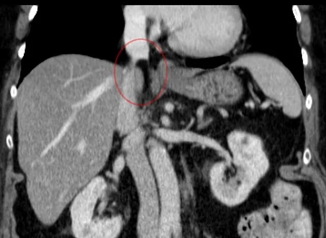
- VCI çevresindeki anatomik yapılar, aksiyel düzlemde lümen içi patoloji ile karışabilir; çoklu düzlem rekonstrüksiyonlar bu ayrımı sağlar.
- VCI çevresindeki yapılar yalancı lezyon olasılığı açısından değerlendirilmelidir.
- Gerçek trombüs lehine, gecikmiş görüntüde de süren ve damar duvarına oturan kontrastlanmayan intraluminal materyal
Normalde
VCI lümen opaklığındaki faz değişimleri ve çevre yapı ilişkileri, çokfazlı ve çoklu düzlem görüntüleme ile birlikte değerlendirilmelidir. Şüpheli venöz bulgular, komşu kesitler, ortogonal düzlemler ve gecikmiş seri ile karşılaştırılarak yalancı lezyonlardan ayırılmalıdır.
Normal BT ile kıyasla
Aynı bölgenin, kaynak veri setinde patoloji içermediği belirtilen bir BT incelemesini kesit kesit kaydırın; organ sınırlarını açarak anatomiyi hasta görüntüsüyle karşılaştırın. Küçük rastlantısal bulgular tümüyle dışlanmamıştır.
Kaynak: TotalSegmentator v2.01 veri seti (Wasserthal ve ark., Radiology: Artificial Intelligence 2023), CC BY 4.0, vaka s0119; organ sınırları veri setinin otomatik segmentasyonundan, birleştirilerek sadeleştirildi. Görüntüler 2,5 mm kesit aralığına yeniden örneklendi.
Ayırıcı tanı
- Bland VCI trombozu
- kontrastlı serilerde, özellikle gecikmiş görüntülerde bile kalıcı intraluminal dolum kusuru yapar ve homojenleşmez.
- Tümör trombüsü
- komşu tümörle süreklilik, damar lümeninde genişleme ve trombüs içinde kontrastlanma görülebilir; basit akım karışımı gecikmiş seride kaybolur.
- VCI'ye bası yapan kitle
- lümen içinde defekt yerine damar ekseninde itilme, düzleşme veya daralma yapar; böbrek, karaciğer, retroperitoneum ve pelviste bası kaynağı aranır.
- Komşu retroperitoneal yağ
- aksiyel kesitte lümene projekte olabilir, ancak ardışık kesit ve ortogonal düzlemde VCI duvarının dışında izlenir.
- Kontrastlanmayan damar içi cihaz veya cerrahi materyal
- yerleşik filtre/greft bileşenleri kesintili lümen izlenimi oluşturabilir; cihaz geometrisi ve metal artefaktı ile tanınır.
Tuzaklar
- Portal fazdaki iki tonlu kan kolonu gerçek trombüs gibi görünebilir; gecikmiş seride VCI opaklığı homojenleşiyorsa erken dolum kusurunu tromboz diye adlandırma.
- VCI'deki şüpheli dolum alanlarında yalancı lezyon olasılığını göz önünde bulundur.
- Sağ kalp yetmezliği gibi durumlarda venöz dolgunluk ve akım paternleri değişebilir; bu durumlar VCI değerlendirilirken klinik bağlamla ilişkilendirilmelidir.
- VCI çevresindeki yumuşak doku ve yağ yapıları, lümen içi patolojilerle karıştırılmamalı; çoklu düzlem rekonstrüksiyonlar bu ayrımı netleştirir.
Kendini dene
Portal fazda renal venlerin hemen kaudalinde VCI içinde keskin sınırlı iki yoğunluklu kan kolonu görülüyor; gecikmiş seride lümen eşit opaklaşıyor. En olası açıklama nedir?
Cevabı göster
Akım karışım artefaktı. Gecikmiş seride dolum farklılığının tamamen kaybolması geçici kan-kontrast karışımını destekler. Bland trombüs kalıcı intraluminal defekt yapar; tümör trombüsü genellikle kitleyle süreklidir ve kontrastlanabilir; leiomyosarkom damar duvarından kaynaklanan kitle görünümündedir.
VCI görüntülemede yanlış tanıyı önlemek ve doğru yönetim planı oluşturmak açısından hangi bulgunun ayırt edilmesi kritik önem taşır?
Cevabı göster
VCI yalancı lezyonları (pseudo-lesion). Doğru ayırım, gereksiz antikoagülasyon veya invaziv girişimin önlenmesi ile hayatı tehdit eden trombozun zamanında tedavisini sağlar.
Kaynaklar
Bu sayfadaki 35 cümle ve 2 quiz sorusu aşağıdaki kaynaklardan alıntılarla tek tek karşılaştırıldı; 19 cümle bu karşılaştırmada düzeltildi (2026-10-09).
- Intracranial venous thrombosis imaging by anatomical location: an educational review with mimics and diagnostic pitfallspmc.ncbi.nlm.nih.gov
- Abstracts from the 11th Annual Meeting of Arthroplasty Society In Asia (ASIA), the 4th Annual Meeting of the Malaysian Society for Hip and Knee Surgeons (MSHKS), and the 17th Annual Meeting of the ASEAN Arthroplasty Association (AAA)pmc.ncbi.nlm.nih.gov
- SNMICON 2024 Abstractspmc.ncbi.nlm.nih.gov
- Abstracts - IANCON 2024pmc.ncbi.nlm.nih.gov
- Additively manufactured metallic biomaterialspmc.ncbi.nlm.nih.gov
- The Initial Diagnostic Approach to Adult Congenital Heart Disease: A Practical Imaging Pathway for General Cardiologists and Cardiac Imagers.pubmed.ncbi.nlm.nih.gov
- Aortic Injury and Hemopericardium: Key Findings and Evaluation.pubmed.ncbi.nlm.nih.gov
- From congenital variants to critical clues: A radiologic review of inferior vena cava pathologies.pubmed.ncbi.nlm.nih.gov
- Intracranial venous thrombosis imaging by anatomical location: an educational review with mimics and diagnostic pitfalls.pubmed.ncbi.nlm.nih.gov
- Cardiac lipoma of the right atrial appendage: Multimodality imaging of a rare tumor at an uncommon site.pubmed.ncbi.nlm.nih.gov
- Hemorrhagic posterior reversible encephalopathy syndrome with pontine infarction in early-onset eclampsia: An atypical MRI spectrum.pubmed.ncbi.nlm.nih.gov
- Imaging insights in veno-venous and veno-arterial extracorporeal membrane oxygenation (ECMO): CT protocols, underlying pathophysiology, and main complications.pubmed.ncbi.nlm.nih.gov
- Consensus recommendations of three-dimensional visualization for diagnosis and management of liver diseases.pubmed.ncbi.nlm.nih.gov
- Special Issue: EACR 2026 Congress: Innovative Cancer Science, 8-11 June 2026.pubmed.ncbi.nlm.nih.gov
- When Cancer Clots: An Extensive Radiologic Analysis of Cancer-Associated Thromboembolism.pubmed.ncbi.nlm.nih.gov
- Diagnostic Challenges of Cerebral Venous Thrombosis Mimicking Brain Tumor: A Case Report.pubmed.ncbi.nlm.nih.gov
- An in-depth examination of variations in cerebral venous sinuses and the occurrence of sinovenous thrombosis.pubmed.ncbi.nlm.nih.gov
- Amorphous Albumin Gadolinium-Based Nanoparticles for Ultrahigh-Resolution Magnetic Resonance Angiography.pubmed.ncbi.nlm.nih.gov
- Prevention of Pulmonary Cement Embolism by Inferior Vena Cava Filter following Vertebroplasty-related Cement Intravasation.pubmed.ncbi.nlm.nih.gov
- Commentary: an eye on PET quantification.pubmed.ncbi.nlm.nih.gov
- Consensus guidelines for the use and interpretation of angiogenesis assays.pubmed.ncbi.nlm.nih.gov
- Guidelines for the use of flow cytometry and cell sorting in immunological studies (second edition).pubmed.ncbi.nlm.nih.gov
- Effect of blending Jersey and Holstein-Friesian milk on Cheddar cheese processing, composition, and quality.pubmed.ncbi.nlm.nih.gov
- Tricuspid regurgitation: standardized stepwise work-up from referral to expert heart valve centre.pubmed.ncbi.nlm.nih.gov
- Long-term safety and effectiveness of the "OptEase" vena cava filter.pubmed.ncbi.nlm.nih.gov
- Society for Cardiovascular Magnetic Resonance 2024 Cases of SCMR case series.pubmed.ncbi.nlm.nih.gov
- Performance of Cardiac MRI in Pediatric and Adult Patients with Fontan Circulation.pubmed.ncbi.nlm.nih.gov
- Value of Multimodality Imaging in the Early Diagnosis of Intravascular and Intracardiac Leiomyomatosis.pubmed.ncbi.nlm.nih.gov
- A duodenocaval fistula due to erosion of a covered vena cava stent after 10 years.pubmed.ncbi.nlm.nih.gov
- Fondaparinux Failure in Heparin-Induced Thrombocytopenia With Acute Limb Ischemia: A Case Report and Literature Review.pubmed.ncbi.nlm.nih.gov
- Idiopathic IgG4-negative perivascular fibrosis with extensive thoracic aortic, coronary artery, and supra-aortic branch involvement: A case report.pubmed.ncbi.nlm.nih.gov
- Splenic pathologies: A pictorial review of common, wandering, twisting and rare presentations.pubmed.ncbi.nlm.nih.gov
- Cardiovascular Computed Tomography Angiographic Assessment of Simple Cardiac Shunts in Adults.pubmed.ncbi.nlm.nih.gov
- Giant Clear Cell Renal Cell Carcinoma With Continuous Tumor Thrombus Extension Into the Left Renal, Gonadal, Ascending Lumbar, and Hemiazygos Veins: A Case Report.pubmed.ncbi.nlm.nih.gov
- Uterine Leiomyomas with Intravenous Leiomyomatosis: A Report of Two Cases.pubmed.ncbi.nlm.nih.gov
- Extreme Variability of the Kidney Hilar Architecture: A Radioanatomical Map to Guide Surgical Approaches.pubmed.ncbi.nlm.nih.gov
- Imaging of inferior vena cava normal variants, anomalies and pathologies, Part 2: Acquired.pubmed.ncbi.nlm.nih.gov
- Case Report: Leiomyosarcoma of the inferior vena cava presenting as a right atrial mass: the value of multimodality imaging.pubmed.ncbi.nlm.nih.gov
- Totally thoracoscopic beating-heart resection of a catheter-related right atrial thrombus: a case report.pubmed.ncbi.nlm.nih.gov
- Laparoscopic resection for retroperitoneum ganglioneuroma with Supine hypotension syndrome.pubmed.ncbi.nlm.nih.gov
- High-affinity alphavbeta3 integrin targeted optical probe as a new imaging biomarker for early atherosclerosis: initial studies in Watanabe rabbits.pubmed.ncbi.nlm.nih.gov
- Evaluating inferior vena cava reflux and RV/LV ratio as predictors of acute pulmonary thromboembolism severity.pubmed.ncbi.nlm.nih.gov
- Quantitative CT Pulmonary Angiography and Echo Cardiography Analysis for Enhanced Cardiovascular Assessment of Right Ventricular Failure in Pulmonary Hypertension.pubmed.ncbi.nlm.nih.gov
- Heterotopic Caval Valve Implantation for Severe Tricuspid Regurgitation: A Systematic Review and Recommendations for Implantation and Futility.pubmed.ncbi.nlm.nih.gov
- Using finite element analysis to obtain the pressure gradient between the left renal vein and the inferior vena cava: a new method for the diagnosis and follow-up of patients with nutcracker syndrome.pubmed.ncbi.nlm.nih.gov
- Feasibility and safety of "filter-thrombus integrated retrieval technique" based on controlled deformation of spindle shaped IVC filters for complex IVC filter retrieval.pubmed.ncbi.nlm.nih.gov
- Therapeutic Nanoparticles for the Different Phases of Ischemic Stroke.pubmed.ncbi.nlm.nih.gov
- 2.5-mm articulated endoluminal forceps using a compliant mechanism.pubmed.ncbi.nlm.nih.gov
- Opportunistic Screening Techniques for Analysis of CT Scans.pubmed.ncbi.nlm.nih.gov
- Ferumoxytol-enhanced cardiovascular magnetic resonance imaging: applications and technical advances.pubmed.ncbi.nlm.nih.gov
- Computed tomography and magnetic resonance imaging for pulmonary embolus evaluation in children: up-to-date review on practical imaging protocols.pubmed.ncbi.nlm.nih.gov
- Blunt Traumatic Isolated Left External Iliac Vein Injury Without Pelvic Fracture.pubmed.ncbi.nlm.nih.gov
- International Society of Paediatric Surgical Oncology (IPSO) Surgical Practice Guidelines.pubmed.ncbi.nlm.nih.gov
- Patterns of injury and violence in Yaoundé Cameroon: an analysis of hospital data.pubmed.ncbi.nlm.nih.gov
- Multimodal ultrasound in the diagnosis and treatment of caudate lobe Klebsiella pneumoniae liver abscess complicated by rare gas-containing inferior vena cava thrombosis: a case report.pubmed.ncbi.nlm.nih.gov
- Pediatric Cardiac Arrest From Tumor-Associated Pulmonary Embolism: Diagnostic Insights From Imaging After Extracorporeal Cardiopulmonary Resuscitation.pubmed.ncbi.nlm.nih.gov
- Acute left ovarian vein thrombosis following percutaneous renal biopsy: A case report.pubmed.ncbi.nlm.nih.gov
- The AFSUMB Consensus Statements and Recommendations for the Clinical Practice of Contrast-Enhanced Ultrasound using Sonazoid.pubmed.ncbi.nlm.nih.gov
- The AFSUMB Consensus Statements and Recommendations for the Clinical Practice of Contrast-Enhanced Ultrasound using Sonazoid.pubmed.ncbi.nlm.nih.gov
- Pediatric emergency medicine point-of-care ultrasound: summary of the evidence.pubmed.ncbi.nlm.nih.gov
- Multimodal imaging in neuro-ophthalmology.pubmed.ncbi.nlm.nih.gov
- Radiologic evaluation of the kidney transplant donor and recipient.pubmed.ncbi.nlm.nih.gov
- Umbilical cord mesenchymal stem cells protect against obstetric deep venous thrombosis in rats by suppressing ferroptosis.pubmed.ncbi.nlm.nih.gov
- A journey full of surprises: surgical insights from pediatric undifferentiated embryonal liver sarcomas.pubmed.ncbi.nlm.nih.gov
- Intraductal Papillary Neoplasms of the Bile Duct: Clinical Case Insights and Literature Review.pubmed.ncbi.nlm.nih.gov
- Lumbar interbody fusion via anterior lumbar interbody fusion versus oblique lumbar interbody fusion versus lateral lumbar interbody fusion: a narrative review for surgeons.pubmed.ncbi.nlm.nih.gov
- Assessing Venous Congestion in Acute and Chronic Heart Failure: A Review of Splanchnic, Cardiac and Pulmonary Ultrasound: Part 1: Conventional B-Mode, Colordoppler, and Vexus Protocol.pubmed.ncbi.nlm.nih.gov
- Transcatheter Edge-to-Edge Repair of Tricuspid Regurgitation With TriClip: A Step-by-Step Guide.pubmed.ncbi.nlm.nih.gov
- Additively manufactured metallic biomaterials.pubmed.ncbi.nlm.nih.gov
- Not Just Another Deep Vein Thrombosis: Unmasking May-Thurner Syndrome.pubmed.ncbi.nlm.nih.gov
- Budd-Chiari syndrome: imaging review.pubmed.ncbi.nlm.nih.gov
- Abdominal vascular emergencies: US and CT assessment.pubmed.ncbi.nlm.nih.gov
- A simple, realistic walled phantom for intravascular and intracardiac applications.pubmed.ncbi.nlm.nih.gov
- Precision surgery for primary liver cancer.pubmed.ncbi.nlm.nih.gov
- Three-Dimensional Coculture Model to Analyze the Cross Talk Between Endothelial and Smooth Muscle Cells.pubmed.ncbi.nlm.nih.gov
- A Stepwise Approach to Computed Tomography Imaging of Pulmonary Hypertension.pubmed.ncbi.nlm.nih.gov
- 2021-2022 state of our JCMR.pubmed.ncbi.nlm.nih.gov
- Acute kidney injury in urological conditions.pubmed.ncbi.nlm.nih.gov
- Practical Step-by-step SYNAPSE VINCENT Rendering of Three-dimensional Graphics in Horseshoe Kidney with Bilateral Varicoceles.pubmed.ncbi.nlm.nih.gov
- Could Pulsed Wave Tissue Doppler Imaging Solve the Diagnostic Dilemma of Right Atrial Masses and Pseudomasses? A Case Series and Literature Review.pubmed.ncbi.nlm.nih.gov
- Right heart thrombi (RHT) and clot in transit with concomitant PE management: Approach and considerations.pubmed.ncbi.nlm.nih.gov
- Initial Clinical Experience with Shape Memory Polymer Plugs for Various Venous Embolization.pubmed.ncbi.nlm.nih.gov
- Primary Renal Ewing Sarcoma Presenting With Divergent Radiological Phenotypes: A Radiology-Focused Case Series Featuring the Mimicking of Renal Cell Carcinoma and Urinary Tract Urothelial Carcinoma.pubmed.ncbi.nlm.nih.gov
- Multifocal autoimmune pancreatitis complicated by pancreatic insufficiency and segmental portal hypertension: A case report and review of the literature.pubmed.ncbi.nlm.nih.gov
- Comparison of virtual to true unenhanced abdominal computed tomography images acquired using rapid kV-switching dual energy imaging.pubmed.ncbi.nlm.nih.gov
- Clinical applications of 4D flow magnetic resonance imaging in hepatic vascular disorders: practical biomarkers, protocol design, and translational implementation.pubmed.ncbi.nlm.nih.gov
- Numerical simulation of gonadal vein hemodynamics in nutcracker syndrome-associated varicocele: effects of Valsalva maneuver and clinical implications.pubmed.ncbi.nlm.nih.gov
- Computational modeling of left ventricular flow using PC-CMR-derived four-dimensional wall motion.pubmed.ncbi.nlm.nih.gov
- Optimal approach to performing and reporting computed tomography angiography for suspected acute pulmonary embolism: a clinical consensus statement of the ESC Working Group on Pulmonary Circulation & Right Ventricular Function, the Fleischner Society, the Association for Acute CardioVascular Care (ACVC) and the European Association of Cardiovascular Imaging (EACVI) of the ESC, endorsed by European Respiratory Society (ERS), Asian Society of Thoracic Radiology (ASTR), European Society of Thoracic Imaging (ESTI), and Society of Thoracic Radiology (STR).pubmed.ncbi.nlm.nih.gov
- Cardiac tamponade following endovascular therapy due to acute aorto-caval leakage after superior vena cava stenting in tuberculosis mediastinal fibrosis.pubmed.ncbi.nlm.nih.gov
- Non-thrombotic pulmonary emboli: imaging findings and differential diagnoses.pubmed.ncbi.nlm.nih.gov
- Prognostic Value of CT-Derived Indicators of Right-Heart Strain and Thrombus Burden for In-Hospital Adverse Events in Acute Pulmonary Embolism.pubmed.ncbi.nlm.nih.gov
- Role of Interventional Radiology in the Management of Venous Trauma.pubmed.ncbi.nlm.nih.gov
- In Situ Pulmonary Artery Thrombosis and Low Flow Stasis Artifact in Parenchymal Lung Disease: An Under-Recognized Phenomenon.pubmed.ncbi.nlm.nih.gov
- Developmental venous anomalies on fetal magnetic resonance imaging: prevalence and reproducible radiological phenotypes.pubmed.ncbi.nlm.nih.gov
- The pseudocirrhotic liver: Hepar lobatum carcinomatosum in a patient with triple-negative breast cancer.pubmed.ncbi.nlm.nih.gov
- Budd-Chiari Syndrome: Presentation, Management, and Prognosis.pubmed.ncbi.nlm.nih.gov
- Liver imaging and pregnancy: what to expect when your patient is expecting.pubmed.ncbi.nlm.nih.gov
- Can Artificial Intelligence Really Help? A Practicing Radiologist's Simplified Guide to AI, with a Critical Appraisal of the Use of AI in Cancer-Associated Thromboembolism.pubmed.ncbi.nlm.nih.gov
- Assessing D-dimer test specificity for pulmonary embolism diagnosis in emergency departments.pubmed.ncbi.nlm.nih.gov
- The inferior vena cava: anatomical variants and acquired pathologies.pubmed.ncbi.nlm.nih.gov
- Embolization in Pediatric Patients: A Comprehensive Review of Indications, Procedures, and Clinical Outcomes.pubmed.ncbi.nlm.nih.gov
Eğitim amaçlıdır; hasta başında tanı ve tedavi kararının yerine geçmez. İçerik yapay zekâ destekli bir süreçle güncel literatüre göre cümle cümle kaynaklarla karşılaştırıldı, ancak uzman radyolog incelemesinden geçmedi. Klinik kararlarda güncel kılavuzları, kurum protokollerini ve radyoloji raporunu esas alın.