Özet
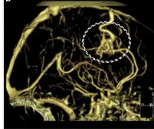
AVM, kapiller yatak olmadan besleyici arterleri drene edici venlere bağlayan parankimal bir nidus içerir. Rüptüre AVM kanamayla; rüptüre olmamış AVM nöbetle veya tesadüfi görüntülemede saptanabilir. BTA’da nidus, besleyici arterler, drene edici venler ve ilişkili anevrizmalar görülebilir; kontrastsız BT rüptürde parankimal hematomu saptar. AVM değerlendirmesinde nidus, besleyici arterler ve drene edici venler birlikte tanımlanır; yalnız serpijinöz damar görünümü dural fistül veya venöz varyanttan ayırmak için yeterli değildir.
Faz ve pencere
- Kontrastsız
- Rüptürde nidus komşuluğunda parankimal hematom görülebilir; intraventriküler veya subaraknoid kanama da eşlik edebilir. Bazı AVM olgularında kontrastsız BT’de eşlik eden kalsifikasyon görülebilir.
- BTA tanısal
- BTA’da parankim içindeki damar yumağı, besleyici arterler ve drene edici venler gösterilebilir; erken venöz dolum şantı destekler, ancak tek zamanlı BTA dolum sırasını kesinleştirmez. Eşlik eden intranidal veya besleyici arter anevrizması ve venöz variks/darlık da değerlendirilebilir.
Önerilen pencereler: Anjiyo (G 600 / M 150), Beyin (G 80 / M 40), Kemik (G 1800 / M 400).
BT bulguları
- Parankimal nidus — arteriyel besleyicilerle venöz drenaj arasındaki, kompakt veya dağınık damar ağı.
- Besleyici arterler — internal karotid veya vertebrobaziler sistemin parankimi besleyen dalları nidusa ulaşabilir.
- Drene edici ven — nidustan çıkan venöz yol yüzeyel kortikal veya derin venöz sisteme açılabilir; erken venöz dolum şantı destekler.
- İntranidal anevrizma — nidus içindeki fokal anevrizmal genişleme ayrıca tanımlanır.
- Venöz variks veya çıkış darlığı — drene edici venin fokal keseleşmesi ya da sinüse yaklaşırken daralması akım çıkış anormalliğini gösterir.
- Kanama — AVM rüptüründe en sık intraparenkimal kanama görülür; subaraknoid veya intraventriküler kanama da görülebilir.
- Kalsifikasyon — bazı AVM olgularında kontrastsız BT’de fokal kalsifikasyon eşlik edebilir.
Ölçütler ve sınıflamalar
- Spetzler–Martin
- Spetzler–Martin sınıflaması cerrahi sonuçları öngörmede kullanılır; nidus boyutu, eloquent yerleşim ve venöz drenaj gibi özellikler AVM sınıflamalarında kullanılan ölçütler arasındadır. Spetzler–Martin sınıflamasında AVM’ler derece I–V arasında sınıflandırılır.
- Spetzler–Ponce
- Spetzler–Martin derece I–II sınıf A, derece III sınıf B, derece IV–V sınıf C olarak gruplanır.
Normalde
Normal parankimde kapiller yatak bulunur; AVM ise parankimal arteriyel besleyiciler ile drene edici ven arasında niduslu bir şanttır. AVM mimarisi değerlendirilirken nidus, besleyici arterler ve drene edici venler ayrı ayrı tanımlanır.
Normal BT ile kıyasla
Aynı bölgenin, kaynak veri setinde patoloji içermediği belirtilen bir BT incelemesini kesit kesit kaydırın; organ sınırlarını açarak anatomiyi hasta görüntüsüyle karşılaştırın. Küçük rastlantısal bulgular tümüyle dışlanmamıştır.
Kaynak: TotalSegmentator v2.01 veri seti (Wasserthal ve ark., Radiology: Artificial Intelligence 2023), CC BY 4.0, vaka s0103; organ sınırları veri setinin otomatik segmentasyonundan, birleştirilerek sadeleştirildi. Görüntüler 2,5 mm kesit aralığına yeniden örneklendi.
Ayırıcı tanı
- Dural arteriovenöz fistül
- meningeal/dural arterlerden beslenen şant dural sinüslere veya kortikal venlere drene olur; bu anatomik yerleşim parankimal AVM’den farklıdır.
- Pial arteriovenöz fistül
- pial arter ile ven arasında doğrudan bağlantı vardır; arada AVM’deki gibi nidus bulunmaz.
- Gelişimsel venöz anomali
- normal beyin venöz drenajını tek bir çıkışta toplayan benign venöz anomalidir.
- Kavernöz malformasyon
- kapiller benzeri sinüzoidal kanallardan oluşur ve arteriyovenöz şant içermez; bu nedenle AVM’nin geniş besleyici arter-nidus-drenaj ağı beklenmez.
Tuzaklar
- Tek zamanlı BTA erken venöz dolumun zamanlamasını güvenle belirleyemez; şant değerlendirmesi nidus, besleyici arter ve venöz drenaj mimarisine dayanmalıdır.
- AVM ilişkili hematom içindeki malforme damarlar çıplak gözle ayrımı zorlaştırabilir; hematom çevresinde genişlemiş venler de eşlik edebilir.
- Spetzler–Martin, nidus çapı, eloquent yerleşim ve derin venöz drenaja dayalı cerrahi risk sınıflamasıdır.
Kendini dene
BTA’da frontal parankim içinde kompakt nidus, nidusa giren geniş MCA besleyicisi ve nidustan çıkan drene edici ven görülüyor. En olası tanı nedir?
Cevabı göster
Beyin AVM'si. Parankim içi nidusla birlikte arteriyel besleyici ve drene edici ven ağı beyin AVM’sinin tipik mimarisidir. Gelişimsel venöz anomalide ışınsal medüller venler toplayıcı vende birleşir; dural fistül dural/meningeal arteriyel besleme ve venöz drenaj şantıdır; kavernöz malformasyon arteriyovenöz şant içermez.
Beyin AVM'leri için kullanılan sınıflama sistemlerinin (Spetzler-Martin, Lawton-Young vb.) temel kullanım amacı nedir?
Cevabı göster
Tedavi stratejisi belirleme ve cerrahi/radyoterapi riskini öngörme. S50, AVM sınıflama sistemlerinin nörocerrahi ve radyoterapi planlamasında tedavi kararlarını iyileştirmek, hastaları sınıflandırmak ve klinik iletişimi güçlendirmek amacıyla kullanıldığını belirtir.
İlgili konular
Kaynaklar
Bu sayfadaki 40 cümle ve 2 quiz sorusu aşağıdaki kaynaklardan alıntılarla tek tek karşılaştırıldı; 30 cümle bu karşılaştırmada düzeltildi (2026-10-08).
- Reporting of angiographic studies in patients diagnosed with a cerebral arteriovenous malformation: a systematic reviewf1000research.com
- Brain arteriovenous malformations: A review of natural history, pathobiology, and interventionspubmed.ncbi.nlm.nih.gov
- The features of computed tomography and digital subtraction angiography images of ruptured cerebral arteriovenous malformationaimspress.com
- Central nervous system vascular malformations: A clinical reviewpmc.ncbi.nlm.nih.gov
- A 3-tier classification of cerebral arteriovenous malformationsresearchworks.creighton.edu
- Radiologic Assessment of Brain Arteriovenous Malformations: What Clinicians Need to Knowpubs.rsna.org
- I saw Medusa’s head sign and turned to stonepubmed.ncbi.nlm.nih.gov
- Cerebral cavernous malformations: Typical and atypical imaging characteristicsonlinelibrary.wiley.com
- Combined transarterial and transvenous embolization of multi-hole pial arteriovenous fistula with large varixjournals.sagepub.com
- Unruptured Arteriovenous Malformations in the Multidetector Computed Tomography Era: Frequency of Detection and Predictable Failuresclinicalimagingscience.org
- Methodological quality assessment of genetic studies on brain arteriovenous malformation related hemorrhage: A cross-sectional studypmc.ncbi.nlm.nih.gov
- Modern Brain Arteriovenous Malformation Models: A Reviewpmc.ncbi.nlm.nih.gov
- Indirect surgical revascularization for management of vascular steal phenomenon in high-grade untreatable brain arteriovenous malformationspmc.ncbi.nlm.nih.gov
- Regression of a symptomatic varix after transarterial embolization of a brain arteriovenous malformationpmc.ncbi.nlm.nih.gov
- Spontaneous Cystic Arteriovenous Malformation-A Novel Treatment Alternative.pubmed.ncbi.nlm.nih.gov
- Contemporary Imaging of Cerebral Arteriovenous Malformations.pubmed.ncbi.nlm.nih.gov
- Present state of microneurosurgery of cerebral arteriovenous malformations.pubmed.ncbi.nlm.nih.gov
- Clinical importance of the occipital artery in vascular lesions: A review of the literature.pubmed.ncbi.nlm.nih.gov
- Trigeminal neuralgia related to arteriovenous malformation of the posterior fossa: three case reports and a review of the literature.pubmed.ncbi.nlm.nih.gov
- Molecular Imaging in Neurovascular Diseases: The Use of Ferumoxytol to Assess Cerebral Aneurysms and Arteriovenous Malformations.pubmed.ncbi.nlm.nih.gov
- Brain arteriovenous malformations and dural arteriovenous fistulas with extensive venous congestive encephalopathy.pubmed.ncbi.nlm.nih.gov
- Segmentation techniques of brain arteriovenous malformations for 3D visualization: a systematic review.pubmed.ncbi.nlm.nih.gov
- Influence of the combination of patient age and deep venous drainage on brain arteriovenous malformation recurrence after surgery.pubmed.ncbi.nlm.nih.gov
- De novo brain arteriovenous malformation after tumor resection: case report and literature review.pubmed.ncbi.nlm.nih.gov
- Regression of a symptomatic varix after transarterial embolization of a brain arteriovenous malformation: A case report and literature review.pubmed.ncbi.nlm.nih.gov
- Targeted Stereotactic Radiosurgery for Arteriovenous Malformation Downgrading Followed by Microsurgical Resection: A Case Report and Review of the Literature.pubmed.ncbi.nlm.nih.gov
- A novel non-contrast-enhanced MRA using silent scan for evaluation of brain arteriovenous malformation: A case report and review of literature.pubmed.ncbi.nlm.nih.gov
- Delayed Fragmentation and Distal Embolization of Retained Microcatheter Causing Lower Limb Ischemia: Case Report and Review of the Literature.pubmed.ncbi.nlm.nih.gov
- Diagnostic and therapeutic dilemmas of asymptomatic intracranial AVMs: A case report and evidence-based review.pubmed.ncbi.nlm.nih.gov
- Central nervous system vascular malformations: A clinical review.pubmed.ncbi.nlm.nih.gov
- Intracerebral atypical calcification in nongalenic pial arteriovenous fistula: a case report.pubmed.ncbi.nlm.nih.gov
- Stroke-Mimic: An Intracranial Dural Arteriovenous Fistula Revealed by New-Onset Seizures and Post-Ictal Deficit.pubmed.ncbi.nlm.nih.gov
- Endovascular Embolization in Neurovascular Disease: Material Science, Multimodal Management, and Future Horizons.pubmed.ncbi.nlm.nih.gov
- Cerebral proliferative angiopathy in pediatric patients: case-based review with an illustrative case.pubmed.ncbi.nlm.nih.gov
- Development and validation of a prediction model for neurological outcomes of brain arteriovenous malformations undergoing microsurgical resection: a nationwide retrospective cohort study.pubmed.ncbi.nlm.nih.gov
- Interobserver Agreement of R<sub>2</sub>eD Brain Arteriovenous Malformation Scoring.pubmed.ncbi.nlm.nih.gov
- Coexistence of a Hemispheric Developmental Venous Anomaly and Two Ipsilateral Arteriovenous Malformations: A Rare Neurovascular Association.pubmed.ncbi.nlm.nih.gov
- Stereotactic Radiosurgery for Spetzler-Martin Grade I and II Arteriovenous Malformations: International Society of Stereotactic Radiosurgery (ISRS) Practice Guideline.pubmed.ncbi.nlm.nih.gov
- A Tractography-Based Grading Scale of Brain Arteriovenous Malformations Close to the Corticospinal Tract to Predict Motor Outcome After Surgery.pubmed.ncbi.nlm.nih.gov
- BrainPath Tubular Retractor System for Subcortical Hemorrhagic Vascular Lesions: A Case Series of Technique and Outcomes.pubmed.ncbi.nlm.nih.gov
- Brainstem arteriovenous malformations: anatomical subtypes, assessment of "occlusion in situ" technique, and microsurgical results.pubmed.ncbi.nlm.nih.gov
- Conjoined Pathways: Unraveling the Coexistence of Developmental Venous Anomaly With Arteriovenous Malformation.pubmed.ncbi.nlm.nih.gov
- Patterns of injury and violence in Yaoundé Cameroon: an analysis of hospital data.pubmed.ncbi.nlm.nih.gov
- International multi-center study to quantify the effect of deep venous drainage after surgical resection of Spetzler-Martin Grade II-III brain arteriovenous malformations.pubmed.ncbi.nlm.nih.gov
- Predictors of complete obliteration and favourable outcome after single-modality treatment for low-grade arteriovenous malformations: a multicentre study.pubmed.ncbi.nlm.nih.gov
- Implementation of the open surgery reporting guidelines in ruptured brain arteriovenous malformations: Feasability and adaptations.pubmed.ncbi.nlm.nih.gov
- Uni-port video-assisted thoracoscopic surgery for pulmonary arteriovenous malformation based on vascular pre-occlusion: a case report and literature review.pubmed.ncbi.nlm.nih.gov
- Co-occurring scalp arteriovenous malformation and dural arteriovenous fistula with diploic vein involvement: illustrative case.pubmed.ncbi.nlm.nih.gov
- Diffuse bilateral cerebral calcification caused by Vein of Galen aneurysmal malformation in a two-year-old child: A rare presentation.pubmed.ncbi.nlm.nih.gov
- Cerebral computed tomography perfusion pattern in Todd's paralysis: A case series, unraveling diagnostic challenges and clinical implications.pubmed.ncbi.nlm.nih.gov
- Meningioangiomatosis with Variable Imaging Feature: Rare Cause of Drug Refractory Epilepsy.pubmed.ncbi.nlm.nih.gov
- Ruptured Cerebral Arteriovenous Malformation Initially Managed As Presumed Eclampsia: A Diagnostic Challenge Following Initial Management in a Resource-Limited Setting.pubmed.ncbi.nlm.nih.gov
- Beyond Resection: Determinants of Outcome After Pediatric Brain Arteriovenous Malformation Surgery.pubmed.ncbi.nlm.nih.gov
- A systematic review and meta-analysis of microsurgery and radiosurgery outcomes in low-grade cerebral arteriovenous malformations: Obliteration rates, morbidity, and mortality.pubmed.ncbi.nlm.nih.gov
- Clinical Presentation, Grading, and Treatment Outcomes of Temporal Arteriovenous Malformations: A Systematic Review and Meta-Analysis.pubmed.ncbi.nlm.nih.gov
- Developmental venous anomaly related to de novo cerebral cavernous malformation: Imaging findings from two cases.pubmed.ncbi.nlm.nih.gov
- Developmental Venous Anomaly Presenting as an Acute Stroke Mimic.pubmed.ncbi.nlm.nih.gov
- Reporting of angiographic studies in patients diagnosed with a cerebral arteriovenous malformation: a systematic review.pubmed.ncbi.nlm.nih.gov
- Cerebral arteriovenous malformations classification systems in comparison with Spetzler-Martin: A comparative review.pubmed.ncbi.nlm.nih.gov
- Surgical outcomes and risk factors for overall mortality in brain arteriovenous malformations patients: a retrospective analysis.pubmed.ncbi.nlm.nih.gov
- Systematic review of brain arteriovenous malformation grading systems evaluating microsurgical treatment recommendation.pubmed.ncbi.nlm.nih.gov
- Developmental venous anomaly associated with dural arteriovenous fistula: Etiopathogenesis and hemorrhagic risk.pubmed.ncbi.nlm.nih.gov
- Thymus and aging: morphological, radiological, and functional overview.pubmed.ncbi.nlm.nih.gov
- Predictors of Functional Outcome After Microsurgical Resection of Brain Arteriovenous Malformations: A Retrospective Single-Center Study.pubmed.ncbi.nlm.nih.gov
- Functional and Hemodynamic Restoration After Microsurgical Resection of Compact High-Flow Temporo-Parieto-Occipital Arteriovenous Malformation.pubmed.ncbi.nlm.nih.gov
- Outcomes of gamma knife treated large symptomatic arteriovenous malformations according to guidelines of Taiwan neurosurgical consensus.pubmed.ncbi.nlm.nih.gov
- Efficacy and Safety of Surgical Management for Cerebral Arteriovenous Malformations: A Study Involving 100 Cases.pubmed.ncbi.nlm.nih.gov
- Somatic mutations in angiogenesis-related pathways and RNA polymerase II activity in sporadic brain arteriovenous malformations.pubmed.ncbi.nlm.nih.gov
- Developmental venous anomalies and cerebral cavernous malformations: Partners in crime.pubmed.ncbi.nlm.nih.gov
- Posterior fossa Hodgkin's lymphoma radiographically mimicking an arteriovenous malformation: illustrative case.pubmed.ncbi.nlm.nih.gov
- Fluid transport in the brain.pubmed.ncbi.nlm.nih.gov
- Brain AVM compactness score in children with hereditary hemorrhagic telangiectasia.pubmed.ncbi.nlm.nih.gov
- Influence of preoperative embolisation on resection of brain arteriovenous malformations: cohort study.pubmed.ncbi.nlm.nih.gov
- frontiersin.orgfrontiersin.org
Eğitim amaçlıdır; hasta başında tanı ve tedavi kararının yerine geçmez. İçerik yapay zekâ destekli bir süreçle güncel literatüre göre cümle cümle kaynaklarla karşılaştırıldı, ancak uzman radyolog incelemesinden geçmedi. Klinik kararlarda güncel kılavuzları, kurum protokollerini ve radyoloji raporunu esas alın.