Özet
Dural arteriovenöz fistül, dura yaprakları içindeki meningeal arterlerle dural sinüs veya kortikal venler arasında oluşan anormal şanttır ve sıklıkla venöz sinüs çevresinde görülür. Kontrastsız BT kanama, venöz konjesyon ve tromboze sinüs ipuçlarını; BTA ise asimetrik sinüs, genişlemiş damarlar ve transosseöz meningeal besleyicileri gösterebilir. Kortikal venöz reflü yalnız görüntüdeki damar kalınlığıyla değil, drenaj yönüyle tanımlanır ve statik BTA bu akım ilişkisini her zaman ortaya koyamaz. Reflülü fistülü gözden kaçırmak venöz hipertansiyona bağlı ödem, infarkt veya intrakraniyal kanama riskinin tanınmasını geciktirebilir.
Faz ve pencere
- Kontrastsız
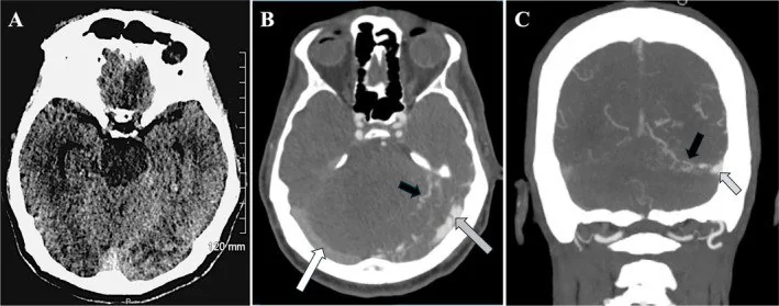
- Kontrastsız BT genellikle normal bulunur; sıklıkla yalnızca venöz konjesyona bağlı ödem veya intrakraniyal kanama tespit edilir. Kontrastsız BT'de sıklıkla yalnızca ödem veya intrakraniyal kanama izlenir; bu bulgular venöz hipertansiyonun bir yansımasıdır.
- BTA tanısal
- Sinüs çevresinde genişlemiş ve kıvrımlı meningeal arterler, asimetrik sinüs kontrastlanması, transosseöz damarlar ve geniş kortikal venöz kanallar aranır. Statik tek faz inceleme şantın besleyici kaynağını gösterebilir ancak reflü yönünü ve venöz drenaj zamanlamasını güvenle belirlemeyebilir.
Önerilen pencereler: Beyin (G 80 / M 40), Anjiyo (G 600 / M 150), Kemik (G 1800 / M 400).
BT bulguları
- Sinüs çevresi damar artışı — dural AV fistüller en sık transvers ve kavernöz sinüslerde görülür; BT anjiyografide bu bölgelerde belirginleşen meningeal arter dalları aranır.
- Asimetrik dural sinüs — karşı sinüse göre geniş, yoğun kontrastlanan ya da çıkış tıkanıklığı nedeniyle zayıf opaklaşan segment.
- Transosseöz damarlar — kafatası iç tabulasını geçen, sinüs çevresinde belirginleşen geniş meningeal kollateraller.
- Genişlemiş kortikal venler — parankim yüzeyinde serpijinöz tübüler yapılar; venöz konjesyonu düşündürür, ancak tek başına reflü yönünü kanıtlamaz.
- Venöz drenaj paterni — fistülün venöz sinüse veya kortikal venlere drenaj yönü; BT anjiyografide asimetrik sinüs kontrastlanması, genişlemiş meningeal arterler ve kortikal venler aranır.
- Venöz konjesyon — drenaj alanında kortikal/subkortikal ödem, sulkus silinmesi veya hemorajik infarkt görünümü.
- Kitle etkisi ya da hemoraji — fistüle komşu intraparenkimal hematom, sulkal kan veya ventriküler kan eşlik edebilir.
- Parankimal nidusun yokluğu — AVM'deki kompakt intraparenkimal damar yumağı yerine şant bulguları dura ve sinüs çevresinde yoğunlaşır.
Ölçütler ve sınıflamalar
- Borden sınıflaması
- Tip I: drenaj yalnız dural sinüse. Tip II: sinüse drenajla birlikte kortikal venöz reflü. Tip III: doğrudan kortikal venöz drenaj.
- Cognard sınıflaması
- Tip I: sinüse antegrad; IIa: sinüs içinde retrograd; IIb: kortikal vene reflü; IIa+b: sinüs ve kortikal ven reflüsü; III: ektazisiz doğrudan kortikal ven; IV: venöz ektazili kortikal ven; V: perimedüller venöz drenaj.
Normalde
Normalde transvers-sigmoid sinüsler ve kortikal venler taraflar arasında makul simetri gösterir; ince iç tabula boyunca belirgin transosseöz meningeal damar ağı izlenmez. Şüpheli taraftaki sinüs lümenini karşı taraf sinüsle, genişlemiş kortikal damarları ise aynı düzeydeki karşı hemisfer yüzey venleriyle kıyaslayın; normal kortikal venlerin yönü ve çapı fistül tanısı koydurmaz.
Normal BT ile kıyasla
Aynı bölgenin, kaynak veri setinde patoloji içermediği belirtilen bir BT incelemesini kesit kesit kaydırın; organ sınırlarını açarak anatomiyi hasta görüntüsüyle karşılaştırın. Küçük rastlantısal bulgular tümüyle dışlanmamıştır.
Kaynak: TotalSegmentator v2.01 veri seti (Wasserthal ve ark., Radiology: Artificial Intelligence 2023), CC BY 4.0, vaka s0103; organ sınırları veri setinin otomatik segmentasyonundan, birleştirilerek sadeleştirildi. Görüntüler 2,5 mm kesit aralığına yeniden örneklendi.
Ayırıcı tanı
- Dural sinüs trombozu, BT venografide sinüs içi dolum kusuru olarak gösterilebilir; dural AVF şüphesinde BTA/BT venografide asimetrik sinüs ile genişlemiş arteriyel veya venöz damarlar ayrıca değerlendirilir.
- Serebral AVM
- parankim içinde nidus ve nidusa giren pial besleyiciler görülür; dural AVF'de şant odağı sinüs/dura yapraklarıyla ilişkilidir.
- Gelişimsel venöz anomali
- medüller venler düzenli biçimde kolektör vene açılır; besleyici meningeal arter veya sinüs çevresi şant paterni yoktur.
- Kavernöz sinüs trombozu orbital konjesyonla ilişkili klinik ayırıcı tanılar arasında yer alabilir; kavernöz sinüs dural fistüllerinde anormal arteriyel bağlantı ve venöz drenaj kateter anjiyografiyle değerlendirilir.
- Normal venöz varyant
- baskın transvers sinüs veya asimetrik juguler çıkış tek başına fistül değildir; serpijinöz besleyici damar ve anormal venöz kanal birlikteliği aranır.
Tuzaklar
- Geniş kortikal veni otomatik olarak kortikal reflü diye sınıflamayın; Borden/Cognard akım yönü kateter anjiyografide veya zamansal anjiyografik görüntülemede gösterilmelidir.
- Tek başına asimetrik transvers sinüs sık anatomik varyant olabilir; çevre meningeal arterler, transosseöz damarlar ve anormal venöz opasifikasyonla birlikte yorumlayın.
- Bulgu takibi — kortikal venlerdeki yoğunluk artışı tek başına fistülü kanıtlamaz; SWI veya kateter anjiyografi ile arteriyovenöz şantın varlığı ve venöz drenaj doğrulanmalıdır.
- Parankimal nidus görülmemesi dural şantı dışlamaz; küçük fistül odağı dura yapraklarında kalabilir ve rutin kesitlerde fark edilmeyebilir.
Kendini dene
BTA'da transvers sinüs çevresinde transosseöz damarlar ve geniş yüzey venleri görülüyor. Tanı hangisidir?
Cevabı göster
Dural AV fistül. Sinüs çevresi meningeal/transosseöz damarlarla geniş kortikal venler dural AV fistül şüphesini doğurur. AVM parankimal nidus yapar; tromboz dolum kusurudur; gelişimsel venöz anomali düzenli medüller ven-kolektör paterni gösterir.
Fistül doğrudan venöz sinüse drenaj yapıyor ve kateter anjiyografide kortikal venöz reflü saptanmıyor. Bu durum Borden sınıflamasının hangi tipine uyar?
Cevabı göster
Tip I. Yalnızca dural sinüse drenaj, Borden Tip I’in tanımıdır. Tip II’de sinüs drenajına kortikal venöz reflü eşlik ederken, Tip III’te drenaj doğrudan kortikal venlere yapılır. Cognard Tip IV ise venöz ektazi ile seyreden direkt kortikal venöz drenajı ifade eder.
İlgili konular
Kaynaklar
Bu sayfadaki 48 cümle ve 2 quiz sorusu aşağıdaki kaynaklardan alıntılarla tek tek karşılaştırıldı; 7 cümle bu karşılaştırmada düzeltildi (2026-10-08).
- Updates in the management of cranial dural arteriovenous fistulapmc.ncbi.nlm.nih.gov
- [Diagnostic imaging of cerebrovascular disease on multi-detector row computed tomography (MDCT)].pubmed.ncbi.nlm.nih.gov
- Intracranial Dural Arteriovenous Fistula as a Reversible Cause of Dementia: Case Series and Literature Review.pubmed.ncbi.nlm.nih.gov
- Diagnostic performance of CT and MRI on the detection of symptomatic intracranial dural arteriovenous fistula: a meta-analysis with indirect comparison.pubmed.ncbi.nlm.nih.gov
- Dural arteriovenous fistula with spinal dural arteriovenous fistula: a case report and review of the literature.pubmed.ncbi.nlm.nih.gov
- Intracranial dural arteriovenous fistula: a comprehensive review of the history, management, and future prospective.pubmed.ncbi.nlm.nih.gov
- Dural Arteriovenous Fistula Mimicking a Brainstem Glioma.pubmed.ncbi.nlm.nih.gov
- Evaluation of dural arteriovenous fistulas with 4D contrast-enhanced MR angiography at 3T.pubmed.ncbi.nlm.nih.gov
- Rapid cognitive decline: not always Creutzfeldt-Jakob disease.pubmed.ncbi.nlm.nih.gov
- Progressive versus Nonprogressive Intracranial Dural Arteriovenous Fistulas: Characteristics and Outcomes.pubmed.ncbi.nlm.nih.gov
- Cervical Myelopathy Caused by Intracranial Dural Arteriovenous Fistula.pubmed.ncbi.nlm.nih.gov
- Utility of silent magnetic resonance angiography in the evaluation and characterisation of intracranial dural arteriovenous fistula.pubmed.ncbi.nlm.nih.gov
- Transarterial treatment with Onyx of intracranial dural arteriovenous fistula with cortical drainage in 17 patients.pubmed.ncbi.nlm.nih.gov
- Congenital Abdominal Aortic Aneurysm in a Four Year Old Girl.pubmed.ncbi.nlm.nih.gov
- The application of susceptibility-weighted MRI in pre-interventional evaluation of intracranial dural arteriovenous fistulas.pubmed.ncbi.nlm.nih.gov
- Single-Session Curative Transarterial Onyx Embolization of a Convexity Dural AVF with a Concomitant Pial AVF Sharing the Vein of Labbé: A Case Report and Literature Review.pubmed.ncbi.nlm.nih.gov
- Transarterial Onyx Embolization of a Hemorrhagic Tentorial Dural Arteriovenous Fistula via a Tortuous Occipital Artery: Technical Tips for Challenging Vascular Access.pubmed.ncbi.nlm.nih.gov
- Long-term Outcomes after Staged and Multimodal Treatment for Diffuse Intracranial Dural Arteriovenous Fistulas Involving Interconnected Dural Sinuses: A Single-center Case Series.pubmed.ncbi.nlm.nih.gov
- Intracranial venous thrombosis imaging by anatomical location: an educational review with mimics and diagnostic pitfalls.pubmed.ncbi.nlm.nih.gov
- A Practical Approach to Intracranial Dural Arteriovenous Fistulas: Pathogenesis, Classification and Management.pubmed.ncbi.nlm.nih.gov
- Narrower jugular bulbs and their tributaries are associated with skull base venous hyperintensity on arterial spin-labeling MRI.pubmed.ncbi.nlm.nih.gov
- Surgical Treatment of Dural Arteriovenous Fistula: A Case Report and Literature Review.pubmed.ncbi.nlm.nih.gov
- Cerebral venous disorders: clinical presentation, diagnostic strategy, and contemporary management.pubmed.ncbi.nlm.nih.gov
- Delayed Intracerebral Hemorrhage Following Transarterial Embolization for a Tentorial Dural Arteriovenous Fistula: Postoperative Venous Outflow Impairment Detected by Susceptibility-Weighted Imaging.pubmed.ncbi.nlm.nih.gov
- Magnetic resonance imaging of the orbit, Part 2: Characterization of orbital pathologies.pubmed.ncbi.nlm.nih.gov
- The Role of Angiographic Imaging in the Treatment of Spinal Vascular Malformations.pubmed.ncbi.nlm.nih.gov
- Dynamic pseudo-continuous arterial spin labeling angiography using a 3D-radial multi-spoke spoiled gradient-recalled sequence.pubmed.ncbi.nlm.nih.gov
- International Society of Paediatric Surgical Oncology (IPSO) Surgical Practice Guidelines.pubmed.ncbi.nlm.nih.gov
- Spontaneous bacteremia and spontaneous bacterial peritonitis share similar prognosis in patients with cirrhosis: a cohort study.pubmed.ncbi.nlm.nih.gov
- Direct Cannulation of a Calvarial Diploic Vein for Embolization of a Symptomatic Intraosseous Arteriovenous Fistula: A Case Report.pubmed.ncbi.nlm.nih.gov
- Incidental vascular findings on brain magnetic resonance angiography.pubmed.ncbi.nlm.nih.gov
- Embolization in Pediatric Patients: A Comprehensive Review of Indications, Procedures, and Clinical Outcomes.pubmed.ncbi.nlm.nih.gov
- Pathophysiology, diagnosis and management of cerebral venous thrombosis: A comprehensive review.pubmed.ncbi.nlm.nih.gov
- Cortical vein involvement and its influence in a cohort of adolescents with cerebral venous thrombosis.pubmed.ncbi.nlm.nih.gov
- Intracerebral Hemorrhage in a Middle-Aged Male With Classical Ehlers-Danlos Syndrome in the Absence of Macrovascular Lesions: A Case Report.pubmed.ncbi.nlm.nih.gov
- Non-contrast CT findings suggestive of secondary intracerebral haemorrhage.pubmed.ncbi.nlm.nih.gov
- Iatrogenic and traumatic Dural arteriovenous fistulas: Illustrative cases and literature review.pubmed.ncbi.nlm.nih.gov
- Dominant Orbitofrontal Pial Supply in Anterior Cranial Fossa Dural Arteriovenous Fistula: Angiographic Differentiation from Mixed Pial-Dural Arteriovenous Malformation and Anatomy-Based Treatment Selection.pubmed.ncbi.nlm.nih.gov
- Stroke-Mimic: An Intracranial Dural Arteriovenous Fistula Revealed by New-Onset Seizures and Post-Ictal Deficit.pubmed.ncbi.nlm.nih.gov
- Co-occurring scalp arteriovenous malformation and dural arteriovenous fistula with diploic vein involvement: illustrative case.pubmed.ncbi.nlm.nih.gov
- Flow control-assisted transarterial embolization of a high-flow superior sagittal sinus dural arteriovenous fistula: An illustrative case.pubmed.ncbi.nlm.nih.gov
- Clinical management of isolated transverse-sigmoid sinus dural arteriovenous fistulas: a three-case illustrative series and narrative review.pubmed.ncbi.nlm.nih.gov
- Endovascular Management of Intracranial Dural AVFs: Principles.pubmed.ncbi.nlm.nih.gov
- Pial arteriovenous fistula with a large intraparenchymal hemorrhage in a 9-year-old child: a case report and case-based mini review.pubmed.ncbi.nlm.nih.gov
- Low-flow vascular malformations without arteriovenous shunting of the central nervous system: a pictorial review.pubmed.ncbi.nlm.nih.gov
- Conjoined Pathways: Unraveling the Coexistence of Developmental Venous Anomaly With Arteriovenous Malformation.pubmed.ncbi.nlm.nih.gov
- Systemic and CNS manifestations of inherited cerebrovascular malformations.pubmed.ncbi.nlm.nih.gov
- Spontaneous Resolution of a Bilateral Barrow Type D Indirect Carotid-Cavernous Fistula: A Rare Case Report and Literature Review.pubmed.ncbi.nlm.nih.gov
- Neuro-Ophthalmic Manifestations of Carotid Cavernous Fistulas: A Systematic Review and Meta-Analysis.pubmed.ncbi.nlm.nih.gov
- Venous Thromboembolism in Neonates, Children, and Adolescents: A Comprehensive Narrative Review of Risk Factors, Diagnosis, Treatment, and Prevention.pubmed.ncbi.nlm.nih.gov
- An Integrated Approach on the Diagnosis of Cerebral Veins and Dural Sinuses Thrombosis (a Narrative Review).pubmed.ncbi.nlm.nih.gov
- Imaging Evaluation of Acute Traumatic Brain Injury.pubmed.ncbi.nlm.nih.gov
- Long-term safety and effectiveness of the "OptEase" vena cava filter.pubmed.ncbi.nlm.nih.gov
- Endovascular treatment of an intraosseous dural arteriovenous fistula of the greater wing of the sphenoid: A case report and literature review.pubmed.ncbi.nlm.nih.gov
- Bilateral Vein of Trolard Thrombosis Presenting with Seizure and Minimal Deficits: A Rare Case Report.pubmed.ncbi.nlm.nih.gov
- Central nervous system vascular malformations: A clinical review.pubmed.ncbi.nlm.nih.gov
- Symptomatic Infratentorial Thrombosed Developmental Venous Anomaly: Case Report and Review of the Literature.pubmed.ncbi.nlm.nih.gov
- Dural Arteriovenous Fistula with Bilateral Agenesia of Intracranial Venous Sinuses: A Case Report.pubmed.ncbi.nlm.nih.gov
- Cerebral proliferative angiopathy in pediatric patients: case-based review with an illustrative case.pubmed.ncbi.nlm.nih.gov
- Intravascular embolization with spinal decompression and vertebral shaping for acute cauda equina syndrome from aggressive vertebral hemangioma: a case report and literature review.pubmed.ncbi.nlm.nih.gov
- A "Mismatched" connection: a rare case of indirect or dural carotid-cavernous fistula: a case based review.pubmed.ncbi.nlm.nih.gov
- Paediatric cranial ultrasound: abnormalities of the brain in term neonates and young infants.pubmed.ncbi.nlm.nih.gov
- Intracranial dural arteriovenous fistulas with and without pial arterial supply: a case series of 227 patients.pubmed.ncbi.nlm.nih.gov
- Imaging for Pediatric Head and Neck Emergencies: Critical Insights.pubmed.ncbi.nlm.nih.gov
- The 'corkscrew' sign: an indirect MRI hint for intracranial venous hypertension.pubmed.ncbi.nlm.nih.gov
- It Looks Like a Spinal Cord Tumor but It Is Not.pubmed.ncbi.nlm.nih.gov
- Cervical Radiculopathy Caused by Epidural Venous Varix Associated with a Dural Arteriovenous Fistula in the Anterior Condylar Confluence: A Case Report.pubmed.ncbi.nlm.nih.gov
- Endovascular and medical management strategies for carotid-cavernous fistulas: A safety and efficacy analysis.pubmed.ncbi.nlm.nih.gov
- Vein of Galen Malformation with Pial Arteriovenous Fistula in a 12-month-old Child.pubmed.ncbi.nlm.nih.gov
- Subarachnoid haemorrhage associated with septic cavernous sinus thrombosis secondary to odontogenic maxillary sinusitis.pubmed.ncbi.nlm.nih.gov
- PHACE syndrome: a systematic literature review and illustrative case report of a patient with severe cerebrovascular and neurodevelopmental sequelae.pubmed.ncbi.nlm.nih.gov
- Carotid Cavernous Fistula: A Case Report of a Rare Diagnosis and Presentation.pubmed.ncbi.nlm.nih.gov
- Syndrome of Inappropriate Antidiuresis and Symptomatic Hyponatremia After Onyx Embolization of a Carotid-Cavernous Fistula: A Case Report.pubmed.ncbi.nlm.nih.gov
- Cerebral venous system stenosis in the sigmoid sinus with resultant dilatation and tortuosity of the cavernous sinus mimicking carotid cavernous fistula.pubmed.ncbi.nlm.nih.gov
- Endovascular Embolization in Neurovascular Disease: Material Science, Multimodal Management, and Future Horizons.pubmed.ncbi.nlm.nih.gov
- Multimodal imaging in neuro-ophthalmology.pubmed.ncbi.nlm.nih.gov
- Prolonged trigeminocardiac reflex following transvenous coil embolization for cavernous sinus dural arteriovenous fistula with brainstem edema: illustrative case.pubmed.ncbi.nlm.nih.gov
- Neuroimaging for neurovascular complications of traumatic brain injury.pubmed.ncbi.nlm.nih.gov
- Intracerebral hemorrhage and carotid-cavernous fistulas: a rare connection.pubmed.ncbi.nlm.nih.gov
Eğitim amaçlıdır; hasta başında tanı ve tedavi kararının yerine geçmez. İçerik yapay zekâ destekli bir süreçle güncel literatüre göre cümle cümle kaynaklarla karşılaştırıldı, ancak uzman radyolog incelemesinden geçmedi. Klinik kararlarda güncel kılavuzları, kurum protokollerini ve radyoloji raporunu esas alın.