Özet
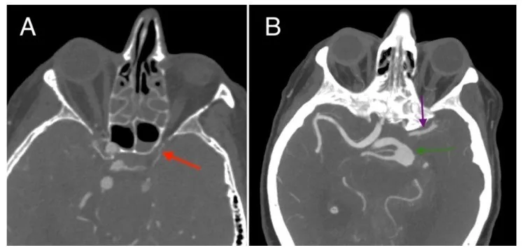
İntrakraniyal arter hipoplazisi, bir segmentin gelişimsel olarak ince kalması; aplazi ise seçilebilir lümenin bulunmamasıdır. Özellikle A1, P1, vertebral ve baziler segmentlerde bu anatomi, karşı hemisfer veya posterior dolaşımın hangi damardan beslendiğini ve akut tıkanıklıkta kollateral kapasiteyi etkiler. BT anjiyografi küçük damar boyunca düzenli kalibreyi, distal devamlılığı ve komşu kollateral yolları göstererek gelişimsel inceliği akut oklüzyondan ayırmaya yardım eder. Fark edilmediğinde hipoplastik damar tıkalı sanılabilir ya da gerçek bir yeni tıkanıklık varyanta bağlanarak gözden kaçabilir.
Faz ve pencere
- Kontrastsız
- Arter hipoplazisinin değerlendirilmesinde yüksek çözünürlüklü CTA ve MRA kullanılır; damar çapı ve gelişimsel varyantlar bu anjiyografik yöntemlerle görüntülenir.
- BTA tanısal
- Arteriyel CTA'da ilgili segmentin başlangıcından distal devamına kadar düzgün ve sürekli ince opaklaşma, karşı tarafla kalibre farkı ve beslediği dal/komünikan arterler birlikte incelenir; ani kesilme, intraluminal dolum kusuru veya distal yeniden dolum yeni oklüzyon lehine ayrıca değerlendirilir.
Önerilen pencereler: Anjiyo (G 600 / M 150), Beyin (G 80 / M 40).
BT bulguları
- Düzgün küçük kalibre — A1, P1, vertebral veya baziler segment boyunca ani omuzlanma olmadan devam eden ince lümen gelişimsel küçüklüğü destekler.
- Hipoplastik vertebral arter ince kalibreli olmakla birlikte patenttir; distal lümen devamlılığı bu bulguyla birlikte değerlendirilir.
- Karşı tarafla kalibre farkı — eş arter segmentlerinin aynı rekonstrüksiyon ve benzer düzlemde kıyası asimetriyi gösterir; tek çap ölçümü bütün dolaşımı tanımlamaz.
- A1 ve ön komünikan arter — bir A1 ince ya da izlenmiyorsa iki A2'nin karşı taraftaki A1 üzerinden dolup dolmadığı kontrol edilir.
- P1 ve PCom ilişkisi — küçük P1 ile geniş posterior komünikan arter birlikteyse fetal tip PCA konfigürasyonu, izole P1 hipoplazisinden daha olasıdır.
- Vertebral-baziler birleşme — bir vertebral arter ince olabilir; iki vertebral arterin baziler gövdeye katılımı ve baziler arterin distal dalları izlenmelidir.
- Eşlik eden damar duvarı bulgusu — fokal plak, diseksiyon şüphesi, trombüs veya anevrizma gelişimsel küçük çap varsayımından bağımsız tarif edilir.
Ölçütler ve sınıflamalar
- A1 segmenti için yayımlanmış hipoplazi eşiği
- <1 mm çap
Normalde
Tipik CTA'da A1 ve P1 segmentleri kendi anatomik yollarında seçilebilir, vertebral arterler baziler arteri oluşturur; çaplar bireyler arasında değişebilir ve tek başına eşitsizlik hastalık anlamına gelmez. Aynı düzlemde başlangıçtan distal dala kadar lümen sürekliliğini, ACom/PCom üzerinden beslenmeyi ve diğer vertebral arterin baziler katkısını kıyaslayın.
Normal BT ile kıyasla
Aynı bölgenin, kaynak veri setinde patoloji içermediği belirtilen bir BT incelemesini kesit kesit kaydırın; organ sınırlarını açarak anatomiyi hasta görüntüsüyle karşılaştırın. Küçük rastlantısal bulgular tümüyle dışlanmamıştır.
Kaynak: TotalSegmentator v2.01 veri seti (Wasserthal ve ark., Radiology: Artificial Intelligence 2023), CC BY 4.0, vaka s0849; organ sınırları veri setinin otomatik segmentasyonundan, birleştirilerek sadeleştirildi. Görüntüler 2,5 mm kesit aralığına yeniden örneklendi.
Ayırıcı tanı
- Akut arter oklüzyonu
- damar bir noktada aniden kesilir veya dolum kusuru içerir; distal dalların yeniden dolması kollateral dolaşımı gösterebilir.
- Aterosklerotik stenoz
- fokal veya eksantrik daralma ve plak/kalsifikasyon eşlik edebilir; segment boyunca düzgün küçüklük beklenmez.
- Diseksiyon
- uzun, düzensiz daralma, eksantrik lümen ya da intimal flep şüphesi doğurur; yalnız çap asimetrisi tanı koydurmaz.
- Fetal PCA
- PCom belirgin, P1 ince olabilir ve PCA ağırlıkla ICA'dan dolar; bu konfigürasyon tek başına P1 aplazisi diye adlandırılmamalıdır.
- CTA'da ışın sertleşmesi artefaktları ve kontras madgesi uygulamalarına bağlı sınırlılıklar dikkat edilmeli; teknik yetersizlik dışlanmalıdır.
Tuzaklar
- Maksimum yoğunluk projeksiyonunda ince A1 veya P1 üst üste binen damarlar arasında kaybolabilir; ham aksiyel kesitlerde ve eğik MPR'de segmenti baştan sona arayın.
- Bir tarafta küçük vertebral arter görülmesi, diğer vertebral ve baziler akımı incelemeden aplazi olarak adlandırılamaz; lümen seçilemiyor olsa bile teknik opasifikasyon yetersizliği dışlanmalıdır.
- Hipoplazi tanımı segment ve çalışmaya göre değişir; yalnızca bu sayfada verilen A1 eşiğini vertebral veya baziler artere uygulamayın.
Kendini dene
CTA'da sağ A1 düzgün biçimde ince ve ACom'a kadar devamlı; iki A2 sol A1 üzerinden doluyor. Bulguyu en iyi açıklayan seçenek?
Cevabı göster
Sağ A1 hipoplazisi. Segment boyunca düzgün incelik ve ACom üzerinden devam eden distal dolum gelişimsel A1 hipoplazisini destekler. Oklüzyon genellikle ani kesilme, diseksiyon düzensiz daralma veya flep, anevrizma ise fokal sakküler genişleme oluşturur.
Sol P1 ince, sol PCom geniş ve sol PCA distal dalları ICA’dan PCom üzerinden doluyor. En uygun anatomik yorum hangisi?
Cevabı göster
Fetal tip PCA. P1'in ince veya absent olması ve PCA'nın ICA'dan PCom üzerinden beslenmesi, S1'e göre fetal tip PCA tanısını destekler. Diğer seçeneklerde bu spesifik kollateral dolaşım paterni gözlenmez.
Kaynaklar
Bu sayfadaki 37 cümle ve 2 quiz sorusu aşağıdaki kaynaklardan alıntılarla tek tek karşılaştırıldı; 3 cümle bu karşılaştırmada düzeltildi (2026-10-08).
- Common and uncommon intracranial arterial anatomic variations in multi-detector computed tomography angiography (MDCTA). What radiologists should be aware ofpmc.ncbi.nlm.nih.gov
- Morphological Variations of the Anterior Cerebral Artery: A Systematic Review with Meta-Analysis of 85,316 Patientspmc.ncbi.nlm.nih.gov
- Congenital Anomalies and Variations of the Carotid and Vertebral Arteries: A Case-Based Imaging Reviewpmc.ncbi.nlm.nih.gov
- Evolving Concept of Small Vessel Disease through Advanced Brain Imaging.pubmed.ncbi.nlm.nih.gov
- Maxillary sinus aplasia.pubmed.ncbi.nlm.nih.gov
- An algorithmic approach to CT of pulmonary arterial disorders.pubmed.ncbi.nlm.nih.gov
- Vertebral artery hypoplasia: an innocent lamb or a disguise?pubmed.ncbi.nlm.nih.gov
- Inner ear malformations: a practical diagnostic approach.pubmed.ncbi.nlm.nih.gov
- Cerebral and occipito-atlanto-axial involvement in mucopolysaccharidosis patients: clinical, radiological, and neurosurgical features.pubmed.ncbi.nlm.nih.gov
- [A case of acute intracranial epidural hematoma caused by chronic nasal sinusitis].pubmed.ncbi.nlm.nih.gov
- Anatomy of the feeding arteries of the cerebral arteriovenous malformations.pubmed.ncbi.nlm.nih.gov
- Abnormal arterial anatomy and an unexpected intracranial haemorrhage.pubmed.ncbi.nlm.nih.gov
- Cerebral developmental venous anomalies.pubmed.ncbi.nlm.nih.gov
- Modeling of internal carotid artery aneurysm and blood flow simulation.pubmed.ncbi.nlm.nih.gov
- Cervical Carotid Pseudo-Occlusions and False Dissections: Intracranial Occlusions Masquerading as Extracranial Occlusions.pubmed.ncbi.nlm.nih.gov
- Considerations on the Relevance of Cerebral Fusiform Aneurysms Observed During HIV Infection.pubmed.ncbi.nlm.nih.gov
- Ovarian inguinal hernia - a possibility in MURCS syndrome.pubmed.ncbi.nlm.nih.gov
- The persistent proatlantal artery: types and subtypes.pubmed.ncbi.nlm.nih.gov
- Foetal disruptive brain injuries: Diagnosing the underlying pathogenetic mechanisms with cranial ultrasonography.pubmed.ncbi.nlm.nih.gov
- Congenital Absence of the Internal Carotid Artery Associated With Contralateral Middle Cerebral Artery Occlusion in a Young Female Patient: Embryology, Multimodal Imaging Findings, and Clinical Implications.pubmed.ncbi.nlm.nih.gov
- Cognitive impairment in asymptomatic carotid artery stenosis is associated with abnormal segments in the Circle of Willis.pubmed.ncbi.nlm.nih.gov
- Effect of age and vascular anatomy on blood flow in major cerebral vessels.pubmed.ncbi.nlm.nih.gov
- Pediatric Stroke Imaging.pubmed.ncbi.nlm.nih.gov
- Long-term safety and effectiveness of the "OptEase" vena cava filter.pubmed.ncbi.nlm.nih.gov
- Total laparoscopic radical hysterectomy for cervical cancer in prolapsed uterus.pubmed.ncbi.nlm.nih.gov
- Vertebral Artery Pseudoaneurysm in a Single-Vessel Posterior Circulation With Chronic Contralateral Occlusion and Recent Methamphetamine Use.pubmed.ncbi.nlm.nih.gov
- Stridor in Bilateral Medial Medullary Infarction: A Case Report, Literature Review, and Pathophysiologic Insights Into a Rare Presentation of an Uncommon Stroke.pubmed.ncbi.nlm.nih.gov
- Advances in Collateral Neuroimaging for Acute Ischemic Stroke: Redefining Time and Tissue Windows in the Reperfusion Era.pubmed.ncbi.nlm.nih.gov
- Patterns of injury and violence in Yaoundé Cameroon: an analysis of hospital data.pubmed.ncbi.nlm.nih.gov
- The Basilar Artery Collateral Simplified Score: a Novel Collateral Circulation Score Associated With Outcomes in Patients With Basilar Artery Occlusion: Results From the ATTENTION Study.pubmed.ncbi.nlm.nih.gov
- Embolization in Pediatric Patients: A Comprehensive Review of Indications, Procedures, and Clinical Outcomes.pubmed.ncbi.nlm.nih.gov
- Update in combined musculoskeletal and vascular injuries of the extremities.pubmed.ncbi.nlm.nih.gov
- The Vulnerable Conduit: Uncovering Segment-Specific Pathological Susceptibility and Bony Co-dependence of the Vertebral Artery in a South Indian Cohort.pubmed.ncbi.nlm.nih.gov
- Association of A1 Segment Morphology with the Rupture Risk and Morphology of Anterior Communicating Artery Aneurysms: A Retrospective, Single-Center Study.pubmed.ncbi.nlm.nih.gov
- Cerebral Venous Outflow Insufficiency: A Study on Symptoms and Venous Stenosis Classification.pubmed.ncbi.nlm.nih.gov
- Multiterritory Bihemispheric Ischemic Stroke in the Setting of Unilateral Internal Carotid Artery Disease.pubmed.ncbi.nlm.nih.gov
- Pre-operative transcranial Doppler ultrasound assessment of cerebral collateral circulation in children undergoing veno-arterial extracorporeal membrane oxygenation or cardiac surgery.pubmed.ncbi.nlm.nih.gov
- Morphological Variability of the P1 Segment of the Posterior Cerebral Artery: An Anatomical and Angiographic Study with Clinical Correlations.pubmed.ncbi.nlm.nih.gov
- Atypical variants of the cerebral arterial circle beyond the Lazorthes classification: a retrospective morphologic and neuroradiologic study.pubmed.ncbi.nlm.nih.gov
- Flow Diversion in Fetal-Type Posterior Communicating Artery Aneurysms: Anatomical Challenges and Treatment Strategy.pubmed.ncbi.nlm.nih.gov
- Vertebral Artery Hypoplasia and Posterior Circulation Vulnerability: A Systematic Review of Epidemiological, Hemodynamic, and Clinical Evidence.pubmed.ncbi.nlm.nih.gov
- Persistent primitive trigeminal artery with carotid-dependent posterior circulation: An incidental but clinically relevant discovery.pubmed.ncbi.nlm.nih.gov
- From intravascular imaging to adaptive vascular care: intelligent photonics and digital twins in panvascular disease.pubmed.ncbi.nlm.nih.gov
- Spontaneous Coronary Artery Dissection and Takotsubo Syndrome: Converging Mechanisms, Diagnostic Pitfalls, and a Unified Clinical Algorithm.pubmed.ncbi.nlm.nih.gov
- Haemodynamic and biomechanical biomarker analysis of carotid fibromuscular dysplasia via fluid-structure interaction.pubmed.ncbi.nlm.nih.gov
- Anatomical study of variations in the configurations of the circle of Willis in relation to age, sex, and diameters of the components.pubmed.ncbi.nlm.nih.gov
- Numerical Modeling of Flow in the Cerebral Vasculature: Understanding Changes in Collateral Flow Directions in the Circle of Willis for a Cohort of Vasospasm Patients Through Image-Based Computational Fluid Dynamics.pubmed.ncbi.nlm.nih.gov
- Geometry and Symmetry of Willis' Circle and Middle Cerebral Artery Aneurysms Development.pubmed.ncbi.nlm.nih.gov
- A novel radiological score on digital subtraction angiography predicts prognosis for acute basilar artery occlusion treated with endovascular thrombectomy: a two-center retrospective study.pubmed.ncbi.nlm.nih.gov
- Azygous anterior cerebral artery occlusion managed with Thrombectomy: a case report and literature review.pubmed.ncbi.nlm.nih.gov
- Distinction between acute and chronic carotid disease based on computed tomography angiography in acute ischemic stroke patients with carotid artery occlusion.pubmed.ncbi.nlm.nih.gov
- Clinical implications of vessel wall imaging-State-of-the-art review.pubmed.ncbi.nlm.nih.gov
- Non-Atherosclerotic and Genetic Intracranial Stenoses: Diagnostic Challenges and Emerging Genetic Insights.pubmed.ncbi.nlm.nih.gov
- Current Insights into the Molecular Mechanisms of Intracranial Atherosclerosis and Their Therapeutic Implications.pubmed.ncbi.nlm.nih.gov
- Structural Restoration of an Unruptured Vertebral Artery Dissection Following Autologous Mesenchymal Stem Cell Therapy: A Case Report.pubmed.ncbi.nlm.nih.gov
- Initial magnetic resonance imaging/magnetic resonance angiography may miss intracranial artery dissection in young adults with stroke: the role of subsequent imaging.pubmed.ncbi.nlm.nih.gov
- Case Report: Three cases involving the use of endovascular intervention for artery of Percheron infarction with a unilateral fetal-type posterior cerebral artery.pubmed.ncbi.nlm.nih.gov
- Accessory middle cerebral artery, hypoplasia of the A1 segment of the left anterior cerebral artery fenestration of the A2 segment of the left anterior cerebral artery and fetal variant of posterior cerebral artery.pubmed.ncbi.nlm.nih.gov
- RAPID CTA versus JLK LVO for large vessel occlusion detection: a pragmatic comparison of performance and common pitfalls.pubmed.ncbi.nlm.nih.gov
- Multimodal imaging in neuro-ophthalmology.pubmed.ncbi.nlm.nih.gov
- Association of middle cerebral artery aneurysms and variation of the A1 segment.pubmed.ncbi.nlm.nih.gov
- [Imaging characteristics of basilar artery hypoplasia].pubmed.ncbi.nlm.nih.gov
- Wall Imaging for Unilateral Intracranial Vertebral Artery Hypoplasia with Three-dimensional High-isotropic Resolution Magnetic Resonance Images.pubmed.ncbi.nlm.nih.gov
- Deep-learning-based extraction of circle of Willis topology with anatomical priors.pubmed.ncbi.nlm.nih.gov
- A Novel Window Into Human Vascular Remodeling and Diagnosing Carotid Flow Impairment: The Petro-Occipital Venous Plexus.pubmed.ncbi.nlm.nih.gov
- The anterior communicating artery variants: a meta-analysis with a proposed classification system.pubmed.ncbi.nlm.nih.gov
- An Atlas of Neonatal Neurovascular Imaging Anatomy as Depicted with Microvascular Imaging: The Intracranial Arteries.pubmed.ncbi.nlm.nih.gov
- Comprehensive review on rhino-neurosurgery.pubmed.ncbi.nlm.nih.gov
- The transnasal approach to the skull base. From sinus surgery to skull base surgery.pubmed.ncbi.nlm.nih.gov
- Evaluation of Circle of Willis variants using magnetic resonance angiography.pubmed.ncbi.nlm.nih.gov
- Emerging insights into cephalic neural crest disorders: A single center experience.pubmed.ncbi.nlm.nih.gov
- Human cerebral arterial morphogenesis explored by 3D microscopic imaging of cleared embryos: Bridging the gap between embryonic and adult anatomy.pubmed.ncbi.nlm.nih.gov
- Congenital internal carotid artery hypoplasia in a symptomatic young adult: A case report.pubmed.ncbi.nlm.nih.gov
- Internal carotid artery agenesis: A narrative review.pubmed.ncbi.nlm.nih.gov
- Intracranial venous thrombosis imaging by anatomical location: an educational review with mimics and diagnostic pitfalls.pubmed.ncbi.nlm.nih.gov
- Extracranial vertebral artery stenosis patients that may benefit from stent placement: post-hoc analysis from randomized controlled trials.pubmed.ncbi.nlm.nih.gov
- Accuracy of large language models in the Turkish dental specialization examination (DUS): a multidimensional evaluation across disciplines and question formats.pubmed.ncbi.nlm.nih.gov
- Performance of DeepSeek in the generation of in-training examination questions in radiology resident education.pubmed.ncbi.nlm.nih.gov
- Cognitive-level analysis of dentomaxillofacial radiology questions in the Turkish dentistry specialization examination: a Bloom's revised taxonomy analysis.pubmed.ncbi.nlm.nih.gov
- Vertebrobasilar artery geometry and quantitative high-resolution vessel wall MRI for stratifying vulnerable plaques in young and middle-aged adults with stroke risk factors.pubmed.ncbi.nlm.nih.gov
- Neurovascular Involvement in Fibromuscular Dysplasia: A Clue for Reappraisal of Old Classifications.pubmed.ncbi.nlm.nih.gov
- Traumatic vertebral artery injury: a review of the screening criteria, imaging spectrum, mimics, and pitfalls.pubmed.ncbi.nlm.nih.gov
Eğitim amaçlıdır; hasta başında tanı ve tedavi kararının yerine geçmez. İçerik yapay zekâ destekli bir süreçle güncel literatüre göre cümle cümle kaynaklarla karşılaştırıldı, ancak uzman radyolog incelemesinden geçmedi. Klinik kararlarda güncel kılavuzları, kurum protokollerini ve radyoloji raporunu esas alın.