Özet
Fenestrasyon, aynı arterin kısa bir bölümde iki kanallı seyredip ileride yeniden tek lümene katıldığı gelişimsel bir damar varyantıdır. BTA'da özellikle vertebrobaziler bileşke ve AComA çevresindeki ince kanallar, tek planda trombüs veya diseksiyon gibi algılanabilir. Damarın tüm seyrini çok düzlemli izlemek, anatomik varyantı gerçek lümen hastalığından ayırmaya yardım eder. Fenestrasyonun kendisi çoğunlukla tesadüfi bulgudur; köşesinde eşlik eden anevrizma bulunup bulunmadığı ayrıca incelenmelidir.
Faz ve pencere
- BTA tanısal
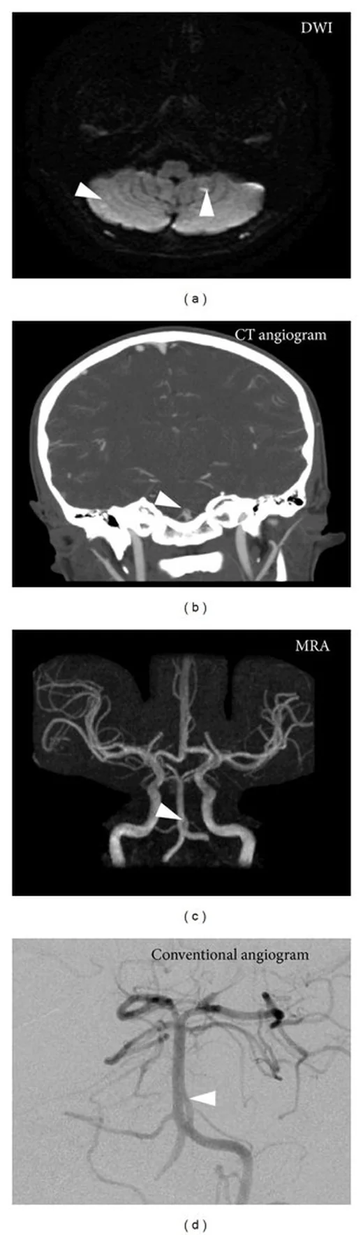
- Vertebral arter fenestrasyonu CTA ve MRA'da iki ayrı lümen olarak görülebilir; vertebral arter fenestrasyonları diseksiyonlara göre daha sık iki ayrı lümen gösterir. Proksimal baziler arterde, arterin iki paralel kanala ayrıldığı ve bu çift lümen yapısının ardışık kesitlerde korunduğu görülür.
Önerilen pencereler G genişlik, M merkez; değerler Hounsfield birimi (HU)
- AnjiyoG 600 / M +150
BT bulguları
- Vertebral arter fenestrasyonu özellikle V4 segmentinde görülebilir ve iki ayrı lümen göstermesiyle diseksiyondan ayrılmasına yardımcı olan bir görüntüleme özelliğine sahiptir.
- Tek arter kökenli olup distalde tek lümene yeniden kavuşan paralel kanallar, her ikisi de düzgün konturlu ve devamlıdır.
- Çok düzlemli ve üç boyutlu görüntülemelerde iki ayrı kanalın ayrılma ve tek lümene katılma ilişkisi tutarlı şekilde izlenir.
- Orta serebral arterde de fenestrasyon görülebilir; bu durumda arterin ana gövdesinde geçici olarak iki kanallı yapı oluşur.
- Ayrılma ya da birleşme köşesinde damarın dış konturundan kese biçimli çıkıntı, komşu anevrizma olasılığını gündeme getirir.
- Vertebral arter fenestrasyonu CTA ve MRA'da değerlendirilebilir; anjiyografik görüntülemede diseksiyondan ayırt edilebilir.
Normalde
Vertebral arterler baziler arteri oluşturmak üzere birleşir; AComA, iki ACA sistemi arasında bağlantı sağlayabilir. Şüpheli damarda kanalların iki uçtaki birleşme ilişkisini göster ve aynı seviyedeki ana lümen konturunu, karşı vertebral arteri ve komşu dalları karşılaştır.
Normal BT ile kıyasla
Aynı bölgenin, kaynak veri setinde patoloji içermediği belirtilen bir BT incelemesini kesit kesit kaydırın; organ sınırlarını açarak anatomiyi hasta görüntüsüyle karşılaştırın. Küçük rastlantısal bulgular tümüyle dışlanmamıştır.
Kaynak: TotalSegmentator v2.01 veri seti (Wasserthal ve ark., Radiology: Artificial Intelligence 2023), CC BY 4.0, vaka s0849; organ sınırları veri setinin otomatik segmentasyonundan, birleştirilerek sadeleştirildi. Görüntüler 2,5 mm kesit aralığına yeniden örneklendi.
Ayırıcı tanı
- Duvar diseksiyonu: düzensiz daralma, duvar boyunca uzanan flep veya dış kontur değişikliği eşlik edebilir; paralel yolların düzenli biçimde ortak gövdede sonlanması beklenmez.
- Vertebral arter diseksiyonundan ayrımda fenestrasyonun daha kısa segmentte görülmesi, daha geniş lümen çapı ve iki ayrı lümen göstermesi yardımcı görüntüleme özellikleridir.
- Tek düzlemli projeksiyonlarda üst üste binen dallar veya görüntü artefaktları yanlışlık verebilir; çok düzlemli rekonstrüksiyon (MPR) ve hacimsel görüntüleme ile gerçek damar bağıntısı doğrulanmalıdır.
- Damar dışı komşuluk üst üste binmesi: özellikle AComA çevresinde dalların projeksiyonda kesişmesi çift gövde sanılabilir; aksiyel kaynak kesit ve eğri düzlem rekonstrüksiyon ilişkiyi netleştirir.
- Duplikasyon
- iki damar ayrı köken ve seyirlerini korur ve distalde yeniden birleşmez; fenestrasyonda tek arter segmenti kısa bir bölümde ikiye ayrılıp distalde yeniden birleşir.
Tuzaklar
- Tek aksiyel kesitte görülen ince baziler çizgiyi gelişimsel varyant diye adlandırma; aynı yapının ardışık kesitlerde ve lümen merkez hattında sürmesini ara.
- AComA çevresindeki küçük damar varyantlarını değerlendirirken MIP görüntülerini dikkatle incele; kısmi hacim görüntüleme doğru tanıya yardımcı olabilir.
- Fenestrasyon saptanması tek başına anevrizma veya diseksiyon tanısı koydurmaz; varyantla ilişkili kese ve damar duvarı bulgularını ayrı tarif et.
- Fenestrasyonla anevrizma birlikteliği bildirilmiştir; vertebral arter fenestrasyonu saptandığında eşlik eden intrakraniyal anevrizma açısından inceleme önerilir. Supraklinoid ICA fenestrasyonunda anevrizma proksimal uçta sık görüldüğünden, bu bölgede anevrizma saptanırsa küçük fenestrasyon kanalı da araştırılmalıdır.
Kendini dene
Baziler gövdede iki opak yol kısa bir mesafe izleniyor ve yeniden tek damarda birleşiyor. En uygun tanı hangisidir?
Cevabı göster
Baziler arter fenestrasyonu. Aynı arterin kısa aralıkta iki kanala ayrılıp tekrar tek gövdede devam etmesi fenestrasyon morfolojisidir. Trombüs opaklaşmayan dolum alanı, diseksiyon duvar kaynaklı düzensizlik oluşturur; persistan trigeminal arter ise farklı damar yatakları arasında bağlantı kurar.
Bir damarın fenestrasyon olarak tanımlanmasını sağlayan temel morfoloji hangisidir?
Cevabı göster
Tek arterin kısa bir segmentte iki kanala ayrılıp yeniden birleşmesi. Fenestrasyonda tek arter iki kanala ayrılır ve bu kanallar yeniden birleşir.
İlgili konular
Kaynaklar
Bu sayfadaki 35 cümle ve 2 quiz sorusu aşağıdaki kaynaklardan alıntılarla otomatik olarak karşılaştırıldı; 14 cümle bu karşılaştırmada düzeltildi (2026-10-09).
- A Case of Horizontal Stent-assisted Coiling for an Aneurysm Arising from Fenestration of the Vertebral Artery: A Technical Case Reportpmc.ncbi.nlm.nih.gov
- Splenic artery fenestration – a previously undescribed arterial variantpmc.ncbi.nlm.nih.gov
- A distal anterior cerebral artery aneurysm arising from the middle of three A2 branches treated by stent-assisted coiling: a case reportpmc.ncbi.nlm.nih.gov
- Internal carotid artery fenestration with contralateral internal carotid artery dissection: a case report and literature reviewpmc.ncbi.nlm.nih.gov
- Internal carotid artery-middle cerebral artery junction small arterial ring: A report of two cases diagnosed by magnetic resonance angiographypmc.ncbi.nlm.nih.gov
- A Case of Horizontal Stent-assisted Coiling for an Aneurysm Arising from Fenestration of the Vertebral Artery: A Technical Case Report.pubmed.ncbi.nlm.nih.gov
- Microvascular Clipping of A1 Segment Aneurysms.pubmed.ncbi.nlm.nih.gov
- Anterior Communicating Artery Aneurysm Morphology and the Risk of Rupture.pubmed.ncbi.nlm.nih.gov
- Robot-assisted endoscopic third ventriculostomy: institutional experience in 9 patients.pubmed.ncbi.nlm.nih.gov
- Intracranial region of the vertebral artery: morphometric study in the context of clinical usefulness.pubmed.ncbi.nlm.nih.gov
- Vascular assessment after clipping surgery using four-dimensional CT angiography.pubmed.ncbi.nlm.nih.gov
- Microsurgical Clip Reconstruction of a Ruptured Internal Carotid Artery Terminus Blister Aneurysm Using a Reverse Picket Fence Technique.pubmed.ncbi.nlm.nih.gov
- Imaging features of vertebral artery fenestration.pubmed.ncbi.nlm.nih.gov
- Cisternostomy for malignant middle cerebral artery infarction: proposed pathophysiological mechanisms and preliminary results.pubmed.ncbi.nlm.nih.gov
- Microneurosurgical Management of Posterior Communicating Artery Aneurysm: A Contemporary Series from Helsinki.pubmed.ncbi.nlm.nih.gov
- Endovascular treatment modalities for basilar artery fenestration aneurysms: experience of two centers and literature review.pubmed.ncbi.nlm.nih.gov
- Basilar tip fenestration giving rise to Percheron's and mesencephalic arteries.pubmed.ncbi.nlm.nih.gov
- Unilateral Supraorbital Approach to Clip Bilateral Posterior Communicating Artery Aneurysms: 2-Dimensional Operative Video.pubmed.ncbi.nlm.nih.gov
- Vascular Neuroembryology: The Genesis of the Intracranial Arterial System and the Circle of Willis.pubmed.ncbi.nlm.nih.gov
- Aplastic or twig-like middle cerebral artery with the RNF213 variant: illustrative cases.pubmed.ncbi.nlm.nih.gov
- Double vertebral artery-connected persisting proatlantal artery coexisting with a persisting carotid duct in an adult case: first evidence.pubmed.ncbi.nlm.nih.gov
- Anatomic variations in basilar artery termination: a systematic review and meta-analysis with presentation of an illustrative case.pubmed.ncbi.nlm.nih.gov
- Complications of catheter-directed interventions in acute pulmonary embolism: a narrative expert review with guidance on management.pubmed.ncbi.nlm.nih.gov
- Picket-Fence Technique in Surgical Treatment of Cerebral Aneurysms and Role of Intraoperative Videoangiography in Aneurysm Surgery.pubmed.ncbi.nlm.nih.gov
- Cortical Venous Approach for Transvenous Embolization of a Greater Sphenoid Wing Dural Arteriovenous Fistula: A Case Report.pubmed.ncbi.nlm.nih.gov
- Carotid Web: An Update Focusing on Its Relationship With Fibromuscular Dysplasia and Therapeutic Strategy.pubmed.ncbi.nlm.nih.gov
- Diagnosis of Unruptured Intracranial Aneurysms Using Proton-Density Magnetic Resonance Angiography: A Comparison With High-Resolution Time-of-Flight Magnetic Resonance Angiography.pubmed.ncbi.nlm.nih.gov
- Internal carotid artery fenestration with contralateral internal carotid artery dissection: a case report and literature review.pubmed.ncbi.nlm.nih.gov
- Congenital external carotid-internal carotid artery anastomosis associated with moyamoya disease diagnosed by magnetic resonance angiography.pubmed.ncbi.nlm.nih.gov
- Anatomic Variants of the Anterior Cerebral Artery and Anterior Communicating Artery: A Digital Subtraction Angiographic Study.pubmed.ncbi.nlm.nih.gov
- Added diagnostic values of three-dimensional high-resolution proton density-weighted magnetic resonance imaging for unruptured intracranial aneurysms in the circle-of-Willis: Comparison with time-of-flight magnetic resonance angiography.pubmed.ncbi.nlm.nih.gov
- Normal anatomical features and variations of the vertebrobasilar circulation and its branches: an analysis with 64-detector row CT and 3T MR angiographies.pubmed.ncbi.nlm.nih.gov
- Long-term safety and effectiveness of the "OptEase" vena cava filter.pubmed.ncbi.nlm.nih.gov
- Radiological anatomy of the intracranial vertebral artery in a select South African cohort of patients.pubmed.ncbi.nlm.nih.gov
- International Society of Paediatric Surgical Oncology (IPSO) Surgical Practice Guidelines.pubmed.ncbi.nlm.nih.gov
- A distal anterior cerebral artery aneurysm arising from the middle of three A2 branches treated by stent-assisted coiling: a case report.pubmed.ncbi.nlm.nih.gov
- Congenital Anomalies and Variations of the Carotid and Vertebral Arteries: A Case-Based Imaging Review.pubmed.ncbi.nlm.nih.gov
- A Study on Branching Pattern of Middle Cerebral Artery and its Variations: A Systematic Review and Meta-analysis.pubmed.ncbi.nlm.nih.gov
- Treatment of Kommerell's diverticulum in rare aberrant aortic arch anatomy.pubmed.ncbi.nlm.nih.gov
- Middle cerebral artery fenestration presenting with cerebral ischemia: a case report and review of the literature.pubmed.ncbi.nlm.nih.gov
- A patient-specific CTA-CFD framework deciphers hemodynamic heterogeneity after fenestrated TEVAR: a pilot study.pubmed.ncbi.nlm.nih.gov
- The Initial Diagnostic Approach to Adult Congenital Heart Disease: A Practical Imaging Pathway for General Cardiologists and Cardiac Imagers.pubmed.ncbi.nlm.nih.gov
- Vertebrobasilar artery geometry and quantitative high-resolution vessel wall MRI for stratifying vulnerable plaques in young and middle-aged adults with stroke risk factors.pubmed.ncbi.nlm.nih.gov
- Frontobasal bridging vein rerouting for microsurgical clipping of a posterior cerebral artery aneurysm through anterior temporal approach: A technical note.pubmed.ncbi.nlm.nih.gov
- Vertebral Artery Pseudoaneurysm in a Single-Vessel Posterior Circulation With Chronic Contralateral Occlusion and Recent Methamphetamine Use.pubmed.ncbi.nlm.nih.gov
- Beyond the "string of beads": case-based exploration of diagnostic pitfalls and solutions in reversible cerebral vasoconstriction syndrome.pubmed.ncbi.nlm.nih.gov
- Expert consensus on intracranial vessel wall MRI in cerebrovascular disease: Society for Magnetic Resonance Angiography recommendations.pubmed.ncbi.nlm.nih.gov
- Contrast opacification on thoracic CT angiography: challenges and solutions.pubmed.ncbi.nlm.nih.gov
- Left Atrial Appendage: Mere Spectator or Protagonist? A Narrative Review of Its Multimodal Imaging Assessment and Its Closure Techniques.pubmed.ncbi.nlm.nih.gov
- Duropathies as Unifying Concept-Part Two: A Narrative Overview of Clinical and Neuroradiological Features.pubmed.ncbi.nlm.nih.gov
- Meningeal lymphatic architecture and drainage dynamics surrounding the human middle meningeal artery.pubmed.ncbi.nlm.nih.gov
- 2022 ACC/AHA guideline for the diagnosis and management of aortic disease: A report of the American Heart Association/American College of Cardiology Joint Committee on Clinical Practice Guidelines.pubmed.ncbi.nlm.nih.gov
- Society for Cardiovascular Magnetic Resonance/European Society of Cardiovascular Imaging/American Society of Echocardiography/Society for Pediatric Radiology/North American Society for Cardiovascular Imaging Guidelines for the Use of Cardiac Magnetic Resonance in Pediatric Congenital and Acquired Heart Disease: Endorsed by The American Heart Association.pubmed.ncbi.nlm.nih.gov
- Society for Cardiovascular Magnetic Resonance/European Society of Cardiovascular Imaging/American Society of Echocardiography/Society for Pediatric Radiology/North American Society for Cardiovascular Imaging Guidelines for the use of cardiovascular magnetic resonance in pediatric congenital and acquired heart disease : Endorsed by The American Heart Association.pubmed.ncbi.nlm.nih.gov
- AI-based detection and sizing of saccular intracranial aneurysms: a single-center retrospective validation study using computed tomography angiography.pubmed.ncbi.nlm.nih.gov
- Vessel Wall Imaging of Intracranial Arteries: Fundamentals and Clinical Applications.pubmed.ncbi.nlm.nih.gov
- Imaging in Infective Endocarditis-Current Opinions and Trends in Cardiac Computed Tomography.pubmed.ncbi.nlm.nih.gov
- Branch-Critical Clipping of a Ruptured Carotid-Posterior Communicating Aneurysm with Fetal PCA Configuration.pubmed.ncbi.nlm.nih.gov
- Unstented large fenestration for close target vessel ostia: Long-term follow-up.pubmed.ncbi.nlm.nih.gov
- Comparative Evaluation of Clinical Outcomes Following Endovascular and Hybrid Repair of Aortic Arch Aneurysms.pubmed.ncbi.nlm.nih.gov
- Medium-term clinical and imaging outcomes of SilverFlow covered bridging stent implantation using <i>in situ</i> fenestration for aortic arch lesions: a single-centre retrospective cohort study.pubmed.ncbi.nlm.nih.gov
- Case Report: A case of atypical persistent proatlantal intersegmental artery.pubmed.ncbi.nlm.nih.gov
- Bilateral persistence of the proatlantal intersegmental arteries in moyamoya syndrome: illustrative case.pubmed.ncbi.nlm.nih.gov
- The Vulnerable Conduit: Uncovering Segment-Specific Pathological Susceptibility and Bony Co-dependence of the Vertebral Artery in a South Indian Cohort.pubmed.ncbi.nlm.nih.gov
- From bench to bedside: relevant animal models across the migraine attack phases.pubmed.ncbi.nlm.nih.gov
- Chronic Subdural Hematoma: Pathophysiology, Diagnosis, and the Emerging Role of Middle Meningeal Artery Embolization.pubmed.ncbi.nlm.nih.gov
- Dysautonomia and Postural Orthostatic Tachycardia Syndrome (POTS) in the ENT Clinic: Differentiating Orthostatic Dizziness From Vestibular Migraine and Persistent Postural-Perceptual Dizziness (PPPD).pubmed.ncbi.nlm.nih.gov
- Clinical outcomes of external fenestration TEVAR in Stanford type B aortic dissection involving the left subclavian artery: a retrospective study.pubmed.ncbi.nlm.nih.gov
- Mechanisms of aortic dissection: From pathological changes to experimental and <i>in silico</i> models.pubmed.ncbi.nlm.nih.gov
- Adhesion G protein-coupled receptors.pubmed.ncbi.nlm.nih.gov
- Splenic artery fenestration - a previously undescribed arterial variant.pubmed.ncbi.nlm.nih.gov
- Morphological Variability of the P1 Segment of the Posterior Cerebral Artery: An Anatomical and Angiographic Study with Clinical Correlations.pubmed.ncbi.nlm.nih.gov
- A Functional Perspective on the Embryology and Anatomy of the Cerebral Blood Supply.pubmed.ncbi.nlm.nih.gov
- Prevalence of suboccipital and intradural vertebral artery variants: a systematic review with meta-analysis.pubmed.ncbi.nlm.nih.gov
- Persistent primitive lateral basilovertebral anastomosis-rethinking vertebrobasilar junction fenestration.pubmed.ncbi.nlm.nih.gov
- Endovascular treatment of fenestration of the posterior communicating artery with an aneurysm at the same site: case report and review of the literature.pubmed.ncbi.nlm.nih.gov
- Stent-assisted coil embolization of a fenestration-related aneurysm of an infraoptic anterior cerebral artery: A case report.pubmed.ncbi.nlm.nih.gov
- Internal carotid artery-middle cerebral artery junction small arterial ring: A report of two cases diagnosed by magnetic resonance angiography.pubmed.ncbi.nlm.nih.gov
- Fenestrations of cerebral arteries and their correlation with brain aneurysms.pubmed.ncbi.nlm.nih.gov
- Pulsatile Tinnitus: A Comprehensive Clinical Approach to Diagnosis and Management.pubmed.ncbi.nlm.nih.gov
- Non-Thrombotic Filling Defects in Cerebral Veins and Sinuses: When Normal Structures Mimic a Disease.pubmed.ncbi.nlm.nih.gov
- Microvascular decompression for hemifacial spasm associated with large fenestration of the vertebral artery: illustrative case.pubmed.ncbi.nlm.nih.gov
- Endovascular treatment of posterior circulation aneurysms with flow diverters with hydrophilic polymer coating in patients receiving prasugrel single antiplatelet therapy: a multicenter case series presenting complication and occlusion rates.pubmed.ncbi.nlm.nih.gov
- Extra-Carotid Arterial Webs: A Single-Center Series and Systematic Review.pubmed.ncbi.nlm.nih.gov
- Middle cerebral artery dissection: pathophysiology, diagnosis, and therapeutic options.pubmed.ncbi.nlm.nih.gov
- Perforator-Sparing Microsurgical Clipping of Tandem Dominant-Hemisphere Middle Cerebral Artery Aneurysms: Geometry-Guided Reconstruction of a Wide-Neck Bifurcation and Dorsal M1 Fusiform Lesion.pubmed.ncbi.nlm.nih.gov
- Cerebral venous blood flow regulates intracerebral pressure and brain clearance via meningeal lymphatic vessels.pubmed.ncbi.nlm.nih.gov
- Functional Arterial Anatomy of the Brain.pubmed.ncbi.nlm.nih.gov
- Common and uncommon intracranial arterial anatomic variations in multi-detector computed tomography angiography (MDCTA). What radiologists should be aware of.pubmed.ncbi.nlm.nih.gov
- Exploring the anatomical configurations of the cerebral arteries in a cohort of South African patients.pubmed.ncbi.nlm.nih.gov
- Is fetal-type posterior cerebral artery a risk factor for intracranial aneurysm as analyzed by multislice CT angiography?pubmed.ncbi.nlm.nih.gov
- 2022 ACC/AHA Guideline for the Diagnosis and Management of Aortic Disease: A Report of the American Heart Association/American College of Cardiology Joint Committee on Clinical Practice Guidelines.pubmed.ncbi.nlm.nih.gov
- The persistent proatlantal artery: types and subtypes.pubmed.ncbi.nlm.nih.gov
- Callosomarginal artery: an in-depth of anatomical characteristics, clinical significance, neurosurgical considerations, and surgical applications.pubmed.ncbi.nlm.nih.gov
- Unusual anatomical variations of cervical and cranial arteries with surgical interest: case report.pubmed.ncbi.nlm.nih.gov
- MDCT Imaging of Non-Traumatic Thoracic Aortic Emergencies and Its Impact on Diagnosis and Management-A Reappraisal.pubmed.ncbi.nlm.nih.gov
- Human cerebral arterial morphogenesis explored by 3D microscopic imaging of cleared embryos: Bridging the gap between embryonic and adult anatomy.pubmed.ncbi.nlm.nih.gov
- Subarachnoid cisterns as surgical corridors: Integrating microsurgical anatomy and neuroimaging for intracranial navigation.pubmed.ncbi.nlm.nih.gov
- Multiterritory Bihemispheric Ischemic Stroke in the Setting of Unilateral Internal Carotid Artery Disease.pubmed.ncbi.nlm.nih.gov
Eğitim amaçlıdır; hasta başında tanı ve tedavi kararının yerine geçmez, tıbbi tavsiye değildir. Metinler yapay zekâ desteğiyle yazıldı ve otomatik bir adımda kaynak alıntılarıyla karşılaştırıldı; bu bir tıbbi doğrulama değildir ve içerik uzman radyolog incelemesinden geçmedi. Klinik kararlarda güncel kılavuzları, kurum protokollerini ve radyoloji raporunu esas alın.