Özet
Willis poligonu varyantları, anterior ve posterior dolaşım arasındaki damarların kalibresini veya sürekliliğini ve ACA dallanma örüntüsünü değiştirebilir. BT anjiyografi, Willis poligonu bağlantı varyantlarını ve ACA dallanma örüntülerini gösterebilir. Willis poligonu varyantları kollateral akımı ve beynin besleyici arterlere ait perfüzyon alanlarının yorumunu etkileyebilir. Willis poligonu varyantlarının oklüzyon veya hipoplaziyle karıştırılması damar anatomisinin ve inme mekanizmasının yanlış yorumlanmasına yol açabilir.
Faz ve pencere
- BTA tanısal
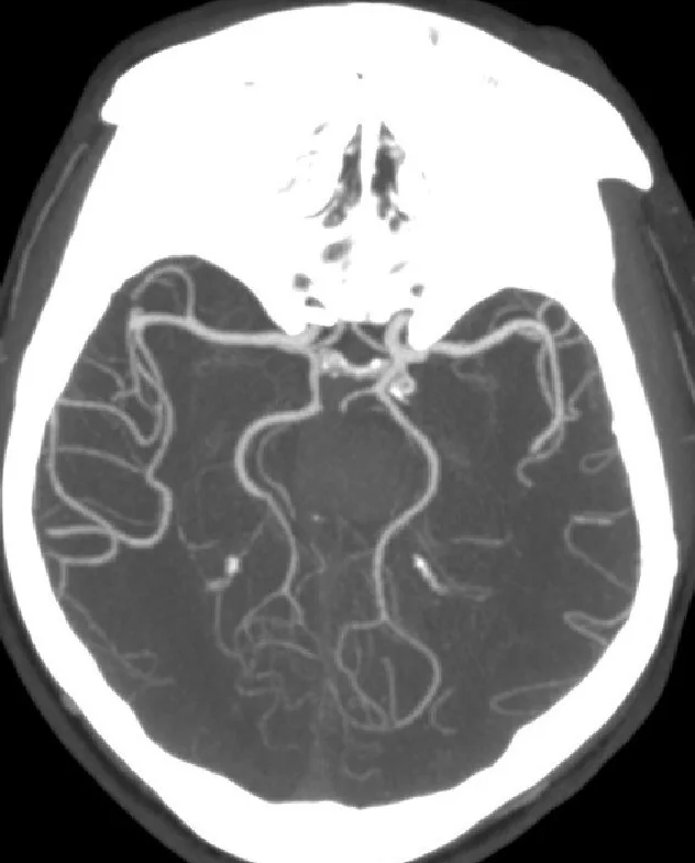
- Arteriyel BTA'da A1 segmentleri, AComA, PComA'lar, P1-P2 PCA geçişi ve distal ACA dalları opak lümen olarak izlenir. Fetal PCA'da ipsilateral PCA'nın PComA üzerinden ICA'dan beslendiği ve P1'in hipoplastik ya da izlenmediği; azygos ACA'da tek A2 gövdesinin iki medial hemisfere uzandığı görülür.
Önerilen pencereler: Anjiyo (G 600 / M 150).
BT bulguları
- Fetal tip PCA'da ipsilateral PComA belirgin bağlantıyı oluşturur ve PCA'nın P2 sonrası akımı internal karotid arterden gelir.
- Parsiyel fetal PCA’da PCA’nın beslenmesine ICA katkı sağlarken, tam fetal örüntüde PCA ağırlıklı ya da yalnızca ICA’dan beslenir.
- Fetal tip PCA'da ipsilateral PCA alanı, hipoplastik veya bulunmayan P1 ile birlikte, ağırlıklı olarak dominant PComA üzerinden ICA'dan beslenir.
- Azygos ACA'da A1 segmentleri tek orta hat A2 gövdesine katılır; distal perikallozal damar tek gövdeden iki medial hemisfer yönüne dallanır.
- ACA triplikasyonunda AComA distalinde üç ACA gövdesi izlenir; orta hatta seyreden ek dal median ACA örüntüsünü oluşturabilir. Her dalın distal gidişi takip edilir ve tek gövdeli azygos ACA ile karıştırılmaz.
- Bihemisferik ACA örüntüsünde bir taraftaki ACA ana gövdesi iki hemisferin medial yüzüne dallar verirken karşı A1 hipoplastik olabilir.
- Görüntülenemeyen A1, P1 veya PComA segmenti anatomik yoklukla eş tutulmamalı; Willis poligonu varyantı ve kollateral düzen bağlamında değerlendirilmelidir.
Normalde
Tipik erişkin bağlantı düzeninde AComA, sağ ve sol ACA sistemleri arasında anastomoz oluşturur; PCA’lar baziler arterin terminal dallarıdır ve PComA, ICA ile PCA arasındaki bağlantıyı sağlar. Willis poligonu damarlarının çap ve bağlantı varyantları kollateral akımı ve perfüzyon alanı yorumunu etkileyebileceğinden, şüpheli segmentin çapını ve ilgili alanın besleyici arterini görüntülerde birlikte değerlendirin.
Normal BT ile kıyasla
Aynı bölgenin, kaynak veri setinde patoloji içermediği belirtilen bir BT incelemesini kesit kesit kaydırın; organ sınırlarını açarak anatomiyi hasta görüntüsüyle karşılaştırın. Küçük rastlantısal bulgular tümüyle dışlanmamıştır.
Kaynak: TotalSegmentator v2.01 veri seti (Wasserthal ve ark., Radiology: Artificial Intelligence 2023), CC BY 4.0, vaka s0849; organ sınırları veri setinin otomatik segmentasyonundan, birleştirilerek sadeleştirildi. Görüntüler 2,5 mm kesit aralığına yeniden örneklendi.
Ayırıcı tanı
- A1 segment oklüzyonu değerlendirilirken, azygos veya bihemisferik ACA varyantlarının distal ACA dallarının tek ya da baskın bir gövdeden beslenmesine yol açabileceği göz önünde tutulmalıdır.
- P1 segment oklüzyonunda P1 başlangıcında tıkanıklık görülebilir; fetal tip PCA’da distal PCA alanı ağırlıklı olarak dominant PComA üzerinden ICA’dan beslenir ve ipsilateral P1 hipoplastik ya da bulunmayabilir.
- Azygos ACA, tek bir A2 ana gövdesi bulunan bir anterior serebral arter varyantıdır.
- ACA triplikasyonu veya median ACA
- AComA distalinde üç ACA gövdesi izlenir; tek gövdeli azygos örüntüsünden farklıdır.
- Arter hipoplazisi, Willis poligonu damarlarında görülen bir kalibre varyantıdır; görüntüde seçilememe anatomik yoklukla aynı kabul edilmemelidir.
- Arteriyovenöz şant, arter ile ven arasında doğrudan bağlantı bulunmasıdır; yalnız arter kalibresindeki farklılık şantı tanımlamak için yeterli değildir.
Tuzaklar
- Fetal PCA'yı P1 oklüzyonu diye yorumlamadan önce PComA'nın ICA'dan PCA'ya kesintisiz devamını ve baziler kökenli P1'i ayrı ayrı izleyin.
- Hipoplastik P1'in ince lümeni kalın MIP slabında kaybolabilir; ince kaynak kesit veya oblik MPR'de baziler tepe bağlantısını arayın.
- Azygos ACA'da tek distal A2 gövdesi, karşı ACA'nın kayıp olduğu izlenimini verebilir; tek gövdenin iki tarafa verdiği distal dalları takip edin.
- PComA veya A1'in ince olması akım yönünü tek başına göstermez; tek fazlı BTA anatomik çapı gösterir, fizyolojik kollateral yönünü doğrudan ölçmez.
Kendini dene
PCA, ICA’dan dominant PComA üzerinden ağırlıklı olarak besleniyor ve ipsilateral P1 hipoplastik ancak açık. En uygun tanımlama hangisidir?
Cevabı göster
Parsiyel fetal PCA. PCA’nın ICA’dan ağırlıklı, ancak yalnızca ICA’dan beslenmediği örüntü parsiyel fetal PCA olarak tanımlanır; tam fetal PCA’da beslenme yalnızca ICA kaynaklıdır.
CTA’da tek bir A2 gövdesi izleniyor ve distalinde dallanıyor. En olası varyant?
Cevabı göster
Azygos ACA. Tek A2 gövdesi azygos ACA örüntüsüyle uyumludur. ACA triplikasyonunda üç ayrı A2 dalı bulunur; fetal PCA ise PCA’nın ICA’dan beslendiği posterior dolaşım varyantıdır.
İlgili konular
Kaynaklar
Bu sayfadaki 39 cümle ve 2 quiz sorusu aşağıdaki kaynaklardan alıntılarla otomatik olarak karşılaştırıldı; 24 cümle bu karşılaştırmada düzeltildi (2026-10-09).
- Rare circle of Willis variants associated with multiple intracranial aneurysms and subarachnoid hemorrhagepmc.ncbi.nlm.nih.gov
- A distal anterior cerebral artery aneurysm arising from the middle of three A2 branches treated by stent-assisted coiling: a case reportpmc.ncbi.nlm.nih.gov
- Human cerebral arterial morphogenesis explored by 3D microscopic imaging of cleared embryos: Bridging the gap between embryonic and adult anatomypmc.ncbi.nlm.nih.gov
- Preoperative planning for endovascular aortic arch repairpmc.ncbi.nlm.nih.gov
- Absent internal carotid artery during neck exploration for penetrating traumapmc.ncbi.nlm.nih.gov
- Duplication of Intracavernous Internal Carotid Artery.pubmed.ncbi.nlm.nih.gov
- Atypical posterior circulation strokes: a case-based review of rare anatomical variations involved.pubmed.ncbi.nlm.nih.gov
- Congenital and acquired anomalies of the basilar artery: A pictorial essay.pubmed.ncbi.nlm.nih.gov
- A rare variant of accessory posterior cerebral artery.pubmed.ncbi.nlm.nih.gov
- The Coexistence of Accessory Anterior Cerebral and Anterior Communicating Arteries.pubmed.ncbi.nlm.nih.gov
- Rare Sighting of Posterior Communicating Artery Infundibulum With Posterior Cerebral Artery Origin.pubmed.ncbi.nlm.nih.gov
- Preoperative planning for endovascular aortic arch repair.pubmed.ncbi.nlm.nih.gov
- Absent internal carotid artery during neck exploration for penetrating trauma.pubmed.ncbi.nlm.nih.gov
- A rare coexistence of hypoplasia of the middle cerebral artery with aortic arch origin of the vertebral artery: a case report and analysis of hemodynamic vulnerability.pubmed.ncbi.nlm.nih.gov
- Computational Framework for Modeling Effects of Brain Collateral Circulation.pubmed.ncbi.nlm.nih.gov
- A distal anterior cerebral artery aneurysm arising from the middle of three A2 branches treated by stent-assisted coiling: a case report.pubmed.ncbi.nlm.nih.gov
- Morphometric and Clinical Analysis of the Azygos Anterior Cerebral Artery: Insights From a Cadaveric Case Report Study.pubmed.ncbi.nlm.nih.gov
- Anatomical variations of the intracranial arteries and their association with intracranial aneurysms: Insights from digital subtraction angiographies.pubmed.ncbi.nlm.nih.gov
- Multiterritory Bihemispheric Ischemic Stroke in the Setting of Unilateral Internal Carotid Artery Disease.pubmed.ncbi.nlm.nih.gov
- Endovascular thrombectomy for subterminal internal carotid artery occlusions: clinical and radiological heterogeneity in a nationwide registry.pubmed.ncbi.nlm.nih.gov
- Posterior Communicating Artery Configuration and Laterality of Thalamic and Lenticulostriate Infarction.pubmed.ncbi.nlm.nih.gov
- Interaction between vertebral artery hypoplasia and circle of Willis variants in posterior circulation stroke and TIA.pubmed.ncbi.nlm.nih.gov
- Vertebral Artery Hypoplasia and Posterior Circulation Vulnerability: A Systematic Review of Epidemiological, Hemodynamic, and Clinical Evidence.pubmed.ncbi.nlm.nih.gov
- Human cerebral arterial morphogenesis explored by 3D microscopic imaging of cleared embryos: Bridging the gap between embryonic and adult anatomy.pubmed.ncbi.nlm.nih.gov
- An Atlas of Neonatal Neurovascular Imaging Anatomy as Depicted with Microvascular Imaging: The Intracranial Arteries.pubmed.ncbi.nlm.nih.gov
- Clinical and Imaging Characteristics of Thalamic Infarction Combined With Moderate-to-Severe Stenosis of the Posterior Cerebral Artery: A Single-Center Experience.pubmed.ncbi.nlm.nih.gov
- Atypical variants of the cerebral arterial circle beyond the Lazorthes classification: a retrospective morphologic and neuroradiologic study.pubmed.ncbi.nlm.nih.gov
- Patterns of Circle of Willis Variants among Patients Undergoing 3 Tesla Magnetic Resonance Angiography at a Tertiary Care Hospital: An Observational Study.pubmed.ncbi.nlm.nih.gov
- Fetal-type posterior cerebral artery (fPCA): association with posterior communicating artery aneurysm rupture and treatment outcomes, with practical surgical risk-mitigation strategies.pubmed.ncbi.nlm.nih.gov
- Rare circle of Willis variants associated with multiple intracranial aneurysms and subarachnoid hemorrhage: A case report.pubmed.ncbi.nlm.nih.gov
- Ischemic complication in the anterior thalamoperforating artery territory following flow diverter stent placement for an internal carotid-posterior communicating artery aneurysm.pubmed.ncbi.nlm.nih.gov
- Flow Diversion in Fetal-Type Posterior Communicating Artery Aneurysms: Anatomical Challenges and Treatment Strategy.pubmed.ncbi.nlm.nih.gov
- Posterior Communicating Artery Aneurysm Microsurgery: PComA-CORE, an Anatomy-Informed Explainable AI Framework for Complexity, Neurovascular Risk, Oculomotor Recovery and Functional Outcome.pubmed.ncbi.nlm.nih.gov
- Predictors of Outcome After Endovascular Thrombectomy in Acute Basilar Artery Occlusion and the 6hr Time Window to Recanalization.pubmed.ncbi.nlm.nih.gov
- Multidetector-row CT angiography of cerebral vasospasm after aneurysmal subarachnoid hemorrhage: comparison of volume-rendered images and digital subtraction angiography.pubmed.ncbi.nlm.nih.gov
- Middle Cerebral Artery Aneurysm and Distal Anterior Cerebral Artery (DACA) Aneurysms Related to Azygos and an Unusual Single Pericallosal Artery Variant: A Case Report.pubmed.ncbi.nlm.nih.gov
- Endovascular Treatment of a Bihemispheric Distal Anterior Cerebral Artery A4 Broad-Necked Bifurcation Aneurysm Using the Woven EndoBridge Device: A Case Report.pubmed.ncbi.nlm.nih.gov
- Potentials of ChatGPT in Anatomy Research: A Conversation With ChatGPT.pubmed.ncbi.nlm.nih.gov
- Distinct Circle of Willis anatomical configurations in healthy preterm born adults: a 3D time-of-flight magnetic resonance angiography study.pubmed.ncbi.nlm.nih.gov
- Branching and nonbranching intracranial aneurysms in the presence of a persistent stapedial artery and an aberrant internal carotid artery assessed with computational hemodynamics: illustrative case.pubmed.ncbi.nlm.nih.gov
- Difficulty and prospects of endovascular treatment for spontaneous intracranial artery dissection.pubmed.ncbi.nlm.nih.gov
- Indirect Flow Diversion for Off-Centered Bifurcation Aneurysms and Distant Small-Vessel Aneurysms, a Retrospective Proof of Concept Study From Five Neurovascular Centers.pubmed.ncbi.nlm.nih.gov
- Prediction of Borderzone Infarction by CTA in Patients Undergoing Carotid Embolization for Carotid Blowout.pubmed.ncbi.nlm.nih.gov
- Anatomical Variations and Morphometric Features of the Anterior Cerebral Artery: A Systematic Review and Meta-Analysis of 24,015 Cases.pubmed.ncbi.nlm.nih.gov
- Case Report: Three cases involving the use of endovascular intervention for artery of Percheron infarction with a unilateral fetal-type posterior cerebral artery.pubmed.ncbi.nlm.nih.gov
- Ischemic Complications After Parent Artery Occlusion for Ruptured Posterior Cerebral Artery Aneurysm With a Fetal-Type Posterior Communicating Artery: A Case Report.pubmed.ncbi.nlm.nih.gov
- Microsurgical clipping for coil-extruded recurrent aneurysms: Case series and technical pearls.pubmed.ncbi.nlm.nih.gov
- Anterior Cerebral Artery Strokepubmed.ncbi.nlm.nih.gov
- Congenital internal carotid artery hypoplasia in a symptomatic young adult: A case report.pubmed.ncbi.nlm.nih.gov
- From imaging to impact: A case of renal arteriovenous malformation.pubmed.ncbi.nlm.nih.gov
- Transportal Embolization of the Dominant Outflow Vein for a Pancreatic Head Arteriovenous Malformation: A Case Report.pubmed.ncbi.nlm.nih.gov
- Balloon-assisted endovascular embolization of a subcutaneous penile arteriovenous fistula using a quadruple coaxial system: A case report.pubmed.ncbi.nlm.nih.gov
- The Effectiveness of the Multimodal Language Model, Google Gemini 2.5 Pro, in Solving the Specialization Exam in Gynecology and Obstetrics.pubmed.ncbi.nlm.nih.gov
- Current state and guidance on arterial spin labeling perfusion MRI in clinical neuroimaging.pubmed.ncbi.nlm.nih.gov
- Misinterpretation of ischaemic infarct location in relationship to the cerebrovascular territories.pubmed.ncbi.nlm.nih.gov
- Angiography of primary central nervous system angiitis of childhood: conventional angiography versus magnetic resonance angiography at presentation.pubmed.ncbi.nlm.nih.gov
- Overlapping Flow Diverter with Coiling for Fetal-type Posterior Communicating Artery Aneurysms with Triple Antithrombotic Therapy: A Case Series.pubmed.ncbi.nlm.nih.gov
- Estimating Flow Direction of Circle of Willis Using Dynamic Arterial Spin-Labeling MR Angiography.pubmed.ncbi.nlm.nih.gov
- Morphological Variations of the Anterior Cerebral Artery: A Systematic Review with Meta-Analysis of 85,316 Patients.pubmed.ncbi.nlm.nih.gov
- Current state of endovascular treatment of anterior cerebral artery aneurysms.pubmed.ncbi.nlm.nih.gov
- Functional Arterial Anatomy of the Brain.pubmed.ncbi.nlm.nih.gov
- Microneurosurgical anatomy of the basal cisterns: A brief review for cisternostomy.pubmed.ncbi.nlm.nih.gov
- Microsurgical Neurovascular Anatomy of the Brain: The Posterior Circulation (Part II).pubmed.ncbi.nlm.nih.gov
- Accessory middle cerebral artery, hypoplasia of the A1 segment of the left anterior cerebral artery fenestration of the A2 segment of the left anterior cerebral artery and fetal variant of posterior cerebral artery.pubmed.ncbi.nlm.nih.gov
- A primer to vascular anatomy of the brain: an overview on anterior compartment.pubmed.ncbi.nlm.nih.gov
- Microsurgical Neurovascular Anatomy of the Brain: The Anterior Circulation (Part I).pubmed.ncbi.nlm.nih.gov
- Microsurgical Anatomy of the Anterior Circulation of the Brain Adjusted to the Neurosurgeon's Daily Practice.pubmed.ncbi.nlm.nih.gov
- Single-Center Retrospective Case Series of 33 Patients with Ophthalmic-Cerebral Embolic Stroke Following Cosmetic Facial Injection, 2017-2025.pubmed.ncbi.nlm.nih.gov
- Motor Improvements Following Non-Invasive Brain Stimulation with kTMP: A Case Study of Cerebellar Stroke.pubmed.ncbi.nlm.nih.gov
- Rewiring the disordered connectome with circuit-based paired stimulation after stroke-a randomized, double-blind and controlled Phase II trial.pubmed.ncbi.nlm.nih.gov
- Middle Interhemispheric Variant of Holoprosencephaly With Septo-Optic Dysplasia: A Rare Association.pubmed.ncbi.nlm.nih.gov
- Unusual anatomical variations of cervical and cranial arteries with surgical interest: case report.pubmed.ncbi.nlm.nih.gov
- Morphological aspects of the vasculogenesis and angiogenesis during prenatal edification of the circle of Willis: a review.pubmed.ncbi.nlm.nih.gov
- Posterior communicating artery variation is linked to compromised cerebral hemodynamics in middle-aged and older adults.pubmed.ncbi.nlm.nih.gov
- Anatomic variations in basilar artery termination: a systematic review and meta-analysis with presentation of an illustrative case.pubmed.ncbi.nlm.nih.gov
- Congenital Anomalies and Variations of the Carotid and Vertebral Arteries: A Case-Based Imaging Review.pubmed.ncbi.nlm.nih.gov
- A comprehensive study of the anatomical variations of the circle of willis in adult human brains.pubmed.ncbi.nlm.nih.gov
- Morphology and Variations of the Posterior Cerebral Artery: A Literature Review.pubmed.ncbi.nlm.nih.gov
- The Anatomical Variation of the Distal Anterior Cerebral Artery: An Angiographic Study in a Greek Population Sample.pubmed.ncbi.nlm.nih.gov
- Circle of Willis Configuration and Thrombus Localization Impact on Ischemic Stroke Patient Outcomes: A Systematic Review.pubmed.ncbi.nlm.nih.gov
- Association between the posterior part of the circle of Willis and the vertebral artery hypoplasia.pubmed.ncbi.nlm.nih.gov
- High-Resolution MRA Cerebrovascular Findings in a Tri-Ethnic Population.pubmed.ncbi.nlm.nih.gov
- The persistent proatlantal artery: types and subtypes.pubmed.ncbi.nlm.nih.gov
- Diagnostic Clues to Platypnoea Orthodexia Syndrome.pubmed.ncbi.nlm.nih.gov
- A giant challenge: Hybrid management of a scalp and neck cirsoid aneurysm.pubmed.ncbi.nlm.nih.gov
- Computational fluid dynamics assessment of altered hemodynamics in the Circle of Willis during acute ischemic stroke and the impact of cerebral collateral development.pubmed.ncbi.nlm.nih.gov
- The detour technique: transcirculation stent coiling for complex cerebellar artery aneurysms. Illustrative cases.pubmed.ncbi.nlm.nih.gov
- Incidental vascular findings on brain magnetic resonance angiography.pubmed.ncbi.nlm.nih.gov
- Persistent trigeminal artery linked with partial duplication of the anterior communicating artery.pubmed.ncbi.nlm.nih.gov
- The anterior communicating artery variants: a meta-analysis with a proposed classification system.pubmed.ncbi.nlm.nih.gov
- Association of the Middle Accessory Cerebral Artery With Bihemispheric Anterior Cerebral Artery and Median Artery of the Corpus Callosum.pubmed.ncbi.nlm.nih.gov
- Planning-free cerebral blood flow territory mapping in patients with intracranial arterial stenosis.pubmed.ncbi.nlm.nih.gov
- Spontaneous bacteremia and spontaneous bacterial peritonitis share similar prognosis in patients with cirrhosis: a cohort study.pubmed.ncbi.nlm.nih.gov
- Operative Procedures for Ultrasound Assessment of Extracranial Artery Disease: A Narrative Review by the Italian Society for Vascular Investigation (SIDV).pubmed.ncbi.nlm.nih.gov
- Spontaneous Intracranial Vertebral Artery Dissection: A Rare Cause of Ischemic Stroke.pubmed.ncbi.nlm.nih.gov
- Cerebral proliferative angiopathy in pediatric patients: case-based review with an illustrative case.pubmed.ncbi.nlm.nih.gov
- The Role of Angiographic Imaging in the Treatment of Spinal Vascular Malformations.pubmed.ncbi.nlm.nih.gov
- Pulmonary arteriovenous fistula: diagnosis, interventional therapy and clinical progress.pubmed.ncbi.nlm.nih.gov
- Fetal-Type Variants of the Posterior Cerebral Artery and Concurrent Infarction in the Major Arterial Territories of the Cerebral Hemisphere.pubmed.ncbi.nlm.nih.gov
- Retrospective study on hemodynamic and morphological characteristics of posterior communicating artery aneurysms associated with fetal-type posterior cerebral artery and their correlation with rupture risk.pubmed.ncbi.nlm.nih.gov
- Azygous anterior cerebral artery occlusion managed with Thrombectomy: a case report and literature review.pubmed.ncbi.nlm.nih.gov
- Multiphase CT angiography increases detection of anterior circulation intracranial occlusion.pubmed.ncbi.nlm.nih.gov
- Multimodal imaging in neuro-ophthalmology.pubmed.ncbi.nlm.nih.gov
- Cerebral Venous Outflow Insufficiency: A Study on Symptoms and Venous Stenosis Classification.pubmed.ncbi.nlm.nih.gov
- Moyamoya Vasculopathy and Atypical Moyamoya-like Patterns: Insights into Diagnosis and Therapeutic Implications.pubmed.ncbi.nlm.nih.gov
- Cirrhosis-associated intracranial shunting lesions: illustrative case.pubmed.ncbi.nlm.nih.gov
- Adult intracranial pial arteriovenous fistulas: a case report and literature review.pubmed.ncbi.nlm.nih.gov
- Stroke-Mimic: An Intracranial Dural Arteriovenous Fistula Revealed by New-Onset Seizures and Post-Ictal Deficit.pubmed.ncbi.nlm.nih.gov
- The Fetal Type of Posterior Cerebral Artery.pubmed.ncbi.nlm.nih.gov
- Beyond visual inspection: the deep learning revolution in quantitative cerebrovascular imaging.pubmed.ncbi.nlm.nih.gov
- A Novel Patient-Specific Computational Fluid Dynamics Study of the Activation of Primary Collateral Pathways in the Circle of Willis During Vasospasm.pubmed.ncbi.nlm.nih.gov
- Proceedings of the 15th International Congress on Circumpolar Health.pubmed.ncbi.nlm.nih.gov
- Virtual test occlusion for assessing ischemic tolerance using computational fluid dynamics.pubmed.ncbi.nlm.nih.gov
- Chinese expert consensus on the diagnosis and treatment of coronary microvascular diseases (2023 Edition).pubmed.ncbi.nlm.nih.gov
- Angiographic characteristics and endovascular treatment of posterior communicating aneurysms: A single-center experience.pubmed.ncbi.nlm.nih.gov
Eğitim amaçlıdır; hasta başında tanı ve tedavi kararının yerine geçmez, tıbbi tavsiye değildir. Metinler yapay zekâ desteğiyle yazıldı ve otomatik bir adımda kaynak alıntılarıyla karşılaştırıldı; bu bir tıbbi doğrulama değildir ve içerik uzman radyolog incelemesinden geçmedi. Klinik kararlarda güncel kılavuzları, kurum protokollerini ve radyoloji raporunu esas alın.