Özet
Transvers sinüs hipoplazisi veya aplazisi anatomik varyant olabilir ve MR venografide sinyal kaybı trombozu taklit edebilir. MR venografide ince veya sinyal vermeyen transvers sinüsün trombozdan ayrımında, BT’de sigmoid çentiğin yokluğu ya da asimetrisi gelişimsel atreziyi veya ağır hipoplaziyi destekler. BT venografide kontrast dolum kusuru trombüsü gösterebilir; BT’de sigmoid çentiğin yokluğu veya asimetrisi atrezik ya da ağır hipoplastik transvers sinüsü destekler. Dural sinüs varyasyonları trombozu taklit ederek yanlış tanıya yol açabilir.
Faz ve pencere
- Kontrastsız
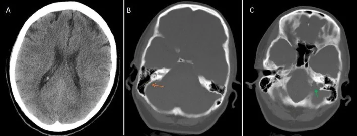
- Kontrastsız BT’de transvers sinüs lümeninde kontrast opasifikasyonu değerlendirilemez; kemik penceresinde küçük sinüs tarafındaki sigmoid çentiğin karşı tarafa göre yokluğu veya asimetrisi gelişimsel atrezi ya da ağır hipoplaziyi destekler.
- BTV tanısal
- BT venografide tromboz, sinüs düzeyinde kontrast dolumunun kesintiye uğramasıyla görülebilir; dolum bulguları kontrastsız BT ile birlikte değerlendirilir.
Önerilen pencereler G genişlik, M merkez; değerler Hounsfield birimi (HU)
- AnjiyoG 600 / M +150
- KemikG 1800 / M +400
BT bulguları
- Uzun segmentte düzgün küçük kalibre — transvers sinüs torculadan sigmoid geçişe kadar ince seyreder; tek noktadaki ani daralma veya düzensizlik varyantı tek başına açıklamaz.
- Aynı tarafta sığ sigmoid çentik — oksipital kemikteki sigmoid oluk karşı tarafa göre küçük gelişmiş olabilir; konjenital düşük venöz kapasite lehine destek bulgusudur.
- Küçük juguler foramen veya bulb — sinüs hipoplazisine aynı tarafta eşlik edebilir ve kemik gelişiminin uzun süreli asimetrisini destekler.
- Karşı transvers sinüs baskınlığı — superior sagittal sinüsün daha geniş sinüse yönelmesi sık bir drenaj örüntüsüdür; torcular akım paylaşımı kişiden kişiye değişebilir.
- Sigmoid sinüse distal devam — transvers sinüsün sigmoid sinüse uzanan kesiminde hipoplazi ile tromboz ayrımında sigmoid çentiğin BT’deki görünümü yardımcı olabilir.
- Trombozda venöz ödem, infarkt veya hemoraji gelişebilir; ancak parankimal lezyon yokluğu trombozu dışlamaz.
Normalde
Venöz faz BT’de sağ ve sol transvers sinüsler torculadan sigmoid sinüslere doğru opaklaşır; çapları eşit olmak zorunda değildir ve taraf baskınlığı kişiden kişiye değişir. Küçük görünen sinüsün tüm uzunluğunu, ipsilateral sigmoid çentik ve juguler foramen boyutunu karşı tarafla kıyaslayın; damar içi dolum kusurunu ayrıca değerlendirin.
Normal BT ile kıyasla
Aynı bölgenin, kaynak veri setinde patoloji içermediği belirtilen bir BT incelemesini kesit kesit kaydırın; organ sınırlarını açarak anatomiyi hasta görüntüsüyle karşılaştırın. Küçük rastlantısal bulgular tümüyle dışlanmamıştır.
Kaynak: TotalSegmentator v2.01 veri seti (Wasserthal ve ark., Radiology: Artificial Intelligence 2023), CC BY 4.0, vaka s0103; organ sınırları veri setinin otomatik segmentasyonundan, birleştirilerek sadeleştirildi. Görüntüler 2,5 mm kesit aralığına yeniden örneklendi.
Ayırıcı tanı
- Akut transvers sinüs trombozu
- sinüs içinde kontrast dolum kusuru veya kesilme, komşu venöz konjesyon ve parankimal venöz infarkt eşlik edebilir.
- Kronik sinüs trombozu
- düzensiz rekanalizasyon, ağsı kanallar veya duvar boyunca kollateral venler oluşturabilir; kemik oluğu tek başına kronik tromboz kanıtı değildir.
- Araknoid granülasyon
- sinüs içinde sınırlı, yuvarlak veya oval dolum defekti yapar; transvers sinüsün bütün uzunluğunu küçültmez.
- Akım-zamanlama artefaktı
- erken ya da asimetrik venöz opasifikasyon bir sinüsü yalancı boş gösterebilir; gecikmeli venöz dolum ve kaynak kesitler kontrol edilir.
Tuzaklar
- TOF-MRV’de yavaş akıma bağlı sinyal kaybı trombozu taklit edebilir; bulguyu tek başına tromboz olarak yorumlamayın, BT venografi veya kontrastlı MR venografi ile sinüs dolumunu doğrulayın.
- Sigmoid çentik bulgusu gelişimsel hipoplaziyi destekler fakat eşzamanlı yeni trombozu dışlamaz; sinüsün opaklaşmasını ve parankimi ayrıca okuyun.
- Torcula bölgesindeki normal drenaj asimetrisi bir sinüsü az dolu gösterebilir; transvers ve sigmoid segmentlerin devamını farklı rekonstrüksiyonlarda izleyin.
- BT’de sigmoid çentiklerin asimetrik olması atrezik veya ağır hipoplastik transvers sinüsü destekler; sinüs lümeni venografiyle ayrıca değerlendirilir.
Kendini dene
Venöz görüntülemede sol transvers sinüs ince; BT’de sol sigmoid çentik karşı tarafa göre asimetrik. En olası açıklama?
Cevabı göster
Konjenital sinüs hipoplazisi. BT'de sigmoid çentiğin yokluğu veya asimetrisi atrezik ya da ağır hipoplastik transvers sinüsü destekler. Sigmoid çentiklerin simetrisi trombozu destekler; venöz dolum bulguları ve kontrastsız BT birlikte değerlendirilmelidir. Tromboz venografide sinüs dolum kusuru veya kesilmesiyle görülebilir; araknoid granülasyonlar tipik yerleşimlerinde daha yuvarlak görünür.
MRV'de sol transvers sinüs sinyali yok; kontrastsız BT'de iki sigmoid çentik benzer büyüklükte. Bu kemik bulgusu neyi düşündürür?
Cevabı göster
Tromboz olasılığını artırır. Sigmoid çentiğin küçük veya asimetrik olması gelişimsel atrezik ya da ağır hipoplastik sinüsü destekler; çentiklerin simetrik olması trombozu destekler. İki çentiğin benzer büyüklükte olması konjenital sol sinüs hipoplazisi/aplazisini desteklemez ve tromboz olasılığını artırır. Sigmoid çentik bulgusu tek başına kesin tanı koydurmaz; sigmoid çentik değerlendirmesi ile venöz görüntüleme bulguları birlikte yorumlanmalıdır.
Kaynaklar
Bu sayfadaki 37 cümle ve 2 quiz sorusu aşağıdaki kaynaklardan alıntılarla otomatik olarak karşılaştırıldı; 20 cümle bu karşılaştırmada düzeltildi (2026-10-09).
- Congenital Anomalies and Variations of the Carotid and Vertebral Arteries: A Case-Based Imaging Reviewpmc.ncbi.nlm.nih.gov
- Intracranial venous thrombosis imaging by anatomical location: an educational review with mimics and diagnostic pitfallspmc.ncbi.nlm.nih.gov
- CraniofacialMorphometric Associations with Frontal Sinus Hypoplasia/Aplasia in Adults: Orbital and Upper Facial Differences on CTpmc.ncbi.nlm.nih.gov
- Congenital and Breed-Associated Vertebral Anomalies in Dogs and Cats: Developmental Anatomy, Genetic Determinants, and Biological Significancepmc.ncbi.nlm.nih.gov
- Morphological variations of the dural venous sinuses: a cadaveric studypmc.ncbi.nlm.nih.gov
- Maxillary sinus aplasia.pubmed.ncbi.nlm.nih.gov
- Tracheobronchial Branching Abnormalities: Lobe-based Classification Scheme.pubmed.ncbi.nlm.nih.gov
- Congenital Thoracic Aortic Disease.pubmed.ncbi.nlm.nih.gov
- Quadrifurcation Variants of the Celiac Trunk.pubmed.ncbi.nlm.nih.gov
- Inner ear malformations: a practical diagnostic approach.pubmed.ncbi.nlm.nih.gov
- Anatomy of extrahepatic portosystemic shunts in dogs as determined by computed tomography angiography.pubmed.ncbi.nlm.nih.gov
- Frontotemporal dementia: an unusual cause.pubmed.ncbi.nlm.nih.gov
- Bilateral parotid glands aplasia: a case report and literature review.pubmed.ncbi.nlm.nih.gov
- CT of the neonatal head.pubmed.ncbi.nlm.nih.gov
- The stylohyoid chain: CT imaging.pubmed.ncbi.nlm.nih.gov
- CT of blunt diaphragmatic rupture.pubmed.ncbi.nlm.nih.gov
- Ovarian inguinal hernia - a possibility in MURCS syndrome.pubmed.ncbi.nlm.nih.gov
- Isolated left pulmonary artery hypoplasia.pubmed.ncbi.nlm.nih.gov
- Pseudoaortic dissection.pubmed.ncbi.nlm.nih.gov
- Malformation of the eighth cranial nerve in children.pubmed.ncbi.nlm.nih.gov
- Intracranial venous thrombosis imaging by anatomical location: an educational review with mimics and diagnostic pitfalls.pubmed.ncbi.nlm.nih.gov
- Non-Thrombotic Filling Defects in Cerebral Veins and Sinuses: When Normal Structures Mimic a Disease.pubmed.ncbi.nlm.nih.gov
- Pearls and Pitfalls in the Magnetic Resonance Diagnosis of Dural Sinus Thrombosis: A Comprehensive Guide for the Trainee Radiologist.pubmed.ncbi.nlm.nih.gov
- Anatomical Reasons for an Impaired Internal Jugular Flow.pubmed.ncbi.nlm.nih.gov
- A Practical Approach to Intracranial Dural Arteriovenous Fistulas: Pathogenesis, Classification and Management.pubmed.ncbi.nlm.nih.gov
- Pulsatile Tinnitus: A Comprehensive Clinical Approach to Diagnosis and Management.pubmed.ncbi.nlm.nih.gov
- Anatomical Variations of the Jugular Bulb: A Critical and Comprehensive Review.pubmed.ncbi.nlm.nih.gov
- Anatomy imaging and hemodynamics research on the cerebral vein and venous sinus among individuals without cranial sinus and jugular vein diseases.pubmed.ncbi.nlm.nih.gov
- "Liberation treatment" for chronic cerebrospinal venous insufficiency in multiple sclerosis: the truth will set you free.pubmed.ncbi.nlm.nih.gov
- Morphological variations of the dural venous sinuses: a cadaveric study.pubmed.ncbi.nlm.nih.gov
- A review of cerebrospinal fluid circulation with respect to Chiari-like malformation and syringomyelia in brachycephalic dogs.pubmed.ncbi.nlm.nih.gov
- Congenital Anomalies and Variations of the Carotid and Vertebral Arteries: A Case-Based Imaging Review.pubmed.ncbi.nlm.nih.gov
- Endovascular treatment of pulsatile tinnitus associated with venous sinus diverticulum: a scoping review.pubmed.ncbi.nlm.nih.gov
- Cerebral Venous Outflow Insufficiency: A Study on Symptoms and Venous Stenosis Classification.pubmed.ncbi.nlm.nih.gov
- Patterns of injury and violence in Yaoundé Cameroon: an analysis of hospital data.pubmed.ncbi.nlm.nih.gov
- Cerebral Venous Thrombosis: Pathophysiologic Insights, Clinical Evaluation Tools, and Novel Therapeutic Strategies.pubmed.ncbi.nlm.nih.gov
- Pathophysiology, diagnosis and management of cerebral venous thrombosis: A comprehensive review.pubmed.ncbi.nlm.nih.gov
- Central nervous system vascular malformations: A clinical review.pubmed.ncbi.nlm.nih.gov
- Venous-significant intracranial hemorrhage in neonatal HIE treated with therapeutic hypothermia: imaging phenotypes, diagnostic pitfalls, and prognostic implications.pubmed.ncbi.nlm.nih.gov
- Can occipital lobe bending, Gibraltar sign of superior sagittal sinus groove and jugular foramen dimensions predict transverse sinus dominance?pubmed.ncbi.nlm.nih.gov
- Oblique occipital sinus in neurosurgical practice: A retrospective analysis of imaging studies.pubmed.ncbi.nlm.nih.gov
- Mastoid emissary vein, mastoid emissary foramen, and mastoid emissary canal: anatomy, variability, imaging, and clinical implications.pubmed.ncbi.nlm.nih.gov
- Jugular foramen dural arteriovenous fistula with bridging vein at the foramen magnum level successfully treated by surgery: illustrative case.pubmed.ncbi.nlm.nih.gov
- Cerebral venous disorders: clinical presentation, diagnostic strategy, and contemporary management.pubmed.ncbi.nlm.nih.gov
- Endovascular Embolization in Neurovascular Disease: Material Science, Multimodal Management, and Future Horizons.pubmed.ncbi.nlm.nih.gov
- Evaluation of Arachnoid Granulations in Cranial Dural Sinuses with Contrast-Enhanced 3-Dimensional T1-Weighted Magnetic Resonance Imaging.pubmed.ncbi.nlm.nih.gov
- Multimodal imaging in neuro-ophthalmology.pubmed.ncbi.nlm.nih.gov
- When Cancer Clots: An Extensive Radiologic Analysis of Cancer-Associated Thromboembolism.pubmed.ncbi.nlm.nih.gov
- Total laparoscopic radical hysterectomy for cervical cancer in prolapsed uterus.pubmed.ncbi.nlm.nih.gov
- Magnetic resonance imaging of the orbit, Part 2: Characterization of orbital pathologies.pubmed.ncbi.nlm.nih.gov
- Polycythemia Vera, Thrombophilia, CTEPH, Cerebral Venous Sinus Thrombosis and Vertebral Artery Occlusion: A Case-Illustrated Narrative Review of Competing Thrombotic and Hemorrhagic Risks.pubmed.ncbi.nlm.nih.gov
- Cerebral Venous Thrombosis Involving the Vein of Labbé and Left Sigmoid Sinus in a Patient With Immune Thrombocytopenia Treated With Romiplostim: A Case Report and Literature Review.pubmed.ncbi.nlm.nih.gov
- "Leave me alone": anatomical structures and variations seen on computed tomography of the temporal bone.pubmed.ncbi.nlm.nih.gov
- Congenital anomalies of the tubular gastrointestinal tract.pubmed.ncbi.nlm.nih.gov
- Standardization of pediatric uroradiological terms: a multidisciplinary European glossary.pubmed.ncbi.nlm.nih.gov
- Role of ethnicity in the determination of the transverse sigmoid sinus Junction (TSSJ): A prospective hospital-based radiological study in adult Sabah, East Malaysia.pubmed.ncbi.nlm.nih.gov
- CraniofacialMorphometric Associations with Frontal Sinus Hypoplasia/Aplasia in Adults: Orbital and Upper Facial Differences on CT.pubmed.ncbi.nlm.nih.gov
- State of the art: noninvasive imaging and management of neurovascular trauma.pubmed.ncbi.nlm.nih.gov
- Transverse Sinus Hypoplasia as a Differential Diagnosis for Cerebral Vein Thrombosis: A Case Report.pubmed.ncbi.nlm.nih.gov
- Can the sigmoid notch sign be used on cranial computed tomography to differentiate between thrombosis and hypoplasia of the cerebral transverse venous sinus?pubmed.ncbi.nlm.nih.gov
- Occipital transtentorial approach selected based on venous anatomy for a vermian cavernous malformation: illustrative case.pubmed.ncbi.nlm.nih.gov
- Case Report: Intracranial hypertension occurring in association with subacute thyroiditis.pubmed.ncbi.nlm.nih.gov
- Cerebral Vein Malformations Result from Loss of Twist1 Expression and BMP Signaling from Skull Progenitor Cells and Dura.pubmed.ncbi.nlm.nih.gov
- International Consensus Statement on Obstructive Sleep Apnea.pubmed.ncbi.nlm.nih.gov
- International Harmonization of Nomenclature and Diagnostic Criteria (INHAND): Non-proliferative and Proliferative Lesions of the Non-human Primate ( M. fascicularis ).pubmed.ncbi.nlm.nih.gov
- Case Report: Pediatric MOG/NMDAR overlap syndrome with delayed cortical MRI evolution and incidental venous sinus hypoplasia.pubmed.ncbi.nlm.nih.gov
- Long-term safety and effectiveness of the "OptEase" vena cava filter.pubmed.ncbi.nlm.nih.gov
- Posterior Fossa Venous Drainage.pubmed.ncbi.nlm.nih.gov
- The cavernous sinus: An anatomic study with clinical implication.pubmed.ncbi.nlm.nih.gov
- Transarterial Treatment of Cranial Dural Arteriovenous Fistulas: The Role of Transarterial and Transvenous Balloon-Assisted Embolization.pubmed.ncbi.nlm.nih.gov
- Radiological assessment of dementia: the Italian inter-society consensus for a practical and clinically oriented guide to image acquisition, evaluation, and reporting.pubmed.ncbi.nlm.nih.gov
- Absent Filling of Ipsilateral Superficial Middle Cerebral Vein Is Associated With Poor Outcome After Reperfusion Therapy.pubmed.ncbi.nlm.nih.gov
- Obstruction of Venous Drainage Linked to Transient Global Amnesia.pubmed.ncbi.nlm.nih.gov
- International Society of Paediatric Surgical Oncology (IPSO) Surgical Practice Guidelines.pubmed.ncbi.nlm.nih.gov
- Rethinking the Origin of the Primary Respiratory Mechanism.pubmed.ncbi.nlm.nih.gov
- High-resolution susceptibility-weighted imaging for detecting cerebral venous thrombosis across various venous segments and disease stages.pubmed.ncbi.nlm.nih.gov
- Anatomical Variability of the Transverse and Sigmoid Sinus Grooves: Implications for Skull Base Surgery.pubmed.ncbi.nlm.nih.gov
- The Mastoid Foramen and Mastoid Emissary Vein Canal as Surgical Landmarks for Mastoid Pneumatization During Retrosigmoid Approach.pubmed.ncbi.nlm.nih.gov
- Two pediatric cases of epidural hematoma in the posterior fossa with extension along the sigmoid sinus groove: MR evaluation.pubmed.ncbi.nlm.nih.gov
- Factors influencing therapy choice and clinical outcome in cerebral venous sinus thrombosis.pubmed.ncbi.nlm.nih.gov
- Cerebral Venous Sinus Thrombosis Revealing ALK-Positive Anaplastic Large-Cell Lymphoma in a Patient with Inherited Thrombophilia and Concomitant Infection: Case Report and Narrative Review.pubmed.ncbi.nlm.nih.gov
- Otogenic Sigmoid Sinus Thrombosis in a 34-Year-Old Woman: A Rare Intracranial Complication.pubmed.ncbi.nlm.nih.gov
- Bony prominence and variations of the transverse sinus groove: novel anatomical findings in relation to sinus pathologies.pubmed.ncbi.nlm.nih.gov
- Imaging of Cerebral Venous Thrombosis.pubmed.ncbi.nlm.nih.gov
Eğitim amaçlıdır; hasta başında tanı ve tedavi kararının yerine geçmez, tıbbi tavsiye değildir. Metinler yapay zekâ desteğiyle yazıldı ve otomatik bir adımda kaynak alıntılarıyla karşılaştırıldı; bu bir tıbbi doğrulama değildir ve içerik uzman radyolog incelemesinden geçmedi. Klinik kararlarda güncel kılavuzları, kurum protokollerini ve radyoloji raporunu esas alın.