Özet
Gelişimsel venöz anomali (DVA), normal beyin dokusunun medüller venöz kanını bir toplayıcı vene aktaran yaygın bir drenaj varyantıdır ve çoğunlukla tesadüfen saptanır. DVA'nın kendisi çoğu kez klinik sorun yaratmaz; komşu kanama, venöz enfarkt veya toplayıcı ven trombozu ayrıca araştırılmalıdır. Drenaj damarını kitle veya anormal yüksek akımlı şant sanmak gereksiz müdahaleye, tromboz ya da ilişkili kavernomu atlamak ise komplikasyonun nedenini kaçırmaya yol açar.
Okuma sırası
- Derin medüller venler. Derin beyaz cevherde ince kontrastlı venleri arayın; venlerin ortak bir çıkışa doğru yelpaze gibi dizilmesi DVA'yı tanıtan örüntüdür.
- Toplayıcı ven. Radiküllerin birleştiği kalın damarı kesitler boyunca izleyin ve kortikal vene, derin vene ya da dural sinüse ulaştığı yeri saptayın.
- Venöz radiküller arası parankim. Venöz radiküller arasındaki parankimin korunmuş olması ve DVA'da arteriyel bileşen bulunmaması, DVA'yı AVM'den ayırmaya yardımcı olur.
- DVA drenaj alanı ve toplayıcı ven. Drenaj alanında hipodens ödem, hematom veya belirgin hiperdens toplayıcı ven arayın; bu bulgular venöz enfarkt, kanama ya da tromboz komplikasyonunu gündeme getirir.
Faz ve pencere
- Kontrastsız
- Küçük DVA genellikle görünmez; büyük ve patent toplayıcı ven hafif hiperdens seçilebilir. Belirgin hiperdens toplayıcı ven tromboz kuşkusunu doğurur ve kontrastlı venöz incelemeyle açıklanmalıdır.
- BTA tanısal
- İnce kesitli venöz opasifikasyon/BT venografide beyaz cevherden gelen radyal ince venler kontrastlanarak tek bir lineer veya kıvrımlı toplayıcı vene birleşir; bu 'caput medusae' drenaj paternidir. Venöz faz opasifikasyonu yetersizse toplayıcı ven tam dolmaz ve DVA saptanamayabilir.
Önerilen pencereler: Anjiyo (G 600 / M 150), Beyin (G 80 / M 40).
BT bulguları
- Caput medusae görünümü — radyal medüller venler şemsiye biçiminde ortak bir toplayıcı vende birleşir.
- Toplayıcı ven — derin beyaz cevherden kortikal vene, derin vene veya dural sinüse uzanan lineer/kıvrımlı kontrastlı damar.
- Korunmuş aradaki parankim — damar radikülleri arasındaki beyin dokusu normal görünür; kitleleşme beklenmez.
- Arteriyel besleyici ve nidus yokluğu — tipik DVA yüksek akımlı arteriovenöz şanttan ayrılır.
- Toplayıcı ven lümeninde yoğunluk artışı — kontrastsız BT'de belirgin hiperdensite trombozu düşündürebilir.
- Drenaj alanında ödem veya hipodensite — venöz konjesyon ya da enfarkt olasılığını artırır.
- Komşu hematom — DVA ile birlikte bulunan kavernöz malformasyon veya venöz komplikasyon için ayrıca incelenir.
Normalde
Normal medüller venler tek tek BT'de belirginleşmez; kontrastlı venöz görüntüde kortikal venler ve dural sinüsler beklenen drenaj yolunda opasifiye olur. Şüpheli bölgede radyal venlerin parankimi bozmadan birleşip birleşmediğini, toplayıcı venin hangi sinüse veya derin vene ulaştığını ve karşı hemisferdeki venöz anatomiyle asimetriyi kıyaslayın.
Normal BT ile kıyasla
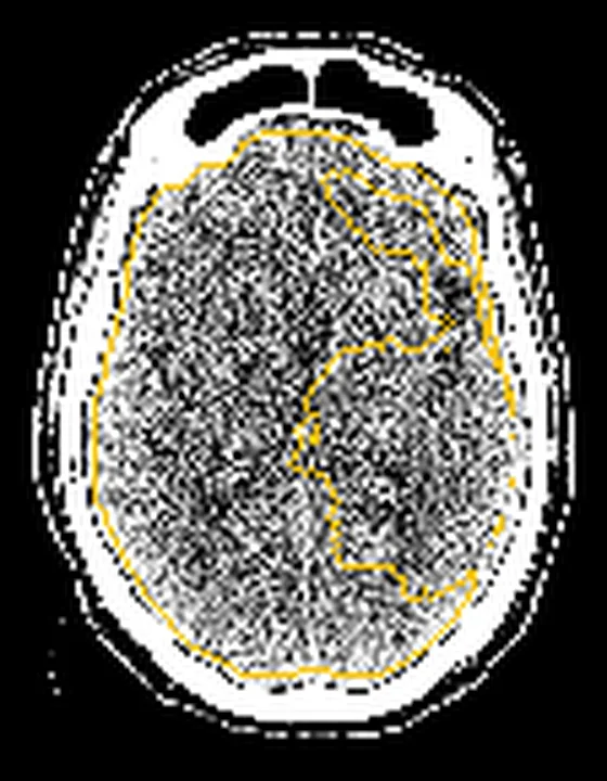
Aynı bölgenin, kaynak veri setinde patoloji içermediği belirtilen bir BT incelemesini kesit kesit kaydırın; organ sınırlarını açarak anatomiyi hasta görüntüsüyle karşılaştırın. Küçük rastlantısal bulgular tümüyle dışlanmamıştır.
Kaynak: TotalSegmentator v2.01 veri seti (Wasserthal ve ark., Radiology: Artificial Intelligence 2023), CC BY 4.0, vaka s0849; organ sınırları veri setinin otomatik segmentasyonundan, birleştirilerek sadeleştirildi. Görüntüler 2,5 mm kesit aralığına yeniden örneklendi.
Ayırıcı tanı
- Arteriovenöz malformasyon
- kontrastlı incelemede besleyici arter, nidus ve erken drene olan ven içeren şant kompleksi bulunur.
- Serebral ven trombozu
- sinüs veya kortikal ven dolum defekti, venöz drenaj alanında ödem ya da hemorajik enfarkt görülebilir.
- Kavernöz malformasyon
- MR görüntülerde hemosiderin kenarlı popkorn benzeri bir odak oluşturur; angiografik olarak sessizdir ve normal parankimi yok sayan daraltıcı kapiller yapılar içerir.
- Gelişmiş kollateral venöz ağ
- dural sinüs tıkanıklığı ya da venöz hipertansiyonla birlikte çok sayıda genişlemiş ven ve alternatif drenaj yolları görülür.
- Kontrastlanmış kortikal damar
- tek bir normal kortikal ven olabilir; medüller radyal kolların ortak toplayıcı vene birleşmesi DVA lehinedir.
Tuzaklar
- Venöz faz görüntülemesi yetersizse toplayıcı ven tam dolmaz; venöz opasifikasyonun yeterliliğini teyit etmeden DVA yok denilmemeli.
- Kontrastsız BT'deki hafif hiperdens toplayıcı veni otomatik olarak normal varyant saymayın; belirgin yoğunluk ve çevre ödem varsa tromboz araştırılmalıdır.
- DVA içindeki normal parankim görünümünü damar yumağıyla karıştırmayın; gerçek AVM'de nidus ve arteriyel beslenme beklenir.
- DVA yakınındaki akut kanamayı doğrudan DVA'ya atfetmeyin; eşlik eden kavernom veya venöz çıkış tıkanıklığı aranmalıdır.
Kendini dene
Venöz faz BT'de derin beyaz cevher venleri yelpaze biçiminde tek toplayıcı vene açılıyor. En olası yapı nedir?
Cevabı göster
Gelişimsel venöz anomali. Radyal medüller venlerin tek toplayıcıya birleşmesi DVA'nın tipik drenaj düzenidir. DVA'da radyal venler tek bir geniş venöz kanalda birleşir ve arteriyel bileşen bulunmaz; bu özellikler AVM'den ayrımını destekler.
DVA olarak bilinen toplayıcı ven kontrastsız BT'de belirgin hiperdens ve çevresinde ödem var. Hangi olasılık önceliklidir?
Cevabı göster
Toplayıcı ven trombozu. Belirgin hiperdens toplayıcı ven tromboz işareti olabilir; eşlik eden ödem venöz konjesyon veya enfarktı destekler. Patent DVA genellikle komplikasyonsuz drenaj varyantıdır; diğer seçenekler bu venöz bulguyu açıklamaz.
Kaynaklar
Bu sayfadaki 38 cümle ve 2 quiz sorusu aşağıdaki kaynaklardan alıntılarla tek tek karşılaştırıldı; 5 cümle bu karşılaştırmada düzeltildi (2026-10-08).
- Developmental venous anomaly (DVA) mimicking thrombosed cerebral veinpmc.ncbi.nlm.nih.gov
- Developmental venous anomalies.pubmed.ncbi.nlm.nih.gov
- Symptomatic developmental venous anomalies.pubmed.ncbi.nlm.nih.gov
- Vascular malformations of the brain.pubmed.ncbi.nlm.nih.gov
- Cerebral capillary telangiectasias: a meta-analysis and review of the literature.pubmed.ncbi.nlm.nih.gov
- Developmental venous anomalies of the brain in children -- imaging spectrum and update.pubmed.ncbi.nlm.nih.gov
- Hemorrhage owing to cerebral cavernous malformation: imaging, clinical, and histopathological considerations.pubmed.ncbi.nlm.nih.gov
- Epilepsy and cerebral developmental venous anomalies (DVAs): A systematic review.pubmed.ncbi.nlm.nih.gov
- Extra-Axial Cavernoma of the Cerebellopontine Angle: A Case Study and Review of Literature.pubmed.ncbi.nlm.nih.gov
- CNS cavernous haemangioma: "popcorn" in the brain and spinal cord.pubmed.ncbi.nlm.nih.gov
- Symptomatic Developmental Venous Anomaly: State-of-the-Art Review on Genetics, Pathophysiology, and Imaging Approach to Diagnosis.pubmed.ncbi.nlm.nih.gov
- Atypical Intracerebral Developmental Venous Anomalies in Sturge-Weber Syndrome: A Case Series and Review of Literature.pubmed.ncbi.nlm.nih.gov
- De novo cavernous angiomas associated with developmental venous anomaly: a mini-series and literature review.pubmed.ncbi.nlm.nih.gov
- Association of venous varix and developmental venous anomaly: report of a case and review of literature.pubmed.ncbi.nlm.nih.gov
- Developmental venous anomalies and cerebral cavernous malformations: Partners in crime.pubmed.ncbi.nlm.nih.gov
- Idiopathic Syringomyelia: A Systematic Scoping Review.pubmed.ncbi.nlm.nih.gov
- Cerebral Venous Outflow Insufficiency: A Study on Symptoms and Venous Stenosis Classification.pubmed.ncbi.nlm.nih.gov
- Incidental noninvasive diagnosis of isolated persistent left superior vena cava in asymptomatic adults: A case report.pubmed.ncbi.nlm.nih.gov
- Postpartum Vertebral Artery Dissection After Neck Massage Resulting in a Right Cerebellar Infarct: A Case Report and Literature Review.pubmed.ncbi.nlm.nih.gov
- Low-flow vascular malformations without arteriovenous shunting of the central nervous system: a pictorial review.pubmed.ncbi.nlm.nih.gov
- PHACE syndrome: a systematic literature review and illustrative case report of a patient with severe cerebrovascular and neurodevelopmental sequelae.pubmed.ncbi.nlm.nih.gov
- Developmental venous anomalies on fetal magnetic resonance imaging: prevalence and reproducible radiological phenotypes.pubmed.ncbi.nlm.nih.gov
- Aeromedical decision-making for asymptomatic cerebral cavernous malformations in military pilots.pubmed.ncbi.nlm.nih.gov
- Papillary thyroid carcinoma with coexisting venous hemangioma mimicking locally invasive disease: A case report.pubmed.ncbi.nlm.nih.gov
- Posterior fossa Hodgkin's lymphoma radiographically mimicking an arteriovenous malformation: illustrative case.pubmed.ncbi.nlm.nih.gov
- Cerebral cavernous malformation in a child leading to a fatal subarachnoid hemorrhage - "silent but sinister:" A case report and literature review.pubmed.ncbi.nlm.nih.gov
- Spontaneous regression of an incidental brain venous aneurysm in a child and literature review.pubmed.ncbi.nlm.nih.gov
- Neurocysticercosis and developmental venous anomaly in close proximity: a case report.pubmed.ncbi.nlm.nih.gov
- Effectiveness of super-selective digital subtraction angiography and 3D rotational digital subtraction venography for a developmental venous anomaly with an arteriovenous malformation: A case report and literature review.pubmed.ncbi.nlm.nih.gov
- New MR sequences in daily practice: susceptibility weighted imaging. A pictorial essay.pubmed.ncbi.nlm.nih.gov
- Intracranial venous thrombosis imaging by anatomical location: an educational review with mimics and diagnostic pitfalls.pubmed.ncbi.nlm.nih.gov
- Balloon-assisted retrograde cerebral venography for enhanced visualization of the posterior and lateral dural venous sinuses: a feasibility study.pubmed.ncbi.nlm.nih.gov
- Imaging features, diagnosis, and clinical management of hepatic involvement in hereditary hemorrhagic telangiectasia: a case series report and literature review.pubmed.ncbi.nlm.nih.gov
- Coexistence of a Hemispheric Developmental Venous Anomaly and Two Ipsilateral Arteriovenous Malformations: A Rare Neurovascular Association.pubmed.ncbi.nlm.nih.gov
- Congenital Intracranial Vascular Malformations in Children : Radiological Overview.pubmed.ncbi.nlm.nih.gov
- Vein of Galen Malformation-Experience of the Last 13 Years in a Reference Center from South-Eastern Europe.pubmed.ncbi.nlm.nih.gov
- Clinical utility of BOLD fMRI in preoperative work-up of epilepsy.pubmed.ncbi.nlm.nih.gov
- Long-term safety and effectiveness of the "OptEase" vena cava filter.pubmed.ncbi.nlm.nih.gov
- Ruptured cerebral arteriovenous malformation associated with enlarged parietal foramina: a rare surgical case with pathogenetic implications.pubmed.ncbi.nlm.nih.gov
- Cirrhosis-associated intracranial shunting lesions: illustrative case.pubmed.ncbi.nlm.nih.gov
- De novo twig-like middle cerebral artery after suspected M1 dissection in early childhood with coexisting KRAS-mutated brain arteriovenous malformation: illustrative case.pubmed.ncbi.nlm.nih.gov
- Developmental venous anomaly coexisting with arteriovenous malformation: a case report.pubmed.ncbi.nlm.nih.gov
- Cerebellar cavernoma excision with a preserved venous anomaly: A case report in girl 28-year-old.pubmed.ncbi.nlm.nih.gov
- Cerebral venous disorders: clinical presentation, diagnostic strategy, and contemporary management.pubmed.ncbi.nlm.nih.gov
- Developmental venous anomaly related to de novo cerebral cavernous malformation: Imaging findings from two cases.pubmed.ncbi.nlm.nih.gov
- Spontaneous hemorrhage of cavernous malformation in the region of the cochlear nucleus: A case report.pubmed.ncbi.nlm.nih.gov
- Progression of a pontine cavernous malformation and delayed bilateral hypertrophic olivary degeneration: illustrative case.pubmed.ncbi.nlm.nih.gov
- Selective Bridging Vein Embolization of a Transverse-Sigmoid Sinus Dural Arteriovenous Fistula Draining via the Jugular Foramen: A Case Report.pubmed.ncbi.nlm.nih.gov
- Decreased Visibility of Deep Medullary Vein Associated with Cerebral Small Vessel Disease Burden through Increased Interstitial Fluid in White Matter.pubmed.ncbi.nlm.nih.gov
- A Practical Approach to Intracranial Dural Arteriovenous Fistulas: Pathogenesis, Classification and Management.pubmed.ncbi.nlm.nih.gov
- Prevalence of Developmental Venous Anomalies in Association with Sporadic Cavernous Malformations on 7T MRI.pubmed.ncbi.nlm.nih.gov
- Vascular Malformations.pubmed.ncbi.nlm.nih.gov
- Infantile Spasms (West Syndrome): Integrating Genetic, Neurotrophic, and Hormonal Mechanisms Toward Precision Therapy.pubmed.ncbi.nlm.nih.gov
- Endovascular Embolization in Neurovascular Disease: Material Science, Multimodal Management, and Future Horizons.pubmed.ncbi.nlm.nih.gov
- Arteriovenous Malformations (AVMs): Molecular Pathogenesis, Clinical Features, and Emerging Therapeutic Strategies.pubmed.ncbi.nlm.nih.gov
- Perfusion imaging abnormalities in acute cerebral venous sinus thrombosis: A case series.pubmed.ncbi.nlm.nih.gov
- Case Report: Structured MRI assessment of posterior thalamic infarction in a distribution compatible with posterior choroidal artery territory presenting as Déjerine-Roussy syndrome in an adolescent: differentiating arterial ischemia from venous thrombosis and thalamic neoplasm.pubmed.ncbi.nlm.nih.gov
- Bilateral Thalamic Infarction Secondary to Deep Cerebral Venous Thrombosis: A Diagnostic Challenge Associated With Hormonal Therapy.pubmed.ncbi.nlm.nih.gov
- Intraventricular giant cavernous malformation mimicking central neurocytoma characterized by digital subtraction angiography: illustrative case.pubmed.ncbi.nlm.nih.gov
- Obstructive hydrocephalus secondary to an anterior mesencephalic cavernous malformation with familial cerebral cavernous malformation syndrome: A case report.pubmed.ncbi.nlm.nih.gov
- Direct superior ophthalmic vein puncture for cavernous sinus dural arteriovenous fistula with dual orbital venous outflow: illustrative case.pubmed.ncbi.nlm.nih.gov
- Treatment of iliac arteriovenous malformation associated with trisomy 21: a case report.pubmed.ncbi.nlm.nih.gov
- Beyond the Usual Suspects: Rare Causes of Hemoptysis.pubmed.ncbi.nlm.nih.gov
- Long-term outcomes in pediatric brain AVMs: beyond radiological cure.pubmed.ncbi.nlm.nih.gov
- Dual-energy CT angiography in detecting underlying causes of intracerebral hemorrhage: an observational cohort study.pubmed.ncbi.nlm.nih.gov
- Non-Thrombotic Filling Defects in Cerebral Veins and Sinuses: When Normal Structures Mimic a Disease.pubmed.ncbi.nlm.nih.gov
- The accuracy of non-contrast brain CT scan in predicting the presence of a vascular etiology in patients with primary intracranial hemorrhage.pubmed.ncbi.nlm.nih.gov
- Microsurgical obliteration of a superior petrosal dural arteriovenous fistula presenting with a symptomatic brainstem cavernous malformation.pubmed.ncbi.nlm.nih.gov
- Comprehensive review of the cerebral aqueduct: anatomy, embryology, imaging, genetics, molecular, and pathology.pubmed.ncbi.nlm.nih.gov
- Moyamoya Vasculopathy and Atypical Moyamoya-like Patterns: Insights into Diagnosis and Therapeutic Implications.pubmed.ncbi.nlm.nih.gov
- Masses of developmental and genetic origin affecting the paediatric craniofacial skeleton.pubmed.ncbi.nlm.nih.gov
- Twenty classic signs in neuroradiology: A pictorial essay.pubmed.ncbi.nlm.nih.gov
- Lymphatic vessel: origin, heterogeneity, biological functions, and therapeutic targets.pubmed.ncbi.nlm.nih.gov
- Role of Transcriptional and Epigenetic Regulation in Lymphatic Endothelial Cell Development.pubmed.ncbi.nlm.nih.gov
- Meningeal Lymphatics in Central Nervous System Diseases.pubmed.ncbi.nlm.nih.gov
- Combined surgical and interventional treatment of a giant superior vena cava aneurysm.pubmed.ncbi.nlm.nih.gov
- Oculomotor Nerve Palsy Induced by a Cerebral Developmental Venous Anomaly: A Case Report and Comprehensive Review.pubmed.ncbi.nlm.nih.gov
- Developmental Venous Anomaly: Benign or Not Benign.pubmed.ncbi.nlm.nih.gov
- Cerebrovascular Malformations Associated With Hereditary Hemorrhagic Telangiectasia and HHT-Like Syndromes: A Comparative Overview.pubmed.ncbi.nlm.nih.gov
- Controversies in Cavernous Malformation Management: A Comprehensive Review of Current Literature.pubmed.ncbi.nlm.nih.gov
- Spontaneous Right Thalamic Hemorrhage With Intraventricular Extension in a Two-Month-Old Preterm Infant Initially Suspected of Having Cow's Milk Protein Allergy.pubmed.ncbi.nlm.nih.gov
- Computed tomography of renal emergencies: A comprehensive diagnostic guide for radiology residents.pubmed.ncbi.nlm.nih.gov
- Multimodal imaging of infantile scimitar syndrome with Eisenmenger physiology: A case report of complex intracardiac and extracardiac shunts.pubmed.ncbi.nlm.nih.gov
- Identification of the Genomic Etiology of Unexplained Congenital Problems in Pediatric Patients: First Reported Case With Coffin-Siris Syndrome and Sialuria From India.pubmed.ncbi.nlm.nih.gov
- Genome sequencing-based CNV analysis in an infant with concurrent partial trisomies 9p and 12p due to maternal translocation: A case report.pubmed.ncbi.nlm.nih.gov
- Fused right S-shaped kidney incidentally detected on multidetector CT during staging of periampullary adenocarcinoma: A case report.pubmed.ncbi.nlm.nih.gov
- Segmental Aplasia and Right Azygos Continuation of the Caudal Vena Cava in a Cat (Felis catus).pubmed.ncbi.nlm.nih.gov
- Transvenous Embolization for Dural Arteriovenous Fistula through the Persistent Left Superior Vena Cava: A Case Report.pubmed.ncbi.nlm.nih.gov
- Central nervous system vascular malformations: A clinical review.pubmed.ncbi.nlm.nih.gov
- Direct Cerebral Revascularization: Extracranial-intracranial Bypass.pubmed.ncbi.nlm.nih.gov
- Intracranial meningeal disease: comparison of contrast-enhanced MR imaging with fluid-attenuated inversion recovery and fat-suppressed T1-weighted sequences.pubmed.ncbi.nlm.nih.gov
- Mastoid emissary vein, mastoid emissary foramen, and mastoid emissary canal: anatomy, variability, imaging, and clinical implications.pubmed.ncbi.nlm.nih.gov
- A giant challenge: Hybrid management of a scalp and neck cirsoid aneurysm.pubmed.ncbi.nlm.nih.gov
- A systematic approach to detection of epileptogenic foci on imaging: protocols, entities, search pattern, and reporting tools.pubmed.ncbi.nlm.nih.gov
- Intracranial dural arteriovenous fistulas with and without pial arterial supply: a case series of 227 patients.pubmed.ncbi.nlm.nih.gov
- Incidental Detection of a Posterior Inferior Cerebellar Artery (PICA)-Terminating Vertebral Artery Variant During CT Evaluation of Venous Sinus Thrombosis: A Case Report.pubmed.ncbi.nlm.nih.gov
- Patterns of injury and violence in Yaoundé Cameroon: an analysis of hospital data.pubmed.ncbi.nlm.nih.gov
- Non-Invasive Retinal Vessel Analysis as a Predictor for Cardiovascular Disease.pubmed.ncbi.nlm.nih.gov
- A proposed framework for cerebral venous congestion.pubmed.ncbi.nlm.nih.gov
- Spontaneous bacteremia and spontaneous bacterial peritonitis share similar prognosis in patients with cirrhosis: a cohort study.pubmed.ncbi.nlm.nih.gov
- Conduction Aphasia in a Case of Left Cortical Veins and Left Lateral Sinus Thrombosis Due to Multiple Risk Factors: A Case Report and Review of the Literature.pubmed.ncbi.nlm.nih.gov
- Morphological variations of the dural venous sinuses: a cadaveric study.pubmed.ncbi.nlm.nih.gov
- Hemodynamic and developmental biomarkers enhance prenatal coarctation prediction: a validated multiparametric ultrasound model.pubmed.ncbi.nlm.nih.gov
- Pathogenesis and Surgical Treatment of Dextro-Transposition of the Great Arteries (D-TGA): Part II.pubmed.ncbi.nlm.nih.gov
- Paediatric cranial ultrasound: abnormalities of the brain in term neonates and young infants.pubmed.ncbi.nlm.nih.gov
- Review of imaging modalities and radiological findings of calvarial lesions.pubmed.ncbi.nlm.nih.gov
- Phakomatoses: A pictorial review.pubmed.ncbi.nlm.nih.gov
- Pitfall in necrotizing pancreatitis: Organized hematoma with arteriovenous fistula mimicking a splenic artery pseudoaneurysm.pubmed.ncbi.nlm.nih.gov
- From imaging to impact: A case of renal arteriovenous malformation.pubmed.ncbi.nlm.nih.gov
- Balloon-Assisted Squid 12 Embolization of a Renal Angiomyolipoma With Imaging Findings Suggestive of Arteriovenous Communication: A Technical Report.pubmed.ncbi.nlm.nih.gov
- Clinical management of isolated transverse-sigmoid sinus dural arteriovenous fistulas: a three-case illustrative series and narrative review.pubmed.ncbi.nlm.nih.gov
Eğitim amaçlıdır; hasta başında tanı ve tedavi kararının yerine geçmez. İçerik yapay zekâ destekli bir süreçle güncel literatüre göre cümle cümle kaynaklarla karşılaştırıldı, ancak uzman radyolog incelemesinden geçmedi. Klinik kararlarda güncel kılavuzları, kurum protokollerini ve radyoloji raporunu esas alın.