
Özet
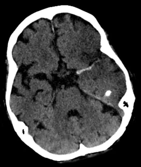
Ani fokal nörolojik bulgusu olan hastada kontrastsız kafa BT'sinde tek bir arterin çevresine ve karşı eş damarına göre belirgin yoğun görünmesi akut lümen içi trombüsü düşündürür. Kontrastsız BT erken kanamayı ve parankim değişikliğini tararken, BT anjiyografi şüpheli damarda gerçek oklüzyon veya dolum kusurunu ve distal akımı doğrudan değerlendirir. Bulguyu doğrulamadan trombüs saymak kalsifik plak, yüksek hematokrit veya yalancı hiperdens görünümün yanlış yorumlanmasına neden olabilir; işaret yokluğu da oklüzyonu dışlamaz. Büyük damar tıkanıklığı atlanırsa akut inme değerlendirmesi eksik kalabilir.
Faz ve pencere
- Kontrastsız tanısal
- M1 segmentinde karşı tarafa göre belirgin hiperdensite veya baziler arterde artmış dansite aranır; damar duvarı kalsifikasyonu ve genel damar yoğunluğu kıyaslanır.
- BTA tanısal
- CTA'da hiperdens arterin bulunduğu yerde kontrast dolum kusuru veya ani damar sonlanması izlenir; gelişmiş kollateral akım kesikliği maskeleyebilir.
Önerilen pencereler: Beyin (G 80 / M 40), İnme (G 40 / M 40), Anjiyo (G 600 / M 150).
BT bulguları
- Hiperdens MCA işareti — M1 boyunca Sylvian fissürde karşı M1'den daha belirgin lineer dansite; trombüs lümen içinde damar boyunca uzanır.
- MCA segmentinde karşı tarafa göre belirgin hiperdensite aranır; noktasal yoğunluklar kalsifikasyon veya venöz yapılarla karışabilir.
- Kontrastsız BT'de baziler arterde hiperdens bulgu saptandığında, posterior sirkülasyon büyük damar tıkanıklığını doğrulamak amacıyla CT anjiyografiye başvurulur.
- Fokal tek taraflı hiperdensite akut trombüsü işaret ederken, sistemik nedenlerle oluşan iki taraflı ve simetrik damar dansitesi artışından ayrılmalıdır.
- İntraluminal trombüs, damar ekseni boyunca uzayan lineer hiperdensite olarak görünür; kalsifik plaklar genellikle eksantrik ve noktasaldır.
- CT anjiografide, kontrastsız BT'de işaret görülen düzeyde kontrast dolum kusuru veya ani damar sonlanması ile oklüzyon doğrulanır.
- Eşlik eden erken iskemi — insular şerit silinmesi, lentiform çekirdek sınır kaybı veya sulkal silinme tromboze arterin alanında görülebilir.
Ölçütler ve sınıflamalar
- Önerilmiş hiperdens MCA nesnel eşiği
- Karşı eş damara göre belirgin şekilde artmış mutlak veya göreceli lümen dansitesi aranır.
- Karşı taraf eş arterle yoğunluk oranı
- Şüpheli arterin yoğunluğu, aynı seviyedeki karşı taraf eş damarla karşılaştırılarak farkın tek taraflı olduğu doğrulanır.
Normalde
Fizyolojik koşullarda eş seviyedeki eş damarlar benzer dansitededir; tek taraflı veya asimmetrik hiperdensite trombüs yönünden değerlendirilir. HU ölçümleri trombozu destekleyebilir; ancak iki taraflı ve yaygın artış sistemik hematokrit değişikliğini düşündürür.
Normal BT ile kıyasla
Aynı bölgenin, kaynak veri setinde patoloji içermediği belirtilen bir BT incelemesini kesit kesit kaydırın; organ sınırlarını açarak anatomiyi hasta görüntüsüyle karşılaştırın. Küçük rastlantısal bulgular tümüyle dışlanmamıştır.
Kaynak: TotalSegmentator v2.01 veri seti (Wasserthal ve ark., Radiology: Artificial Intelligence 2023), CC BY 4.0, vaka s0103; organ sınırları veri setinin otomatik segmentasyonundan, birleştirilerek sadeleştirildi. Görüntüler 2,5 mm kesit aralığına yeniden örneklendi.
Ayırıcı tanı
- Intrakraniyal kalsifikasyonlar, özellikle damar duvarında eksantrik ve noktasal parlak odaklar oluşturarak hiperdens arter işareti ile karışabilir.
- Sistemik yüksek hematokrit durumlarında damarlar iki taraflı ve simetrik yoğunlaşabilir; bu durum tek bir damarda beklenen lokal oklüzyon paterninden farklıdır.
- Kontrastsız BT'deki hiperdens arter işareti intralüminal trombüsle ilişkili olabilir; büyük damar tıkanıklığının doğrulanmasında BT anjiyografi altın standarttır.
- Önceki incelemede kullanılan iyotlu kontrastın kalıntısı, damarların genel olarak hiperdens görünmesine neden olabilir; bu durum akut trombozdan ayırt edilmelidir.
- Arter diseksiyonunda yüksek çözünürlüklü damar duvarı MR'ı (VW-MRI) intramural hematom, intimal flep ve duvar kalınlaşmasını gösterebilir; yalnızca hiperdensiteyle trombüs tanımlanamaz.
- Damar kalsifikasyonları noktasal veya oval, çok yüksek dansitede görünür; lümen içi trombüsten ayırt edilebilmek için konumu ve dansite özellikleri dikkatle incelenmelidir.
Tuzaklar
- Damar duvarı kalsifikasyonları noktasal veya eksantrik yerleşimli olabilir ve hiperdens arter işaretiyle karışabilir; konum ve ek anjiyografi bulguları ayrım sağlar.
- Hematokrit yüksekliğinde yalnız MCA değil, iki taraflı arterler ve venöz sinüsler de yoğunlaşabilir; karşı taraf kontrolü şarttır.
- Çevresindeki ödemli/hipodens parankim, arterin mutlak yoğunluğu artmadan yalancı hiperdens işaret oluşturabilir; HU ve eş damar kıyasını birlikte kullan.
- Hiperdens arter işareti duyarlılığı sınırlı olduğundan işaretin görülmemesi CTA'da damar açıklığını kanıtlamaz.
Kendini dene
Kontrastsız BT'de hiperdens arter işareti görülüyor ve BT anjiyografide büyük damar oklüzyonu saptanıyor. En olası açıklama nedir?
Cevabı göster
Akut intralüminal trombüs. Hiperdens arter işareti kontrastsız BT'de intralüminal trombüsü yansıtır; CTA büyük damar oklüzyonunu doğrulamada temel görüntüleme yöntemidir.
Kontrastsız BT'de hiperdens arter işareti görülüyor. Bu işaret en doğrudan neyi yansıtır?
Cevabı göster
İntralüminal trombüs. Hiperdens arter işareti intralüminal trombüsü yansıtır ve büyük damar oklüzyonu için hemen fark edilebilen bir belirteç olabilir.
Bu konunun yer aldığı turlar
Kaynaklar
Bu sayfadaki 38 cümle ve 2 quiz sorusu aşağıdaki kaynaklardan alıntılarla tek tek karşılaştırıldı; 28 cümle bu karşılaştırmada düzeltildi (2026-10-08).
- Intracranial venous thrombosis imaging by anatomical location: an educational review with mimics and diagnostic pitfallspmc.ncbi.nlm.nih.gov
- Stroke or Seizure? Diagnostic Role of Neuroimaging in Acute Neurologic Mimicspmc.ncbi.nlm.nih.gov
- Automated detection of hyperdense artery sign on non-contrast CT for rapid identification of large vessel occlusion: a multicenter validation studypmc.ncbi.nlm.nih.gov
- Editorial: Emerging fast medical imaging techniques in radiologypmc.ncbi.nlm.nih.gov
- CT and MRI imaging of mass in the carotid space: a pictorial reviewpmc.ncbi.nlm.nih.gov
- Unidentified bright objects of spleen on arterial phase CT: mimicker of splenic vascular injury in blunt abdominal trauma.pubmed.ncbi.nlm.nih.gov
- Noncontrast CT-based habitat analysis for differentiating hemorrhagic transformation from contrast extravasation after endovascular therapy of acute ischemic stroke.pubmed.ncbi.nlm.nih.gov
- Prognostic value of the hyperdense middle cerebral artery sign in patients undergoing mechanical thrombectomy.pubmed.ncbi.nlm.nih.gov
- Effects of Computed Tomography Hyperdense Middle Cerebral Artery Sign on the Efficacy of Endovascular Therapy in Acute Ischemic Stroke With Large Infarcts: A Subgroup Analysis of the ANGEL-ASPECT Trial.pubmed.ncbi.nlm.nih.gov
- Initial Experience With a Deep Learning Algorithm for Detecting Posterior Circulation Large Vessel Occlusion on Noncontrast Computed Tomography.pubmed.ncbi.nlm.nih.gov
- Early Reperfusion in Basilar Artery Occlusion Stroke Managed With Tenecteplase Versus Alteplase Before Endovascular Treatment.pubmed.ncbi.nlm.nih.gov
- Intracranial venous thrombosis imaging by anatomical location: an educational review with mimics and diagnostic pitfalls.pubmed.ncbi.nlm.nih.gov
- Stroke or Seizure? Diagnostic Role of Neuroimaging in Acute Neurologic Mimics.pubmed.ncbi.nlm.nih.gov
- Management of spontaneous abdominal wall hematomas: a narrative review with a focus on CTA-negative endovascular cases.pubmed.ncbi.nlm.nih.gov
- Where Does the Signal Go? Technical Challenges of CT Perfusion in Lacunar and Infratentorial Stroke.pubmed.ncbi.nlm.nih.gov
- Diagnostic Accuracy of the ACT-FAST Tool for Prehospital Stroke Identification: A Systematic Review.pubmed.ncbi.nlm.nih.gov
- Cardiac photon-counting detector computed tomography in acute ischemic stroke: diagnostic accuracy for detection of left atrial appendage thrombus.pubmed.ncbi.nlm.nih.gov
- Segmentation of the Hyperdense Artery Sign on Noncontrast CT in Ischemic Stroke Using Artificial Intelligence.pubmed.ncbi.nlm.nih.gov
- Multicenter Validation of Artificial Intelligence Predicting Anterior Circulation Large Vessel Occlusion Using Noncontrast Head CT.pubmed.ncbi.nlm.nih.gov
- Clinical image analysis to build patient-specific models of acute ischemic stroke patients.pubmed.ncbi.nlm.nih.gov
- Middle cerebral artery fenestration presenting with cerebral ischemia: a case report and review of the literature.pubmed.ncbi.nlm.nih.gov
- Using Multimodal Imaging to Correlate Link Between Intracranial Aneurysms and Acute Ischemic Stroke.pubmed.ncbi.nlm.nih.gov
- Delayed M1 Dissection After Thrombectomy Treated With Rescue Stenting and Tirofiban: A Case Report.pubmed.ncbi.nlm.nih.gov
- The evolution of personalized stroke thrombectomy.pubmed.ncbi.nlm.nih.gov
- A multi-center study of the MicroNET-covered stent in consecutive patients with acute carotid-related stroke: SAFEGUARD-STROKE.pubmed.ncbi.nlm.nih.gov
- Artificial Intelligence in Acute Ischemic Stroke Subtypes According to Toast Classification: A Comprehensive Narrative Review.pubmed.ncbi.nlm.nih.gov
- Prognostic value of CT contrast staining after endovascular therapy in basilar artery occlusion stroke.pubmed.ncbi.nlm.nih.gov
- Current state and advancements of imaging in acute ischemic stroke: a practical review.pubmed.ncbi.nlm.nih.gov
- Predictors of Mortality in Posterior Circulation Ischemic Stroke.pubmed.ncbi.nlm.nih.gov
- Diabetic striatopathy presenting as stroke-mimicking hemiparesis without involuntary movements: A case report.pubmed.ncbi.nlm.nih.gov
- Interocular symmetry and asymmetry in ocular health and disease.pubmed.ncbi.nlm.nih.gov
- Parry-Romberg Syndrome: Radioclinical Dissociation in a Paucisymptomatic Form and a Proposed Diagnostic Framework.pubmed.ncbi.nlm.nih.gov
- Non-Thrombotic Filling Defects in Cerebral Veins and Sinuses: When Normal Structures Mimic a Disease.pubmed.ncbi.nlm.nih.gov
- Sudden Cardiac Arrest Due to Ascending Aortic Thrombus Originating From Penetrating Aortic Ulcer.pubmed.ncbi.nlm.nih.gov
- Imaging assessment of acute ischaemic stroke: a review of radiological methods.pubmed.ncbi.nlm.nih.gov
- Clinical and CT angiography characteristics of Chinese stroke patients with carotid web: a retrospective study.pubmed.ncbi.nlm.nih.gov
- Angiographically occult in-stent protrusion detected by intravascular ultrasound during carotid artery stenting: a case report.pubmed.ncbi.nlm.nih.gov
- Giant cervical internal carotid artery aneurysm associated with multiple intra- and extracranial aneurysms: a case report.pubmed.ncbi.nlm.nih.gov
- Influence of deep learning image reconstruction and adaptive statistical iterative reconstruction-V on automated Alberta stroke program early CT score- evaluation.pubmed.ncbi.nlm.nih.gov
- Insights into the pathophysiology of stroke-heart syndrome: a review of recent literature and clinical trials to illuminate promising therapeutic strategies.pubmed.ncbi.nlm.nih.gov
- Marine Mobile Stroke Unit: A Pilot Study on the Feasibility and Quality of Brain Imaging.pubmed.ncbi.nlm.nih.gov
- Utility of Non-Contrast CT and MRA Fusion Images for Depicting Occluded Vessels in Mechanical Thrombectomy.pubmed.ncbi.nlm.nih.gov
- Attenuation measurements on non-contrast-enhanced CT brain: a re-visit of their role in the diagnosis of cerebral venous thrombosis.pubmed.ncbi.nlm.nih.gov
- Diagnostic Value and Operational Recommendations for Late Iodine Enhancement and ECV Quantification in Single-Energy Computed Tomography: A Narrative Review.pubmed.ncbi.nlm.nih.gov
- Hypoperfusion Intensity Ratio as a Predictive Marker in Pre-Endovascular Treatment Perfusion Magnetic Resonance Image for Acute Middle Cerebral Artery Occlusion with Intracranial Atherosclerosis.pubmed.ncbi.nlm.nih.gov
- A Novel and Simple Score to Predict Embolic or Atherosclerotic Middle Cerebral Artery Occlusion Before Mechanical Thrombectomy: AHOC Score.pubmed.ncbi.nlm.nih.gov
- Incidence, Predictors, and Outcomes of Symptomatic Intracranial Hemorrhage after Tenecteplase in Acute Ischemic Stroke.pubmed.ncbi.nlm.nih.gov
- Acute Infarct Core Volume Estimation on Noncontrast Computed Tomography With a Deep Learning Algorithm.pubmed.ncbi.nlm.nih.gov
- Contrast Injection from an Intermediate Catheter Placed in an Intradural Artery is Associated with Contrast-Induced Encephalopathy following Neurointervention.pubmed.ncbi.nlm.nih.gov
- Vasculitis syndromes: large vessel vasculitides and ANCA-associated vasculitides from a neuroradiologist's perspective.pubmed.ncbi.nlm.nih.gov
- CT and MRI imaging of mass in the carotid space: a pictorial review.pubmed.ncbi.nlm.nih.gov
- Incidental Calcifications of Carotid and Vertebral Arteries: Frequency and Associations in Pediatric Population.pubmed.ncbi.nlm.nih.gov
- Catastrophic Hypoxic-Ischemic Brain Injury Following Influenza A Illness in a Young Woman: A Case Report.pubmed.ncbi.nlm.nih.gov
- Case Report: Renal artery aneurysm rupture, axillary artery pseudoaneurysm, and catastrophic intraperitoneal hemorrhage caused by fibromuscular dysplasia with the involvement of multiple arterial beds.pubmed.ncbi.nlm.nih.gov
- Hyperthyroidism-Induced Dural Venous Sinus Thrombosis: A Case Report.pubmed.ncbi.nlm.nih.gov
- Neuroimaging of Acute Ischemic Stroke: Multimodal Imaging Approach for Acute Endovascular Therapy.pubmed.ncbi.nlm.nih.gov
- Vascular tumors of the liver: A brief review.pubmed.ncbi.nlm.nih.gov
- Imaging Evaluation of Acute Traumatic Brain Injury.pubmed.ncbi.nlm.nih.gov
- Plaque-Adapted Virtual Monoenergetic Image Selection for Carotid Plaque Visualization Using Photon-Counting CT.pubmed.ncbi.nlm.nih.gov
- Prolonged retention of oil-based iodinated contrast medium observed on plain abdominal radiograph after cesarean section: A case report.pubmed.ncbi.nlm.nih.gov
- Spectral Computed Tomography Angiography in Visceral Artery Aneurysms: Technical Principles and Clinical Applications.pubmed.ncbi.nlm.nih.gov
- Coronary Intramural Hematoma in a Young Athlete After Direct Ball Impact to the Chest.pubmed.ncbi.nlm.nih.gov
- CT-Centered Multimodality Imaging of Arterial Wall Fragility in Acute Aortic Syndromes: A Narrative Review of Imaging Markers and Clinical Implications.pubmed.ncbi.nlm.nih.gov
- Craniocervical arterial dissections with cerebral infarction: a five-case series with diagnostic application of vessel wall MRI.pubmed.ncbi.nlm.nih.gov
- Coronary Artery Disease and Atherosclerosis in Other Vascular Districts: Epidemiology, Risk Factors and Atherosclerotic Plaque Features.pubmed.ncbi.nlm.nih.gov
- Phenotyping atherosclerotic plaque and perivascular adipose tissue: signalling pathways and clinical biomarkers in atherosclerosis.pubmed.ncbi.nlm.nih.gov
- Ultrasound-Mediated Thrombolysis: From Mechanistic Insights to Advanced Nanoplatforms and Clinical Translation.pubmed.ncbi.nlm.nih.gov
- Multimodal imaging in neuro-ophthalmology.pubmed.ncbi.nlm.nih.gov
- Unmasking the Imitators: Challenges in Identifying Intracranial Atherosclerosis-Related Large Vessel Occlusion Mimics During Mechanical Thrombectomy.pubmed.ncbi.nlm.nih.gov
- Intracranial aneurysms mimicking third ventricular masses: case series and systematic review.pubmed.ncbi.nlm.nih.gov
- A Mechanistic and Pathophysiological Approach for Stroke Associated with Drugs of Abuse.pubmed.ncbi.nlm.nih.gov
- A Review on Computer Aided Diagnosis of Acute Brain Stroke.pubmed.ncbi.nlm.nih.gov
- Brain Midline Shift Measurement and Its Automation: A Review of Techniques and Algorithms.pubmed.ncbi.nlm.nih.gov
- A review of current imaging methods used in stroke research.pubmed.ncbi.nlm.nih.gov
- Automated detection of hyperdense artery sign on non-contrast CT for rapid identification of large vessel occlusion: a multicenter validation study.pubmed.ncbi.nlm.nih.gov
- The Empty Sylvian Fissure Sign: A Novel Non-contrast CT Finding in Moyamoya Disease.pubmed.ncbi.nlm.nih.gov
- Middle cerebral artery dissection: pathophysiology, diagnosis, and therapeutic options.pubmed.ncbi.nlm.nih.gov
- Extensive Cerebral Venous Sinus Thrombosis With Obstructive Hydrocephalus in a Young Female With Underlying Essential Thrombocythemia: A Case Report.pubmed.ncbi.nlm.nih.gov
- Late-Onset Moyamoya Disease Presenting With Acute Hemiparesis in a 77-Year-Old Patient: A Case Report.pubmed.ncbi.nlm.nih.gov
- Case Report: Endovascular treatment of spontaneous iliac vein rupture complicated with lower extremity deep vein thrombosis.pubmed.ncbi.nlm.nih.gov
- Insights into Angiographic Recanalization of Large Arterial Occlusion: Institutional Experience with Mechanical Thrombectomy for Acute Ischemic Stroke.pubmed.ncbi.nlm.nih.gov
- Admission hyperdense sinus sign predicts poorer outcomes in cerebral venous sinus thrombosis.pubmed.ncbi.nlm.nih.gov
- Modified Alberta Stroke Program Early CT Score (ASPECTS) of Contrast Extravasation on Dual-Energy CT Predicts Haemorrhagic Transformation and Poor Outcome After Endovascular Thrombectomy.pubmed.ncbi.nlm.nih.gov
- Analysis of the baseline multiphase computed tomographic angiography findings to predict clinical outcomes in patients with middle cerebral artery M1 occlusion treated with mechanical thrombectomy.pubmed.ncbi.nlm.nih.gov
- Analysis of 90-day risk factors for poor prognosis in patients with postoperative CT with hyperdense signs after thrombectomy for acute ischemic stroke.pubmed.ncbi.nlm.nih.gov
- Data-Driven Prognostication in Distal Medium Vessel Occlusions Using Explainable Machine Learning.pubmed.ncbi.nlm.nih.gov
- Mechanical thrombectomy for tandem internal carotid artery-middle cerebral artery occlusion in a patient with a persistent hypoglossal artery: illustrative case.pubmed.ncbi.nlm.nih.gov
- Computed tomography perfusion-defined ischemic core predicts functional outcome after basilar artery thrombectomy.pubmed.ncbi.nlm.nih.gov
- MDCT Imaging of Non-Traumatic Thoracic Aortic Emergencies and Its Impact on Diagnosis and Management-A Reappraisal.pubmed.ncbi.nlm.nih.gov
- Imaging More than Skin-Deep: Radiologic and Dermatologic Presentations of Systemic Disorders.pubmed.ncbi.nlm.nih.gov
- MDCT evaluation of acute aortic syndrome (AAS).pubmed.ncbi.nlm.nih.gov
- Prediction of functional outcome after mechanical thrombectomy using radiomics and deep learning features derived from intra-thrombus and peri-thrombus regions.pubmed.ncbi.nlm.nih.gov
- Prediction of hemorrhagic transformation via pre-treatment CT radiomics in acute ischemic stroke patients receiving endovascular therapy.pubmed.ncbi.nlm.nih.gov
- Prediction model of early biomarkers of massive cerebral infarction caused by anterior circulation occlusion: Establishment and evaluation.pubmed.ncbi.nlm.nih.gov
- Diabetic striatopathy: an updated overview of current knowledge and future perspectives.pubmed.ncbi.nlm.nih.gov
- Impact of dose reduction and iterative model reconstruction on multi-detector CT imaging of the brain in patients with suspected ischemic stroke.pubmed.ncbi.nlm.nih.gov
- Association of a High Neutrophil-to-Lymphocyte Ratio with Hyperdense Artery Sign and Unfavorable Short-Term Outcomes in Patients with Acute Ischemic Stroke.pubmed.ncbi.nlm.nih.gov
- Erythrocyte-Rich Thrombus Is Associated with Reduced Number of Maneuvers and Procedure Time in Patients with Acute Ischemic Stroke Undergoing Mechanical Thrombectomy.pubmed.ncbi.nlm.nih.gov
- When Cancer Clots: An Extensive Radiologic Analysis of Cancer-Associated Thromboembolism.pubmed.ncbi.nlm.nih.gov
- Life-threatening Carotid Complications Caused by Extraluminal Migration of Ingested Foreign Bodies; a Case Report and Narrative Review of Literature.pubmed.ncbi.nlm.nih.gov
- Infarcts and ischemia in the abdomen: an imaging perspective with an emphasis on cross-sectional imaging findings.pubmed.ncbi.nlm.nih.gov
- Imaging of the chemotherapy-induced hepatic damage: Yellow liver, blue liver, and pseudocirrhosis.pubmed.ncbi.nlm.nih.gov
- Society for Cardiovascular Magnetic Resonance 2024 Cases of SCMR case series.pubmed.ncbi.nlm.nih.gov
- State-of-the-art CT and MR imaging and assessment of atherosclerotic carotid artery disease: the reporting-a consensus document by the European Society of Cardiovascular Radiology (ESCR).pubmed.ncbi.nlm.nih.gov
- A case of acute traumatic aortic injury of a right-sided aortic arch with rupture of an aberrant left subclavian artery.pubmed.ncbi.nlm.nih.gov
- RAPID CTA versus JLK LVO for large vessel occlusion detection: a pragmatic comparison of performance and common pitfalls.pubmed.ncbi.nlm.nih.gov
- Quantitative multi-slice spiral CT perfusion parameters as predictors of collateral status and 90-day functional outcome in acute ischemic stroke.pubmed.ncbi.nlm.nih.gov
- Differentiating effective salvage from ineffective delayed compensation in anterior circulation stroke: a dynamic quantitative collateral index.pubmed.ncbi.nlm.nih.gov
- Cystic lesions and their mimics involving the intrahepatic bile ducts and peribiliary space: diagnosis, complications, and management.pubmed.ncbi.nlm.nih.gov
- Application of postmortem imaging modalities in cases of sudden death due to cardiovascular diseases-current achievements and limitations from a pathology perspective : Endorsed by the Association for European Cardiovascular Pathology and by the International Society of Forensic Radiology and Imaging.pubmed.ncbi.nlm.nih.gov
- Postpartum ovarian vein thrombosis: plain CT with clinical indicators-diagnostic performance and clinical application.pubmed.ncbi.nlm.nih.gov
- Imaging of synchronous multifocal renal masses: a case report.pubmed.ncbi.nlm.nih.gov
- Machine learning in neuroradiology: Recent developments and applications.pubmed.ncbi.nlm.nih.gov
- Chronic Subdural Hematoma: Pathophysiology, Diagnosis, and the Emerging Role of Middle Meningeal Artery Embolization.pubmed.ncbi.nlm.nih.gov
- A diagnostic approach to mediastinal masses in clinical practice.pubmed.ncbi.nlm.nih.gov
Eğitim amaçlıdır; hasta başında tanı ve tedavi kararının yerine geçmez. İçerik yapay zekâ destekli bir süreçle güncel literatüre göre cümle cümle kaynaklarla karşılaştırıldı, ancak uzman radyolog incelemesinden geçmedi. Klinik kararlarda güncel kılavuzları, kurum protokollerini ve radyoloji raporunu esas alın.