Özet
Geçici iskemik atak, fokal nörolojik işlev bozukluğunun akut infarkt olmadan geçici olarak ortaya çıktığı klinik bir sendromdur; belirtilerin düzelmesi acil damar kaynaklı riski ortadan kaldırmaz. Kontrastsız BT hızlı biçimde intrakraniyal kanama, kitle ve belirgin yerleşmiş infarktı araştırır, ancak erken veya küçük akut iskemiyi dışlayamaz. BTA baş-boyun damarlarındaki semptomatik darlık ya da oklüzyonu gösterir; difüzyon MR akut infarktı saptamada BT'den daha duyarlıdır ve tanımın doku bileşenini aydınlatabilir. BT'nin negatifliğini nörolojik olay yokluğu gibi yorumlamak küçük infarktı ve yüksek riskli damar lezyonunu kaçırabilir.
Faz ve pencere
- Kontrastsız tanısal
- Kontrastsız BT'de erken iskemik değişiklikler arasında sulkus effasmanı, bazal gangliyon/subkortikal hipodensite ve kortikal gri-beyaz cevher ayrımının kaybı değerlendirilebilir.
- BTA tanısal
- Baş-boyun arteriyel lümenlerinde akut büyük damar kesilmesi, semptomla ilişkili stenoz, intraluminal trombüs, kollateral doluş ve anatomik varyantlar incelenir; normal BTA küçük damar veya BT-negatif doku iskemisini dışlamaz.
Önerilen pencereler: Beyin (G 80 / M 40), İnme (G 40 / M 40), Anjiyo (G 600 / M 150).
BT bulguları
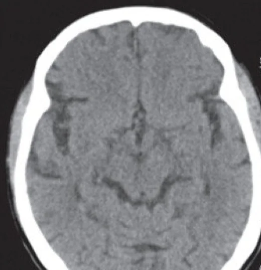
- Kontrastsız BT'de kanama ve kitle etkisinin bulunmaması TIA veya akut iskemi olasılığını ortadan kaldırmaz; inceleme öncelikle önemli alternatifleri hızla dışlar.
- MCA alanında insular ribbon'ın silinmesi, lentiform nükleus kontur kaybı, gri-beyaz cevher ayrımında azalma ve sulkus effasmanı erken iskemi ipuçlarıdır.
- Akut infarkt hipoattenüasyonu ilgili arter alanında gelişir; erken dönemde ince, bölgesel ve karşı hemisferle asimetrik olabilir.
- Kronik enfarkt glioz ve ensefalomalaziye bağlı olarak yan komşu boşluk genişlemesi ve ventriküler dilateasyon gösterebilir.
- BTA'da intrakraniyal ICA veya MCA'da ani kesilme, semptomla uyumlu distal dal dolum kaybı ve kollateral pial damarlar eşlik edebilir.
- Damar duvarı kalsifikasyonu, fokal lümen daralması ve distal doluş azalması semptomatik intrakraniyal ateroskleroz için birlikte yorumlanır.
- Parankim BT'si normal ve BTA açık olsa bile küçük kortikal, laküner veya posterior fossa infarktı olabilir; difüzyon MR daha duyarlı testtir.
Normalde
Normal kontrastsız BT'de hemisferler simetrik, kortikal gri-beyaz cevher sınırı belirgin, insular korteks ve bazal gangliyonlar seçilebilir, sulkuslar doğal açıklıktadır; akut kanama veya fokal kitle etkisi bulunmaz. BTA'da intrakraniyal ICA, MCA ve ana dallar kontrastla devamlı dolar; şüpheli taraftaki lümen ve distal dal opasifikasyonunu karşı hemisferle karşılaştır, fakat normal görünümün BT-negatif akut infarktı dışlamadığını akılda tut.
Normal BT ile kıyasla
Aynı bölgenin, kaynak veri setinde patoloji içermediği belirtilen bir BT incelemesini kesit kesit kaydırın; organ sınırlarını açarak anatomiyi hasta görüntüsüyle karşılaştırın. Küçük rastlantısal bulgular tümüyle dışlanmamıştır.
Kaynak: TotalSegmentator v2.01 veri seti (Wasserthal ve ark., Radiology: Artificial Intelligence 2023), CC BY 4.0, vaka s0103; organ sınırları veri setinin otomatik segmentasyonundan, birleştirilerek sadeleştirildi. Görüntüler 2,5 mm kesit aralığına yeniden örneklendi.
Ayırıcı tanı
- Akut iskemik infarkt
- arter alanına uyan fokal gri-beyaz cevher silinmesi, insular ribbon/lentiform nükleus kaybı veya sulkus effasmanı görülebilir; erken BT normal kalabilir.
- İntraserebral kanama
- kontrastsız BT'de parankimal kan ve çevresinde kitle etkisi ile ortaya çıkar; CTA'da spot veya fırça işareti erken hematoma genişlemesini öngörmede kullanılır.
- Hipoglisemi veya metabolik ensefalopati
- diffüzyon ağırlıklı MR'da iç kapsül arkası, corona radiata ve korpus kallozum splenyumunda çift taraflı, geri döndürülebilir yüksek sinyaller görülebilir ve bu durum akut izkemik inme ile karışabilir.
- Nöbet sonrası Todd parezisi
- BT ve BTA normal olabilir veya geçici bölgesel ödem bulunabilir; klinik olayın nöbet özellikleri ve MR bulguları ayırıcıdır.
- Migrenli hastalarda beyaz madde lezyon yükü kontrol grubuna göre belirgin şekilde daha yüksektir.
- Kronik enfarkt glioz ve ensefalomalaziye bağlı olarak ventriküler ve subaraknoid boşluklarda genişleme gösterebilir; eşlik eden damar anomalileri veya kronik stenoz ayrınot edilir.
Tuzaklar
- Semptomların düzelmesini BT'de infarkt yokluğuyla eşitleme; doku temelli değerlendirmede klinik geçici olay da MR'da akut lezyon gösterebilir.
- Erken iskemi işaretleri silik olabilir; geniş beyin penceresinin yanında gri-beyaz cevher farkını belirginleştiren dar inme penceresini kullan.
- Posterior fossa ve küçük laküner infarktlar kontrastsız BT'de özellikle zor seçilir; normal görüntü şüpheyi sürdürüyorsa difüzyon MR gerekir.
- BTA'da hipoplastik A1/P1 veya vertebral arter varyantını akut oklüzyon sanma; distal devamlılığı, communicating arterleri ve damar alanı bulgularını birlikte izle.
Kendini dene
On dakika süren afazi düzelmiş; kontrastsız BT'de kanama ve belirgin hipodensite yok. En doğru çıkarım hangisidir?
Cevabı göster
BT küçük akut infarktı dışlayamaz. Erken veya küçük akut infarkt kontrastsız BT'de görünmeyebilir; semptomların düzelmesi de iskemik olayı dışlamaz. Migren tanısı yalnız görüntülemeyle konmaz, eski infarkt ise hacim kaybı ve ensefalomalazi gibi kronik bulgu gerektirir.
Geçici sağ kol güçsüzlüğü sonrası BTA'da sol M1 fokal daralmış ve distal dallar zayıf opak. BT'de erken iskemik değişiklikler görülmeyebileceğinden, bunları saptamak için hangi görüntüleme yöntemi daha duyarlıdır?
Cevabı göster
Difüzyon MR. MR görüntülemenin küçük infarktları saptama duyarlılığı BT'den daha yüksektir; başlangıç BT'sinde infarkt görülmemesi inme olasılığını dışlamaz. Kemik ya da sinüs penceresi ve kontrastsız boyun BT'si beyin parankimindeki erken iskemiyi değerlendirmez.
İlgili konular
Kaynaklar
Bu sayfadaki 38 cümle ve 2 quiz sorusu aşağıdaki kaynaklardan alıntılarla tek tek karşılaştırıldı; 8 cümle bu karşılaştırmada düzeltildi (2026-10-08).
- Detection of Early Ischemic Changes in Noncontrast CT Head Improved with “Stroke Windows”pmc.ncbi.nlm.nih.gov
- From seropositivity to neurosyphilis: clinical spectrum and diagnostic classification in hospitalized neurology patients: a 16-year retrospective study.pubmed.ncbi.nlm.nih.gov
- A Toothache That Was a Stroke: Acute Right Parietal Infarction Presenting As Isolated Left-Sided Odontogenic-Like Pain.pubmed.ncbi.nlm.nih.gov
- Quantitative MRI Susceptibility: Mapping of Transient Ischemic Attack-A Preliminary Study.pubmed.ncbi.nlm.nih.gov
- Sensitivity of Non-contrast Computed Tomography Net Water Uptake Versus FLAIR-MRI, with MR-DWI Reference Standard, for Early Detection and Physiological Characterization of Ischemic Edema in Hyperacute Stroke.pubmed.ncbi.nlm.nih.gov
- Cardiac photon-counting detector computed tomography in acute ischemic stroke: diagnostic accuracy for detection of left atrial appendage thrombus.pubmed.ncbi.nlm.nih.gov
- Machine learning in neuroradiology: Recent developments and applications.pubmed.ncbi.nlm.nih.gov
- Multiterritory Acute Ischemic Stroke with Bilateral Middle Cerebellar Peduncle T2/FLAIR Hyperintensities in the Setting of a Fatal Systemic Arterial Thrombotic Process: A Case Report.pubmed.ncbi.nlm.nih.gov
- Bilateral internal carotid artery fibromuscular dysplasia identified during acute ischemic stroke evaluation: A case report.pubmed.ncbi.nlm.nih.gov
- Sequential bilateral common carotid artery occlusion in protein C deficiency and patent foramen ovale: illustrative case.pubmed.ncbi.nlm.nih.gov
- Role of Low-Field MRI in Acute Stroke.pubmed.ncbi.nlm.nih.gov
- Detection of Hemorrhagic Transformation of Cerebral Infarcts with Ultra-low Field MRI.pubmed.ncbi.nlm.nih.gov
- Severe Multisystem Thrombotic Phenotype Associated With Protein C Deficiency in A Young Woman: A Case Report.pubmed.ncbi.nlm.nih.gov
- Current state and advancements of imaging in acute ischemic stroke: a practical review.pubmed.ncbi.nlm.nih.gov
- Regional perfusion parameters as potential indicators of parenchymal hematoma risk following reperfusion therapy for acute ischemic stroke in the extended time window.pubmed.ncbi.nlm.nih.gov
- Neuroimaging Modalities Used for Ischemic Stroke Diagnosis and Monitoring.pubmed.ncbi.nlm.nih.gov
- Hybrid revascularization for acute common carotid artery occlusion after transcarotid transcatheter aortic valve implantation: illustrative case.pubmed.ncbi.nlm.nih.gov
- Direct balloon dilatation angioplasty for intracranial atherosclerotic stenosis-related acute ischemic stroke: a single-arm retrospective study of technical feasibility and safety.pubmed.ncbi.nlm.nih.gov
- Endovascular Revascularization for Chronic Vertebral Artery Occlusion with Poor Collateral Circulation and a Large Perfusion Abnormality Identified by Automated Perfusion Imaging: A Case Report.pubmed.ncbi.nlm.nih.gov
- Central Retinal Artery Occlusion Diagnosed via Ocular Point-of-care Ultrasound: Case Report.pubmed.ncbi.nlm.nih.gov
- Diagnostic evaluation of suspected aneurysmal subarachnoid hemorrhage after negative CT: risk-stratified use of lumbar puncture and CT angiography.pubmed.ncbi.nlm.nih.gov
- Integrating Invasive and Noninvasive Imaging for Coronary Atherosclerosis: A Systematic Review With Pragmatic Algorithms From Anatomy to Physiology.pubmed.ncbi.nlm.nih.gov
- Evaluation of Nonlinear Pixel-Wise Squaring for Enhancing Low-Contrast Resolution in CT Imaging.pubmed.ncbi.nlm.nih.gov
- Ischemic infarct detection, localization, and segmentation in noncontrast CT human brain scans: review of automated methods.pubmed.ncbi.nlm.nih.gov
- Malignant middle cerebral artery infarction revealing atrial fibrillation: A Multimodal imaging case of M1 occlusion treated with decompressive craniectomy.pubmed.ncbi.nlm.nih.gov
- A systematic approach to detection of epileptogenic foci on imaging: protocols, entities, search pattern, and reporting tools.pubmed.ncbi.nlm.nih.gov
- Synchronous subarachnoid haemorrhage and ischaemic stroke as a result of complete internal carotid artery occlusion.pubmed.ncbi.nlm.nih.gov
- Pathophysiology-driven neuroimaging: decoding brain injury in sepsis.pubmed.ncbi.nlm.nih.gov
- Fetal intracranial hemorrhage and infarct: Main sonographic and MRI characteristics: A review article.pubmed.ncbi.nlm.nih.gov
- Perforator-Sparing Microsurgical Clipping of Tandem Dominant-Hemisphere Middle Cerebral Artery Aneurysms: Geometry-Guided Reconstruction of a Wide-Neck Bifurcation and Dorsal M1 Fusiform Lesion.pubmed.ncbi.nlm.nih.gov
- Endovascular Treatment Strategies for Acute Intracranial Atherosclerotic Stenosis-Related Large Vessel Occlusion.pubmed.ncbi.nlm.nih.gov
- Post-Traumatic Middle Cerebral Artery Pseudoaneurysm Following Depressed Skull Fracture: Case Report and PRISMA-Guided Literature Review.pubmed.ncbi.nlm.nih.gov
- Azygous anterior cerebral artery occlusion managed with Thrombectomy: a case report and literature review.pubmed.ncbi.nlm.nih.gov
- Multimodal imaging in neuro-ophthalmology.pubmed.ncbi.nlm.nih.gov
- Carotid plaque vulnerability and circle of Willis anatomy predict ipsilateral brain infarcts and long-term mortality in carotid endarterectomy patients.pubmed.ncbi.nlm.nih.gov
- A Case of Bilateral Globus Pallidus Infarction Onset With Mental Disorder.pubmed.ncbi.nlm.nih.gov
- Acute Ischemic Stroke in 2026: From Time to Penumbra-An Updated Narrative Review of Reperfusion Strategies with a Latin American Implementation Perspective.pubmed.ncbi.nlm.nih.gov
- CT perfusion lesions are present in most MRI confirmed lacunar strokes.pubmed.ncbi.nlm.nih.gov
- Diagnosis and Management of Patients with Patent Foramen Ovale: Current Evidence and Future Perspective.pubmed.ncbi.nlm.nih.gov
- Through Massage to the Brain-Neuronal and Neuroplastic Mechanisms of Massage Based on Various Neuroimaging Techniques (EEG, fMRI, and fNIRS).pubmed.ncbi.nlm.nih.gov
- Autoimmune encephalitis: what the radiologist needs to know.pubmed.ncbi.nlm.nih.gov
- Advances in human intracranial electroencephalography research, guidelines and good practices.pubmed.ncbi.nlm.nih.gov
- Human cerebral arterial morphogenesis explored by 3D microscopic imaging of cleared embryos: Bridging the gap between embryonic and adult anatomy.pubmed.ncbi.nlm.nih.gov
- Stent Retriever-Assisted Embolic Protection During Recanalization of Chronic Internal Carotid Artery Occlusion with Contralateral Carotid Stenosis: A Case Report.pubmed.ncbi.nlm.nih.gov
- Initial experience with the antithrombogenic-coated CARESTO stent for acute symptomatic cervical internal carotid artery dissections: multicenter study.pubmed.ncbi.nlm.nih.gov
- Insights into the pathophysiology of stroke-heart syndrome: a review of recent literature and clinical trials to illuminate promising therapeutic strategies.pubmed.ncbi.nlm.nih.gov
- Stroke or Seizure? Diagnostic Role of Neuroimaging in Acute Neurologic Mimics.pubmed.ncbi.nlm.nih.gov
- Rapid spontaneous regression of a massive post-thrombolysis intracerebral hemorrhage: A case report.pubmed.ncbi.nlm.nih.gov
- Contrast-Induced Encephalopathy Due to Contrast Extravasation From a Chronic Post-hemorrhagic Encephalomalacic Region: Prior Hemorrhage as a Potential Risk Factor.pubmed.ncbi.nlm.nih.gov
- The "brush sign": a novel and accessible CT marker for hematoma expansion in hypertensive intracerebral hemorrhage.pubmed.ncbi.nlm.nih.gov
- Stepwise clinical and diagnostic strategy for coma of unknown origin.pubmed.ncbi.nlm.nih.gov
- Reversible Metabolic and Liver Disease in Complex III Deficiency: Novel Variants Expand the Reported <i>UQCRC2</i>-Associated Phenotype.pubmed.ncbi.nlm.nih.gov
- Pediatric endocrine disorders: a review of intracranial findings and appropriate imaging.pubmed.ncbi.nlm.nih.gov
- Immune Thrombotic Thrombocytopenic Purpura Presenting as an Acute Stroke Mimic in a Young Pakistani Man.pubmed.ncbi.nlm.nih.gov
- Postictal Todd's Paralysis Mimicking Acute Ischemic Stroke in Medullary Melanocytoma: A Multimodal Diagnostic Challenge.pubmed.ncbi.nlm.nih.gov
- Bilateral Vein of Trolard Thrombosis Presenting with Seizure and Minimal Deficits: A Rare Case Report.pubmed.ncbi.nlm.nih.gov
- White matter lesions in migraine: a single-center retrospective study of prevalence and risk factors.pubmed.ncbi.nlm.nih.gov
- When headache meets visual disturbance: An evidence-based review of common and critical entities.pubmed.ncbi.nlm.nih.gov
- Pediatric occipital lobe epilepsy: A modern review of etiological classification, management, and outcomes.pubmed.ncbi.nlm.nih.gov
- Advances in cerebrovascular reserve function assessment for moyamoya disease.pubmed.ncbi.nlm.nih.gov
- Acute Ischemic Stroke in an Infant With COVID-19 Infection and Delayed Diagnosis of Neurofibromatosis Type 1.pubmed.ncbi.nlm.nih.gov
- Paediatric cerebrovascular syndromes: imaging clues to a diagnosis.pubmed.ncbi.nlm.nih.gov
- Vertebral artery pseudoaneurysm with recurrent posterior circulation transient ischemic attacks in a post-radiotherapy nasopharyngeal carcinoma survivor: a case report.pubmed.ncbi.nlm.nih.gov
- Disruption of amino acid metabolism in acute cerebrovascular diseases: diagnostic and pathobiochemical perspectives.pubmed.ncbi.nlm.nih.gov
- Carotid Free-Floating Thrombus: A Case Series, Narrative Review, and Proposal for Biology-Guided Treatment Selection.pubmed.ncbi.nlm.nih.gov
- Todd's Paralysis Simulating Acute Ischemic Stroke: A Case Report.pubmed.ncbi.nlm.nih.gov
- Where Does the Signal Go? Technical Challenges of CT Perfusion in Lacunar and Infratentorial Stroke.pubmed.ncbi.nlm.nih.gov
- Integrated diagnostic algorithm for acute vertigo combining TiTrATE, STANDING, and HINTS: a validation study in the emergency department.pubmed.ncbi.nlm.nih.gov
- Lacunar stroke with incidental congenital absence of the internal carotid artery: importance of recognising a rare anatomical variant.pubmed.ncbi.nlm.nih.gov
- Persistence of the hypoglossal artery: A rare case report.pubmed.ncbi.nlm.nih.gov
- Ischemic posterior circulation stroke: a review of anatomy, clinical presentations, diagnosis, and current management.pubmed.ncbi.nlm.nih.gov
- Refined AI-ASPECTS with modified atlas and lesion-load thresholds: advancing acute ischemic stroke imaging and prognostic prediction.pubmed.ncbi.nlm.nih.gov
- Balancing Radiation Dose and Image Quality: Protocol Optimization for Mobile Head CT in Neurointensive Care Unit Patients.pubmed.ncbi.nlm.nih.gov
- Brain microstructural alterations in COVID-19: a systematic review of diffusion weighted imaging studies.pubmed.ncbi.nlm.nih.gov
- Associations Among CTA Collateral Scores, Multimodal MRI Lesion Volumes, and Clinical Severity in Acute Middle Cerebral Artery Infarction.pubmed.ncbi.nlm.nih.gov
- Adjusting input arterial function to improve the accuracy of hypoperfusion assessment in computed tomography perfusion (CTP) for stroke: a preliminary study.pubmed.ncbi.nlm.nih.gov
- The value and limitations of the Oxford Community Stroke Project classification in the hyperacute phase of cerebral infarction.pubmed.ncbi.nlm.nih.gov
- Coronary Intramural Hematoma in a Young Athlete After Direct Ball Impact to the Chest.pubmed.ncbi.nlm.nih.gov
- Ictal laughter induced by repetitive transcranial magnetic stimulation in stroke patients: a case series.pubmed.ncbi.nlm.nih.gov
- Case Report: Structured MRI assessment of posterior thalamic infarction in a distribution compatible with posterior choroidal artery territory presenting as Déjerine-Roussy syndrome in an adolescent: differentiating arterial ischemia from venous thrombosis and thalamic neoplasm.pubmed.ncbi.nlm.nih.gov
- Low-Field and Portable MRI for Acute Ischemic Stroke: A Systematic Review.pubmed.ncbi.nlm.nih.gov
- Role of magnetic resonance in bowel pathologies: Beyond inflammatory bowel disease.pubmed.ncbi.nlm.nih.gov
- The Anticoagulation Dilemma: Concurrent Traumatic Brain Hemorrhage and Cerebral Venous Thrombosis in a Blast Injury Patient.pubmed.ncbi.nlm.nih.gov
- Imaging for Pediatric Head and Neck Emergencies: Critical Insights.pubmed.ncbi.nlm.nih.gov
- Radiologic Characterization of Invasive Fungal Infections of the Paranasal Sinuses and Skull Base: A Prospective Analysis.pubmed.ncbi.nlm.nih.gov
- Automated advanced imaging in acute ischemic stroke. Certainties and uncertainties.pubmed.ncbi.nlm.nih.gov
- Efficacy of Computed Tomography Perfusion - Alberta Stroke Program Early Computed Tomography Score for Identifying Patients with Anterior Circulation Acute Ischemic Stroke that Would Benefit from Endovascular Treatment.pubmed.ncbi.nlm.nih.gov
- Recurrent brief positional vertigo as the initial presentation of medial branch of the posterior inferior cerebellar artery territory cerebellar infarction: a case report.pubmed.ncbi.nlm.nih.gov
- CT - derived fractional flow reserve can predict recurrent ischemia in patients with MCA stenosis.pubmed.ncbi.nlm.nih.gov
- Precision Neuromodulation Treatment Reverses Motor and Cognitive Slowing After Stroke: Clinical and Neurophysiological Evidence.pubmed.ncbi.nlm.nih.gov
- Post-Stroke Recrudescence: A Case Report and Literature Review.pubmed.ncbi.nlm.nih.gov
- Atypical Hypoglycemia-Induced Stroke Mimic: A Case Report and Literature Review.pubmed.ncbi.nlm.nih.gov
- Fulminant Powassan Virus Encephalitis Presenting as Acute Ischemic Stroke: A Case Report.pubmed.ncbi.nlm.nih.gov
- Hypoglycemic Encephalopathy Caused by Insulinoma With Reversible High Signals in the White Matter on Diffusion-Weighted MRI: A Case Report.pubmed.ncbi.nlm.nih.gov
- Neurologic Symptoms and Cerebrovascular Events During Atogepant Therapy: A Case Series with Contextual Comparison with a Non-Gepant-Treated Migraine Cohort.pubmed.ncbi.nlm.nih.gov
- Migraine and Cardiovascular Risk: A Scoping Review of Vascular Outcomes, Risk Assessment, and Endothelial Dysfunction.pubmed.ncbi.nlm.nih.gov
- Ischemic Stroke as a Possible Initial Manifestation of Primary Sjögren's Syndrome: A Case Report.pubmed.ncbi.nlm.nih.gov
- Radiation-Induced Sphenoid Wing Meningioma After Childhood Medulloblastoma: A Case Report.pubmed.ncbi.nlm.nih.gov
- When Stroke Leads to Hydrocephalus: A Case of Unilateral Ventricular Enlargement.pubmed.ncbi.nlm.nih.gov
- Bacterial Meningoencephalitis in Newborns.pubmed.ncbi.nlm.nih.gov
- Pediatric Meningeal Diseases: What Radiologists Need to Know.pubmed.ncbi.nlm.nih.gov
- Alternating Hemispheric Episodes in Bilateral Rasmussen Encephalitis: The Crucial Role of Sequential Multimodal Neuroimaging.pubmed.ncbi.nlm.nih.gov
- Identifying central positional vertigo to prevent diagnostic pitfalls: A case series and review of literature.pubmed.ncbi.nlm.nih.gov
- Retractor-related liver injury after surgery: MRI findings in a case.pubmed.ncbi.nlm.nih.gov
- Targeting Inflammation Across the Myocardial Infarction Continuum: Biomarkers, Imaging, and Emerging Therapies.pubmed.ncbi.nlm.nih.gov
- Moyamoya Vasculopathy and Atypical Moyamoya-like Patterns: Insights into Diagnosis and Therapeutic Implications.pubmed.ncbi.nlm.nih.gov
- Mechanisms of aortic dissection: From pathological changes to experimental and <i>in silico</i> models.pubmed.ncbi.nlm.nih.gov
- Guidelines for evaluation and management of cerebral collateral circulation in ischaemic stroke 2017.pubmed.ncbi.nlm.nih.gov
- Spontaneous bacteremia and spontaneous bacterial peritonitis share similar prognosis in patients with cirrhosis: a cohort study.pubmed.ncbi.nlm.nih.gov
- Brain imaging in stroke: insight beyond diagnosis.pubmed.ncbi.nlm.nih.gov
- Neurological involvement in localized scleroderma: a systematic review of clinical manifestations and neuroimaging findings.pubmed.ncbi.nlm.nih.gov
Eğitim amaçlıdır; hasta başında tanı ve tedavi kararının yerine geçmez. İçerik yapay zekâ destekli bir süreçle güncel literatüre göre cümle cümle kaynaklarla karşılaştırıldı, ancak uzman radyolog incelemesinden geçmedi. Klinik kararlarda güncel kılavuzları, kurum protokollerini ve radyoloji raporunu esas alın.