Özet
Geniş hemisferik infarkt, çoğunlukla proksimal MCA tıkanıklığıyla gelişen ve ödem arttıkça intrakraniyal basıncı yükselten büyük doku hasarıdır. Kontrastsız BT'de geniş alanda hipoattenüasyon ve gri-beyaz sınır kaybının yanı sıra sulkus, ventrikül ve bazal sistern basısı aranır. Orta hat kayması, subfalsin veya unkal herniasyon yönündeki ilerleme acil nörolojik kötüleşmeyi haber verebilir. Kitle etkisini veya sistern kapanmasını kaçırmak beyin sapı basısı ve herniasyon riskinin geç fark edilmesine yol açar.
Okuma sırası
- İnsula, lentiform nükleus ve MCA korteksi. Bazal gangliyon ve insula düzeyinde gri-beyaz sınırı izleyin; hipoattenüasyonun tek küçük odak yerine geniş MCA alanına yayılıp yayılmadığını haritalayın.
- Lateral ventrikül. Lateral ventrikül gövdesine çıkıp ipsilateral frontal boynuz ve gövdenin basılıp basılmadığını karşı tarafla kıyaslayın.
- Septum pellucidum ve orta hat. Anatomik orta hattı belirleyin ve septum pellucidumun karşı tarafa sapmasını ölçün; bu ölçüm kitle etkisini belgeler.
- Bazal sisternler. Tentoryal çentik düzeyinde ambient sisternlerin simetrisinin kaybolup kaybolmadığını izleyin; beyin sapı çevresindeki BOS mesafelerini de değerlendirin.
- Unkus ve ambient sistern. Unkusların konumunu ve ambient sisterni iki tarafta karşılaştırın; medial temporal lobun aşağı-mediale yer değiştirmesini arayın.
Faz ve pencere
- Kontrastsız tanısal
- MCA alanının geniş bölümünde veya komşu ACA/PCA alanlarına taşan hipoattenüasyon, kortikal gri-beyaz ayrım kaybı ve şişme görülür. Aynı tarafta sulkuslar ve ipsilateral lateral ventrikül basılır; septum pellucidum karşıya itilebilir, perimezensefalik/bazal sisternler daralabilir veya silinebilir. Akut ödem etkisini göstermek için kontrastsız BT yeterlidir.
- BTA
- İntrakraniyal arterlerde proksimal MCA veya eşlik eden büyük damar oklüzyonu ve kollateral dolum araştırılır; bu, tıkanıklığın yerini gösterir, parankimal ödem ve herniasyon değerlendirmesinin yerini tutmaz.
Önerilen pencereler: Beyin (G 80 / M 40), İnme (G 40 / M 40).
BT bulguları
- İnsula, lentiform nükleus ve frontotemporoparietal MCA korteksinde yaygın hipoattenüasyon ve gri-beyaz ayrım kaybı.
- Birden fazla arter alanına yayılan ödem; ipsilateral ACA veya PCA korteksinin de etkilenebilmesi.
- Kortikal sulkusların ve sylvian fissürün daralması, hemisferik şişliğin erken kitle etkisi.
- İpsilateral lateral ventrikül gövdesi ve frontal boynuzda bası; septum pellucidumun karşı tarafa yer değiştirmesi.
- Subfalsin yer değiştirme — singulat girusun falx altından karşı hemisfere doğru itilmesi.
- Unkal herniasyon yönünde medial temporal lobun tentoryal çentiğe yaklaşması ve ipsilateral suprasellar/perimezensefalik sisternlerin silinmesi.
- Bazal sisternlerin belirgin daralması veya kaybolması ve beyin sapı çevresindeki BOS aralıklarının kapanması.
Ölçütler ve sınıflamalar
- İlk 6 saatte BT'de geniş MCA alanı erken hipoattenüasyonu (malign ödem riskiyle ilişkili radyolojik belirteç)
- >%50 MCA alanı
Normalde
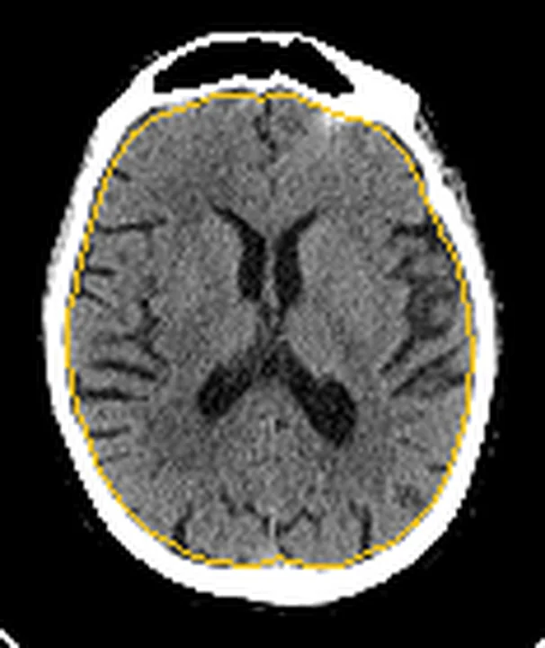
Normal hemisferde kortikal gri-beyaz ayrımı ve sulkuslar korunur; lateral ventriküller orta hatta benzer genişlikte, bazal sisternler açıktır. Septum pellucidumun falxla ilişkisini, ipsilateral ventrikül çapını ve suprasellar/perimezensefalik sisternlerin açıklığını karşı taraf ve önceki BT ile kıyaslayın.
Normal BT ile kıyasla
Aynı bölgenin, kaynak veri setinde patoloji içermediği belirtilen bir BT incelemesini kesit kesit kaydırın; organ sınırlarını açarak anatomiyi hasta görüntüsüyle karşılaştırın. Küçük rastlantısal bulgular tümüyle dışlanmamıştır.
Kaynak: TotalSegmentator v2.01 veri seti (Wasserthal ve ark., Radiology: Artificial Intelligence 2023), CC BY 4.0, vaka s0103; organ sınırları veri setinin otomatik segmentasyonundan, birleştirilerek sadeleştirildi. Görüntüler 2,5 mm kesit aralığına yeniden örneklendi.
Ayırıcı tanı
- Beyin tümörleri ve enfeksiyonlar akut infarktı taklit edebilir; ayırıcı tanı için detaylı klinik öykü, laboratuvar incelemeleri ve çok modlu BT veya MR görüntüleme gereklidir (S50, S51).
- Otoimmun ensefalit ve diğer enfeksiyonlar infarkt bulgularıyla karışabilir; ayırıcı tanı için detaylı klinik öykü, laboratuvar ve manyetik rezonans görüntüleme değerlendirmesi şarttır (S50).
- Postiktal durumlar kortikal şişme ve geçici hipoattenüasyona yol açabilir; damar alanıyla örtüşmeyen perfüzyon değişiklikleri ve arteriyel oklüzyonun yokluğu ayırıcı tanıda önemlidir (S51, S75).
- Hipoksi-iskemi ve metabolik toksik durumlar akut İnme taklitçileri arasında yer alır; çok modlu BT (kontrastsız, BTA, BTP) bu taklitçileri ekarte etmek ve gereksiz revasküler müdahalelerden kaçınmak için yüksek özgüllük sağlar (S51).
- Serebral venöz tromboz
- ödem ve hemoraji venöz drenaj alanını izler, arteriyel dağılımı aşabilir; venöz sinüs/damar trombozu BTV'de araştırılır.
Tuzaklar
- Erken büyük infarkt henüz belirgin şişme yapmayabilir; yalnız orta hat kayması yok diye ciddi iskemiyi dışlamayın, gri-beyaz ayrımı ve sulkusları da gözden geçirin.
- Eski hemisferik ensefalomalazi geniş hipoattenüasyon taklidi yapar; kronik lezyon ventrikül ve sulkusları genişletirken akut ödem bu BOS aralıklarını daraltır.
- Orta hat kaymasını yalnız septum pellucidumda değerlendirmek unkal basıyı kaçırabilir; bazal sisternleri, medial temporal lobu ve beyin sapı çevresini ayrıca inceleyin.
- Kronik lezyonlarda görülen hacim kaybının aksine, akut geniş hemisferik infarkt geniş hipoattenüasyona, orta hat kaymasına ve bazal sistern basısına yol açar.
Kendini dene
Geniş sağ MCA hipoattenüasyonuna sağ ventrikül basısı, sola septum kayması ve sistern silinmesi eşlik ediyor. En önemli görüntüleme yorumu nedir?
Cevabı göster
Malign hemisferik ödem. Geniş arter alanı infarktı ile BOS aralıklarının basılması ve sistern silinmesi ilerleyen kitle etkisini gösterir. Kronik ensefalomalazi hacim kaybı yapar; lakün küçük ve derindir; ince subdural hematom bu yaygın parankim ödemini açıklamaz.
Bir hemisferde hipoattenüasyon var, ancak aynı taraftaki sulkuslar ve lateral ventrikül genişlemiş. Hangi süreç daha olasıdır?
Cevabı göster
Kronik infarkt. Sulkus ve ventrikül genişlemesi doku kaybına bağlı ex vacuo değişikliği destekler. Akut malign ödem bu boşlukları daraltır; ensefalit ve arter oklüzyonu bu kronik hacim kaybı paternini tek başına açıklamaz.
İlgili konular
Kaynaklar
Bu sayfadaki 41 cümle ve 2 quiz sorusu aşağıdaki kaynaklardan alıntılarla tek tek karşılaştırıldı; 5 cümle bu karşılaştırmada düzeltildi (2026-10-08).
- Advances in the Critical Care Management of Ischemic Strokepmc.ncbi.nlm.nih.gov
- Intravenous glibenclamide for cerebral oedema after large hemispheric stroke (CHARM): a phase 3, double-blind, placebo-controlled, randomised trial.pubmed.ncbi.nlm.nih.gov
- Pneumocephalus associated with massive cerebral air embolism.pubmed.ncbi.nlm.nih.gov
- A multiparameter model predicting in-hospital mortality in malignant cerebral infarction.pubmed.ncbi.nlm.nih.gov
- Application of Machine Learning to Automated Analysis of Cerebral Edema in Large Cohorts of Ischemic Stroke Patients.pubmed.ncbi.nlm.nih.gov
- Comparative Analysis of Markers of Mass Effect after Ischemic Stroke.pubmed.ncbi.nlm.nih.gov
- Herniation despite Decompressive Hemicraniectomy in Large Hemispherical Ischemic Strokes.pubmed.ncbi.nlm.nih.gov
- Automated quantification of cerebral edema following hemispheric infarction: Application of a machine-learning algorithm to evaluate CSF shifts on serial head CTs.pubmed.ncbi.nlm.nih.gov
- A serious thrombotic event in a patient with immune thrombocytopenia requiring intravenous immunoglobulin: a case report.pubmed.ncbi.nlm.nih.gov
- Hemicraniectomy and durotomy upon deterioration from infarction-related swelling trial: randomized pilot clinical trial.pubmed.ncbi.nlm.nih.gov
- Stroke Lateralization in Large Hemisphere Infarctions: Characteristics, Stroke-Related Complications, and Outcomes.pubmed.ncbi.nlm.nih.gov
- Cerebral Edema in Patients With Large Hemispheric Infarct Undergoing Reperfusion Treatment: A HERMES Meta-Analysis.pubmed.ncbi.nlm.nih.gov
- Integrating Brain Imaging Volumetrics and Quantitative Pupillometry for Predicting Neurologic Deterioration after Large Hemispheric Stroke.pubmed.ncbi.nlm.nih.gov
- Pupillometry in the follow-up of patients undergoing EVT - prediction of space-occupying hemispheric infarction.pubmed.ncbi.nlm.nih.gov
- Contrast-Induced Neurotoxicity Following Mechanical Thrombectomy for Acute Left MCA Occlusion: A Reversible Radiological Mimic of Intracranial Haemorrhage.pubmed.ncbi.nlm.nih.gov
- From Cervical Trauma and Head Injury to Malignant Cerebral Edema: Blunt Cerebrovascular Injury Causing Large-Vessel Occlusion Treated With Thrombectomy and Neurocritical Care.pubmed.ncbi.nlm.nih.gov
- Decompressive hemicraniectomy as a salvage therapy in the Neuro-ICU: a meta-analysis of neurologic outcomes for malignant middle cerebral artery infarction.pubmed.ncbi.nlm.nih.gov
- Anatomy-Guided Microsurgical Resection of a Dominant Frontal Lobe Tumor Without Intraoperative Adjuncts: A Case Report from a Resource-Limited Context.pubmed.ncbi.nlm.nih.gov
- Fixed, Dilated, and Conversing-Unreactive Pupil With Preserved Consciousness Indicating Acutely Rising Intracranial Pressure due to Traumatic Intraparenchymal Contusions: Case Report and Review of the Literature.pubmed.ncbi.nlm.nih.gov
- Neurological manifestations and neuroimaging findings in patients with SARS-CoV2-a systematic review.pubmed.ncbi.nlm.nih.gov
- Diagnostic Approach to Macrocephaly in Children.pubmed.ncbi.nlm.nih.gov
- Susceptibility-weighted imaging in pediatric arterial ischemic stroke: a valuable alternative for the noninvasive evaluation of altered cerebral hemodynamics.pubmed.ncbi.nlm.nih.gov
- Acute large vessel occlusion stroke as a neurovascular complication of immune thrombotic thrombocytopenic purpura: Mechanical thrombectomy and neurocritical care challenges.pubmed.ncbi.nlm.nih.gov
- Neuroimaging for differential diagnosis of transient neurological attacks.pubmed.ncbi.nlm.nih.gov
- Imaging-based management of acute ischemic stroke patients: current neuroradiological perspectives.pubmed.ncbi.nlm.nih.gov
- Association of 24-Hour Computed Tomography Infarct Density on Functional Outcomes in Stroke: Secondary Analysis From the AcT Trial.pubmed.ncbi.nlm.nih.gov
- Machine learning in neuroradiology: Recent developments and applications.pubmed.ncbi.nlm.nih.gov
- The Effect of Intensive Blood Pressure Reduction on Perihematomal Edema After Intracerebral Hemorrhage: A Systematic Review and Meta-Analysis.pubmed.ncbi.nlm.nih.gov
- Multiterritory Bihemispheric Ischemic Stroke in the Setting of Unilateral Internal Carotid Artery Disease.pubmed.ncbi.nlm.nih.gov
- Stent Retriever-Assisted Embolic Protection During Recanalization of Chronic Internal Carotid Artery Occlusion with Contralateral Carotid Stenosis: A Case Report.pubmed.ncbi.nlm.nih.gov
- Computed Tomography Angiography Recognition of a Hemorrhagic Moyamoya Pattern May Alter Acute Blood Pressure Management.pubmed.ncbi.nlm.nih.gov
- Neuroimaging Modalities Used for Ischemic Stroke Diagnosis and Monitoring.pubmed.ncbi.nlm.nih.gov
- Risk factors for post-traumatic hydrocephalus after decompressive craniectomy in patients with traumatic brain injury.pubmed.ncbi.nlm.nih.gov
- Fetal intracranial hemorrhage and infarct: Main sonographic and MRI characteristics: A review article.pubmed.ncbi.nlm.nih.gov
- Brain Herniationpubmed.ncbi.nlm.nih.gov
- Peritumoral MRI findings and brain herniations in epileptic dogs with prosencephalic brain tumors: a multicentre retrospective study.pubmed.ncbi.nlm.nih.gov
- Optimal Timing for Stereotactic Minimally Invasive Surgery in Supratentorial Spontaneous Intracerebral Hemorrhage With Tentorial Herniation: A Retrospective Study.pubmed.ncbi.nlm.nih.gov
- Brain Shift Patterns: Upward, Lateral and Downward Herniation, Its Correlation with Clinical Patterns in Acute Intracranial Pathologies and Neurosurgical Management.pubmed.ncbi.nlm.nih.gov
- Early isolated subarachnoid hemorrhage versus hemorrhagic infarction in cerebral venous thrombosis.pubmed.ncbi.nlm.nih.gov
- Brain Midline Shift Measurement and Its Automation: A Review of Techniques and Algorithms.pubmed.ncbi.nlm.nih.gov
- Decompressive hemicraniectomy in supra-tentorial malignant infarcts.pubmed.ncbi.nlm.nih.gov
- Long-term safety and effectiveness of the "OptEase" vena cava filter.pubmed.ncbi.nlm.nih.gov
- Minimally invasive interventions for intracranial pathologies using tubular retractors in the pediatric population: Safety, efficacy, technical aspects and outcomes.pubmed.ncbi.nlm.nih.gov
- Development of neonatal-specific sequences for portable ultralow field magnetic resonance brain imaging: a prospective, single-centre, cohort study.pubmed.ncbi.nlm.nih.gov
- Spectrum of Neuroimaging Findings in CNS Lymphoproliferative Disorders.pubmed.ncbi.nlm.nih.gov
- Amyloid-related imaging abnormalities (ARIA) in anti-amyloid therapies for Alzheimer's disease: An update from the Alzheimer's Association ARIA workgroup.pubmed.ncbi.nlm.nih.gov
- Posterior Reversible Encephalopathy Syndrome: A Case Report With Contemporary Literature Review and Neuropathological Evidence From Autopsy.pubmed.ncbi.nlm.nih.gov
- Monogenic Mimics of Neuroinflammatory Phenotypes in Children and Young Adults: An Evolving Landscape.pubmed.ncbi.nlm.nih.gov
- Case report: From misdiagnosis to timely detection: a clinical and imaging guide to neurological presentations of diffuse large B-cell lymphoma-insights from six cases.pubmed.ncbi.nlm.nih.gov
- Mimickers of autoimmune encephalitis: a literature review.pubmed.ncbi.nlm.nih.gov
- Stroke Mimics in the Acute Setting: Role of Multimodal CT Protocol.pubmed.ncbi.nlm.nih.gov
- Overview of Acute Ischemic Stroke Evaluation and Management.pubmed.ncbi.nlm.nih.gov
- Chikungunya-Associated Acute Respiratory Distress Syndrome: A Rare Fatal Case.pubmed.ncbi.nlm.nih.gov
- 소아 신경영상에서 보이는 비선천성 원인의 영상 소견: 임상 화보.pubmed.ncbi.nlm.nih.gov
- Atypical Posterior Reversible Encephalopathy Syndrome with Intraventricular Hemorrhage in a Patient with Eclampsia : A Case Report.pubmed.ncbi.nlm.nih.gov
- Intracranial venous thrombosis imaging by anatomical location: an educational review with mimics and diagnostic pitfalls.pubmed.ncbi.nlm.nih.gov
- Cerebral venous disorders: clinical presentation, diagnostic strategy, and contemporary management.pubmed.ncbi.nlm.nih.gov
- Venous-significant intracranial hemorrhage in neonatal HIE treated with therapeutic hypothermia: imaging phenotypes, diagnostic pitfalls, and prognostic implications.pubmed.ncbi.nlm.nih.gov
- Amyloid-Related Imaging Abnormalities with Emerging Alzheimer Disease Therapeutics: Detection and Reporting Recommendations for Clinical Practice.pubmed.ncbi.nlm.nih.gov
- Multisystem Imaging Manifestations of COVID-19, Part 2: From Cardiac Complications to Pediatric Manifestations.pubmed.ncbi.nlm.nih.gov
- Non-invasive intracranial pressure estimation in the intensive care unit: narrative review of methods and clinical applications.pubmed.ncbi.nlm.nih.gov
- Diagnosis and management of elevated intracranial pressure in the emergency department.pubmed.ncbi.nlm.nih.gov
- Commentary: an eye on PET quantification.pubmed.ncbi.nlm.nih.gov
- Patterns of injury and violence in Yaoundé Cameroon: an analysis of hospital data.pubmed.ncbi.nlm.nih.gov
- Quantitative pupillometry as a sensitive tool for detecting tumor-related mass effect in the posterior fossa: a prospective feasibility study comparing metastatic versus skull base lesions.pubmed.ncbi.nlm.nih.gov
- Deep learning-based prediction of mortality using brain midline shift and clinical information.pubmed.ncbi.nlm.nih.gov
- Fetal Brain Development: Regulating Processes and Related Malformations.pubmed.ncbi.nlm.nih.gov
- Imaging Evaluation of Acute Traumatic Brain Injury.pubmed.ncbi.nlm.nih.gov
- Ruptured Posterior Inferior Cerebellar Artery Aneurysms: Integrating Microsurgical Expertise, Endovascular Challenges, and AI-Driven Risk Assessment.pubmed.ncbi.nlm.nih.gov
- Contrast extravasation on Dyna-CT as a predictor of malignant brain edema after mechanical thrombectomy for acute anterior circulation large vessel occlusion.pubmed.ncbi.nlm.nih.gov
- VEXAS syndrome-associated tumefactive demyelination.pubmed.ncbi.nlm.nih.gov
- Neuroimaging standards for research into small vessel disease and its contribution to ageing and neurodegeneration.pubmed.ncbi.nlm.nih.gov
- Neuroradiology in women of childbearing age.pubmed.ncbi.nlm.nih.gov
- Coexistence of a Hemispheric Developmental Venous Anomaly and Two Ipsilateral Arteriovenous Malformations: A Rare Neurovascular Association.pubmed.ncbi.nlm.nih.gov
- Stroke or Seizure? Diagnostic Role of Neuroimaging in Acute Neurologic Mimics.pubmed.ncbi.nlm.nih.gov
- Radiological Findings of Retrograde Venous Cerebral Air Embolism Infarcts: A Case Report and Literature Review.pubmed.ncbi.nlm.nih.gov
- Progressive multifocal leukoencephalopathy - a diagnostic guide for the clinical neurologist.pubmed.ncbi.nlm.nih.gov
- A systematic approach to MR spectroscopy of the brain: Indications, protocol, metabolites, and spectral analysis in neurologic diseases.pubmed.ncbi.nlm.nih.gov
- Brain perfusion imaging in patients with status epilepticus, seizures, and IIC patterns.pubmed.ncbi.nlm.nih.gov
- Primary Angiitis of the Central Nervous System Mimicking a Cardioembolism: Posterior Circulation Infarction With Subarachnoid Hemorrhage and Obstructive Hydrocephalus.pubmed.ncbi.nlm.nih.gov
- Beyond the embolus: "do not miss" diffusion abnormalities of ischaemic and non-ischaemic neurological disease.pubmed.ncbi.nlm.nih.gov
- Magnetic Resonance Imaging of the Neonatal Brain: Modalities, Scoring Systems, and Clinical Applications.pubmed.ncbi.nlm.nih.gov
- 2.5-mm articulated endoluminal forceps using a compliant mechanism.pubmed.ncbi.nlm.nih.gov
- Unusual images of ischemic stroke with hyperacute spontaneous recanalization: a case report.pubmed.ncbi.nlm.nih.gov
- Prognostic value of computed tomography score in patients after extracorporeal cardiopulmonary resuscitation.pubmed.ncbi.nlm.nih.gov
- Cerebral Edema in Traumatic Brain Injury.pubmed.ncbi.nlm.nih.gov
- Cerebellitis following COVID-19 infection: A case-based systematic review and pooled analysis.pubmed.ncbi.nlm.nih.gov
- Molecular, Pathological, Clinical, and Therapeutic Aspects of Perihematomal Edema in Different Stages of Intracerebral Hemorrhage.pubmed.ncbi.nlm.nih.gov
- Multi-modal lesion mapping of expressive aprosodia.pubmed.ncbi.nlm.nih.gov
- Different diseases, same circuits: lessons from rare and overlooked causes of disorders of consciousness.pubmed.ncbi.nlm.nih.gov
- Does mRNA-based COVID-19 vaccination in the subacute phase lead to microstructural brain changes? A prospective pilot MRI study using T1 relaxometry.pubmed.ncbi.nlm.nih.gov
- Multidimensional analysis of cerebral iron and vascular changes in chronic unilateral middle cerebral artery stenosis with quantitative susceptibility mapping, arterial spin labeling and high-resolution vessel wall imaging-a pilot study.pubmed.ncbi.nlm.nih.gov
- The pathogenesis of cerebral small vessel disease and vascular cognitive impairment.pubmed.ncbi.nlm.nih.gov
- Sulfonylurea Receptor 1 in Central Nervous System Injury: An Updated Review.pubmed.ncbi.nlm.nih.gov
- Stroke recovery beyond initial severity: the complementary roles of brain structure and brain function in acute stroke.pubmed.ncbi.nlm.nih.gov
- Evaluation of ChatGPT-5 for Automated ASPECTS Assessment on Non-Contrast CT in Acute Ischemic Stroke.pubmed.ncbi.nlm.nih.gov
- Cerebral blood flow from arterial spin labeling as an imaging biomarker of outcome after endovascular therapy for ischemic stroke.pubmed.ncbi.nlm.nih.gov
- Development of the SIMPLE scoring system for predicting malignant brain edema in patients with large hemispheric infarction following acute ischemic stroke.pubmed.ncbi.nlm.nih.gov
- Anatomically Precise Microsurgical Resection of a Posterior Fossa Cerebellar Metastasis in an Elderly Patient with Preservation of Venous Outflow, Dentate Nucleus, and Cerebrospinal Fluid Pathways.pubmed.ncbi.nlm.nih.gov
- Development and validation of a nomogram for the risk prediction of malignant cerebral edema after acute large hemispheric infarction involving the anterior circulation.pubmed.ncbi.nlm.nih.gov
- Refined AI-ASPECTS with modified atlas and lesion-load thresholds: advancing acute ischemic stroke imaging and prognostic prediction.pubmed.ncbi.nlm.nih.gov
- Relationship of ASPECTS Lesion Topography with Clinical Outcomes in Acute Ischemic Stroke Treated with Endovascular Thrombectomy: A Single-Center Cohort Study.pubmed.ncbi.nlm.nih.gov
- Motor Outcome After Posterior Insular Resection for Pediatric Epilepsy.pubmed.ncbi.nlm.nih.gov
- Multiplanar MRI approach to the differential diagnosis of central nervous system tumors and tumor-like lesions in patients with HIV: what radiologists need to know.pubmed.ncbi.nlm.nih.gov
- Advances in the prevention and treatment of radiation-induced brain necrosis: a narrative review.pubmed.ncbi.nlm.nih.gov
- Tumor-like Lesions in Primary Angiitis of the Central Nervous System: The Role of Magnetic Resonance Imaging in Differential Diagnosis.pubmed.ncbi.nlm.nih.gov
- Persistent Delirium in the Elderly: A Case Report and Review of Multifactorial Contributors and Management Challenges.pubmed.ncbi.nlm.nih.gov
- Rapidly Progressive Multiple Ischemic Lesions Caused by <i>Cryptococcus neoformans</i> Infection Presenting with Meningoencephalitis and Cerebral Infarction as the Initial Manifestations: A Case Report.pubmed.ncbi.nlm.nih.gov
- The invisible emergencies: metabolic and neuro-critical disorders often missed in the pediatric ED - a narrative review.pubmed.ncbi.nlm.nih.gov
- Primary Ischemic Core Resection (Corectomy) with Immediate Bone Flap Replacement in Malignant Middle Cerebral Artery Infarction: A Case Series and Systematic Comparison with Decompressive Craniectomy Outcomes.pubmed.ncbi.nlm.nih.gov
- Vasculitis and Ischemic Stroke in Lyme Neuroborreliosis-Interventional Management Approach and Literature Review.pubmed.ncbi.nlm.nih.gov
Eğitim amaçlıdır; hasta başında tanı ve tedavi kararının yerine geçmez. İçerik yapay zekâ destekli bir süreçle güncel literatüre göre cümle cümle kaynaklarla karşılaştırıldı, ancak uzman radyolog incelemesinden geçmedi. Klinik kararlarda güncel kılavuzları, kurum protokollerini ve radyoloji raporunu esas alın.