Özet
Akut posterior dolaşım bulgusu olan hastada pons ve serebellum kontrastsız BT'de küçük veya erken iskemi nedeniyle normal görünebilir. Petroz kemikler ve klivus kaynaklı ışın sertleşmesi, foton açlığı ve çizgilenmeler bu bölgelerde yalancı hipoattenüasyon ya da gerçek lezyonun silinmesine yol açar. Kontrastsız BT'nin rolü öncelikle kanamayı dışlamak ve belirgin parankim değişikliğini aramaktır; pencere ayarı gri-beyaz cevher sınırını görünür kılsa da artefaktı ortadan kaldırmaz. Artefaktı infarkt sanmak gereksiz alarm, artefakt arkasındaki gerçek infarktı gözden kaçırmak ise posterior dolaşım inmesinin tanısını geciktirebilir.
Okuma sırası
- Pons ve dördüncü ventrikül. Aksiyel kesitte ponsu petroz kemikler arasında belirle; bu düzeyde yoğun kemik yapıların oluşturduğu artefaktların görüntü kalitesini etkileyebileceğini göz önünde bulundur.
- Petroz kemik çevresi. Petroz kemiklerin yakınındaki posterior fossa artefaktlarını değerlendir; yoğun kemik yapıların oluşturduğu artefaktların görüntü kalitesini düşürebileceğini göz önünde bulundur.
- Pontin parankim. Pons ve serebellum düzeyindeki şüpheli alanları değerlendirirken posterior fossadaki kemik artefaktlarının ince iskemi örüntülerini saptamayı güçleştirebileceğini hesaba katın.
- Serebellum ve dördüncü ventrikül. Kontrastsız BT'de erken dönemdeki posterior dolaşım infarktı belirgin olmayabilir; posterior fossadaki görüntüleme güçlükleri bulgunun gözden kaçmasına veya tanının gecikmesine yol açabilir.
Faz ve pencere
- Kontrastsız tanısal
- Pons ve serebellar hemisferler, beyin penceresinde gri-beyaz cevher dansitesi ve simetri açısından incelenir; petroz kemiklerden uzanan bant biçimli hipo/hiperdens çizgiler ve klivus çevresindeki foton açlığı gerçek fokal infarktla karıştırılabilir. Bu bulgu için kontrast gerekmez.
Önerilen pencereler: Beyin (G 80 / M 40), İnme (G 40 / M 40), Kemik (G 1800 / M 400).
BT bulguları
- Petroz kemiklerin arkasında uzanan parlak-koyu çizgilenmeler kemik kaynaklı ışın sertleşmesi ve foton açlığı örüntüsüdür.
- Artefakt pons tabanında yamalı düşük dansite oluşturabilir; lezyon sınırlarının anatomik damar alanına uymaması ve karşı tarafta benzer bant etkisi taklit lehinedir.
- İskemik değişiklikler BT’de gri-beyaz cevher ayrımında azalma veya şişme şeklinde görülebilir; posterior fossadaki artefaktlar bu değişikliklerin değerlendirilmesini güçleştirebilir.
- Serebellar hemisferlerdeki ince iskemik değişiklikleri değerlendirirken posterior fossada kemik kaynaklı artefaktların saptamayı güçleştirebileceğini göz önünde bulundur.
- NCCT’de erken iskemi bulguları silik olabilir; görüntü değerlendirmesinde pencereleme ve yakınlaştırma ayarlanabilir.
- Kemik penceresinde petroz kemik konturları ve klivus artefaktın olası kaynağını gösterir, fakat parankim iskemisi bu pencerede değerlendirilmez.
Normalde
Normal pons, serebellar pedinküller ve serebellar hemisferler aynı seviyede iki taraflı simetrik dansite ve korunmuş gri-beyaz cevher ayrımı gösterir; petroz kemikler çevresindeki çizgiler beyin dokusunun anatomisini izlemeyen bantlar halinde kalır. Posterior fossada kemik kaynaklı artefaktların ince iskemik değişiklikleri saptamayı güçleştirebileceğini ve erken dönemde incelemenin normal görünebileceğini akılda tut.
Normal BT ile kıyasla
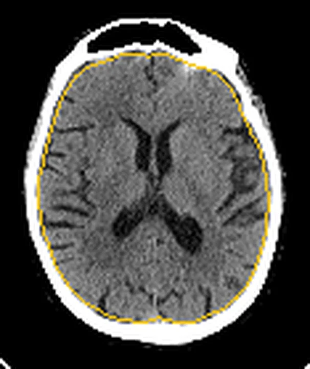
Aynı bölgenin, kaynak veri setinde patoloji içermediği belirtilen bir BT incelemesini kesit kesit kaydırın; organ sınırlarını açarak anatomiyi hasta görüntüsüyle karşılaştırın. Küçük rastlantısal bulgular tümüyle dışlanmamıştır.
Kaynak: TotalSegmentator v2.01 veri seti (Wasserthal ve ark., Radiology: Artificial Intelligence 2023), CC BY 4.0, vaka s0103; organ sınırları veri setinin otomatik segmentasyonundan, birleştirilerek sadeleştirildi. Görüntüler 2,5 mm kesit aralığına yeniden örneklendi.
Ayırıcı tanı
- Akut pontin infarkt kontrastsız BT’de ince hipodensite olarak görülebilir; erken dönemde iskemi bulguları silik olduğundan inceleme normal görünebilir. Serebellar infarkta eşlik eden şişme hidrosefaliye yol açabilir.
- Serebellar infarkt
- hemisfer veya vermiste bölgesel gri-beyaz cevher silinmesi, folial effasman ve ilerleyen olguda dördüncü ventrikül basısı yapar.
- Serebral küçük damar hastalığının görüntüleme özellikleri arasında küçük (<20 mm) infarktlar veya lakünler yer alır. Kontrastsız BT’de erken iskemik değişiklikler silik kalabilir ve posterior fossadaki artefaktlar ince iskemi bulgularının saptanmasını güçleştirebilir.
- Hareket artefaktı
- belirgin hareket artefaktı bulunan incelemeler görüntü kalitesi değerlendirmesini etkileyebilir.
- Kısmi hacim etkisi
- BT görüntülerinde görülebilen ve yorumlamayı etkileyebilen bir görüntüleme artefaktıdır.
- Pontin hipoattenüasyon değerlendirilirken yoğun kemik yapıların oluşturduğu ışın sertleşmesi artefaktı göz önünde bulundurulmalıdır.
Tuzaklar
- Tek bir koyu pontin alanı infarkt kabul etme; petroz kemiklerden aynı hizada çıkan bantları ve komşu kesitlerde devamlılığı kontrol et.
- Metal artefaktın görünümü pencere ayarından etkilenebilir; bir çalışmada geniş pencere metal artefakt ölçümünü azaltmış, ancak bu teknikler yumuşak doku değerlendirmesinde etkili olmamıştır.
- Posterior fossada normal görünen kontrastsız BT küçük veya erken infarktı dışlamaz; klinik şüphe sürerse difüzyon MR ve damar görüntülemesi gerekir.
- Aynı dokudaki HU değerlerini farklı kafa yönelimi, kesit seviyesi veya tarayıcıda mutlak eşik gibi kullanma; ışın sertleşmesi ölçülen dansiteyi değiştirir.
Kendini dene
Posterior fossada yoğun kemik yapıların yakınında görüntü artefaktları görülüyor. Bu tür artefaktların bilinen nedenlerinden biri nedir? En olası açıklama nedir?
Cevabı göster
Işın sertleşmesi. Yoğun kemik yapıların neden olduğu ışın sertleşmesi, posterior fossada görülebilen artefakt nedenlerinden biridir. İskemik lezyonlar hipodens, hemorajik lezyonlar ise genellikle hiperdens görünebilir.
Kontrastsız BT'de posterior fossadaki yoğun kemik yapıların oluşturduğu artefaktlar en çok hangi değerlendirmeyi güçleştirir?
Cevabı göster
İnce iskemik değişikliklerin saptanmasını. Posterior fossada yoğun kemik yapılar, düşük kontrastlı yumuşak dokuların görüntüsünde artefakt oluşturarak ince iskemik değişikliklerin saptanmasını güçleştirebilir.
Kaynaklar
Bu sayfadaki 38 cümle ve 2 quiz sorusu aşağıdaki kaynaklardan alıntılarla otomatik olarak karşılaştırıldı; 19 cümle bu karşılaştırmada düzeltildi (2026-10-09).
- Diagnostic Accuracy of Ischemia and Hemorrhage on Cone-Beam CT Immediately After Endovascular Thrombectomypmc.ncbi.nlm.nih.gov
- Pediatric Neck Impalement Injuries: A Report of Two Cases and Review of the Literaturepmc.ncbi.nlm.nih.gov
- Apparent Cerebellar Volumetric Stabilization and Emergent Cortical Hyperexcitability During Long-Acting Injectable Aripiprazole Maintenance in Adolescent Bipolar Disorderpmc.ncbi.nlm.nih.gov
- Non-contrast photon counting computed tomography of the head: optimized modeling of off-focal radiation to reduce calvarium-related tissue inhomogeneitypmc.ncbi.nlm.nih.gov
- AGTS-Net: Anatomically Guided Two-Stage Network for Actionable Stroke Classification in Complete NCCT Studiespmc.ncbi.nlm.nih.gov
- An optimized computed tomography protocol for metallic gunshot head trauma in a seal model.pubmed.ncbi.nlm.nih.gov
- Transient Ischemic Attack Progressing to Top-of-the-Basilar Syndrome: A Case Report.pubmed.ncbi.nlm.nih.gov
- Integrated predictors of outcome in vertebrobasilar artery occlusion treated With endovascular therapy.pubmed.ncbi.nlm.nih.gov
- Optimization of Posterior Fossa Image Quality Using Virtual Monoenergetic Reconstructions from Dual-Layer Spectral CT.pubmed.ncbi.nlm.nih.gov
- Where Does the Signal Go? Technical Challenges of CT Perfusion in Lacunar and Infratentorial Stroke.pubmed.ncbi.nlm.nih.gov
- Acute Ischemic Stroke in 2026: From Time to Penumbra-An Updated Narrative Review of Reperfusion Strategies with a Latin American Implementation Perspective.pubmed.ncbi.nlm.nih.gov
- Cranial POCUS in Neonates and Infants: Structured Evidence Synthesis and Proposal of the KORE Brain POCUS Protocol.pubmed.ncbi.nlm.nih.gov
- Society for Cardiovascular Magnetic Resonance 2024 Cases of SCMR case series.pubmed.ncbi.nlm.nih.gov
- Duropathies as Unifying Concept-Part Two: A Narrative Overview of Clinical and Neuroradiological Features.pubmed.ncbi.nlm.nih.gov
- Spontaneous Non-Aneurysmal Convexity Subarachnoid Hemorrhage: A Scoping Review of Different Etiologies beyond Cerebral Amyloid Angiopathy.pubmed.ncbi.nlm.nih.gov
- Neuroimaging with photon-counting computed tomography: A review of clinical applications.pubmed.ncbi.nlm.nih.gov
- Tradeoffs between Radiation Exposure to the Lens of the Eyes and Diagnostic Image Quality in Pediatric Brain Computed Tomography.pubmed.ncbi.nlm.nih.gov
- Brain microstructural alterations in COVID-19: a systematic review of diffusion weighted imaging studies.pubmed.ncbi.nlm.nih.gov
- Clinical and genetic spectrum of pediatric mitochondrial disorders in China: insights from a 47-case genetically confirmed cohort.pubmed.ncbi.nlm.nih.gov
- Stroke risk following bee and wasp stings: a systematic review of ischemic and hemorrhagic events.pubmed.ncbi.nlm.nih.gov
- Performance of Deep Learning Reconstruction for Detection of Early Ischemic Changes in NCCT: Comparison with ASIR-V in Acute Stroke.pubmed.ncbi.nlm.nih.gov
- Current state and advancements of imaging in acute ischemic stroke: a practical review.pubmed.ncbi.nlm.nih.gov
- Continuum of Irreversible and Potentially Reversible Traumatic Brain Injury Sequelae: Axonal Injury and Hydrocephalus.pubmed.ncbi.nlm.nih.gov
- Congenital central nervous system malformations in piedmontese calves-part 1. structural brain disorders.pubmed.ncbi.nlm.nih.gov
- The Central Variant of Posterior Reversible Encephalopathy Syndrome: A Systematic Review and Meta-Analysis.pubmed.ncbi.nlm.nih.gov
- Atypical Parkinsonian Syndromes: Structural, Functional, and Molecular Imaging Features.pubmed.ncbi.nlm.nih.gov
- Posterior Circulation Stroke as a Time-Sensitive Systems Failure: A Case of Basilar Artery Occlusion With Multisystem Complications.pubmed.ncbi.nlm.nih.gov
- 'I Can See Letters but Cannot Read Sentences': A Case of Pure Alexia Without Agraphia Due to Left Posterior Cerebral Artery Infarction.pubmed.ncbi.nlm.nih.gov
- Rogers Syndrome and Callosal Disconnection in the Setting of Moyamoya Disease.pubmed.ncbi.nlm.nih.gov
- Fetal intracranial hemorrhage and infarct: Main sonographic and MRI characteristics: A review article.pubmed.ncbi.nlm.nih.gov
- Opioid neurotoxicity: A case series and review from members of the Child Neurology Society Neurocritical Care Special Interest Group.pubmed.ncbi.nlm.nih.gov
- Anatomy-Guided Microsurgical Resection of a Dominant Frontal Lobe Tumor Without Intraoperative Adjuncts: A Case Report from a Resource-Limited Context.pubmed.ncbi.nlm.nih.gov
- Rapidly Progressive Multiple Ischemic Lesions Caused by <i>Cryptococcus neoformans</i> Infection Presenting with Meningoencephalitis and Cerebral Infarction as the Initial Manifestations: A Case Report.pubmed.ncbi.nlm.nih.gov
- The pathogenesis of cerebral small vessel disease and vascular cognitive impairment.pubmed.ncbi.nlm.nih.gov
- Drivers of Chronic Pathology Following Ischemic Stroke: A Descriptive Review.pubmed.ncbi.nlm.nih.gov
- Innovative approaches in precision radiation oncology: advanced imaging technologies and challenges which shape the future of radiation therapy.pubmed.ncbi.nlm.nih.gov
- Comprehensive review of post-treatment imaging in head and neck cancers: from expected to unexpected and beyond.pubmed.ncbi.nlm.nih.gov
- Detection of Collaterals from Cone-Beam CT Images in Stroke.pubmed.ncbi.nlm.nih.gov
- The combined compartmental maxillary resection: technique codification and preliminary multicenter series analysis.pubmed.ncbi.nlm.nih.gov
- Understanding Photon-Counting CT: Physics, Detector Technology, and Image Reconstructions.pubmed.ncbi.nlm.nih.gov
- Neuroimaging in traumatic brain imaging.pubmed.ncbi.nlm.nih.gov
- An agentic AI framework for integrated decision support and surgical planning in intracerebral hemorrhage.pubmed.ncbi.nlm.nih.gov
- Regional Reliability of Combining CT Angiography-source Image and Non-contrast CT in Acute Ischemic Stroke.pubmed.ncbi.nlm.nih.gov
- The imaging-quiescent phenotype in thrombolysed acute ischemic stroke: a diffusion-weighted MRI-based deconstruction.pubmed.ncbi.nlm.nih.gov
- Role of Low-Field MRI in Acute Stroke.pubmed.ncbi.nlm.nih.gov
- Analysis of Pontine cavernous malformation resection based on 3D microanatomical study.pubmed.ncbi.nlm.nih.gov
- Leptomeninges: Anatomy, Mechanisms of Disease and Neuroimaging.pubmed.ncbi.nlm.nih.gov
- Operative Corridors in Endoscopic Skull Base Tumor Surgery.pubmed.ncbi.nlm.nih.gov
- CT perfusion imaging in aneurysmal subarachnoid hemorrhage. State of the art.pubmed.ncbi.nlm.nih.gov
- Commentary: an eye on PET quantification.pubmed.ncbi.nlm.nih.gov
- High-affinity alphavbeta3 integrin targeted optical probe as a new imaging biomarker for early atherosclerosis: initial studies in Watanabe rabbits.pubmed.ncbi.nlm.nih.gov
- Coronary Computed Tomography Angiography for the Diagnosis and Revascularization Guidance of Coronary Bifurcation Lesions: A Contemporary Review.pubmed.ncbi.nlm.nih.gov
- Taxonomy of Acute Stroke: Imaging, Processing, and Treatment.pubmed.ncbi.nlm.nih.gov
- Diagnostic Uncertainty in Rapidly Progressive Brainstem Lesions: A Radiological Diagnosis of Probable CNS Relapse of Diffuse Large B-cell Lymphoma.pubmed.ncbi.nlm.nih.gov
- Posterior reversible encephalopathy syndrome in dengue virus infection with concurrent encephalitis and ischemic stroke: a case report.pubmed.ncbi.nlm.nih.gov
- Cerebellum and neurodegenerative diseases: Beyond conventional magnetic resonance imaging.pubmed.ncbi.nlm.nih.gov
- Diagnostic Accuracy of Ischemia and Hemorrhage on Cone-Beam CT Immediately After Endovascular Thrombectomy.pubmed.ncbi.nlm.nih.gov
- Apparent Cerebellar Volumetric Stabilization and Emergent Cortical Hyperexcitability During Long-Acting Injectable Aripiprazole Maintenance in Adolescent Bipolar Disorder.pubmed.ncbi.nlm.nih.gov
- Pediatric Neck Impalement Injuries: A Report of Two Cases and Review of the Literature.pubmed.ncbi.nlm.nih.gov
- Hashimoto's Encephalopathy With Progressive Cognitive Decline: A Rare Autoimmune Disorder in Central America.pubmed.ncbi.nlm.nih.gov
- Parent Artery Disease-Related Stroke: What Is the Impact on Endovascular Treatment? A Narrative Review.pubmed.ncbi.nlm.nih.gov
- Isolated brainstem death: case presentation demonstrating preserved supratentorial perfusion on <sup>99m</sup>Tc-bicisate brain scintigraphy.pubmed.ncbi.nlm.nih.gov
- Posterior Reversible Encephalopathy Syndrome Following Massive Blood Transfusion in a Case of Pelvic Schwannoma.pubmed.ncbi.nlm.nih.gov
- Neurolymphomatosis: A Comprehensive Review of Clinical and Imaging Features.pubmed.ncbi.nlm.nih.gov
- An Atlas of Neonatal Neurovascular Imaging Anatomy as Depicted with Microvascular Imaging: The Intracranial Veins.pubmed.ncbi.nlm.nih.gov
- The impact of arm position on image quality and radiation dose in pediatric toddler chest computed tomography: a phantom and clinical study.pubmed.ncbi.nlm.nih.gov
- Deep learning image reconstruction technique for improving image quality and radiation dose reduction compared to iterative reconstruction technique in non-contrast CT head imaging.pubmed.ncbi.nlm.nih.gov
- CT Angiography of the Upper Extremities: Review of Acute Arterial Entities.pubmed.ncbi.nlm.nih.gov
- The Genetic Basis of Strokes in Pediatric Populations and Insight into New Therapeutic Options.pubmed.ncbi.nlm.nih.gov
- Imaging the facial nerve: a contemporary review.pubmed.ncbi.nlm.nih.gov
- Research progress on vertebrobasilar dolichoectasia.pubmed.ncbi.nlm.nih.gov
- A Novel 3D-Printed Strut-Type Prosthesis Reconstructs the Hemicortical Defect After Distal Femur Parosteal Osteosarcoma Resection: The Mid-Term Follow-Up Outcome.pubmed.ncbi.nlm.nih.gov
- Daily online adaptation enhances target coverage in prostate cancer radiotherapy: a retrospective analysis.pubmed.ncbi.nlm.nih.gov
- Computed tomography reveals multiple origins of extreme caudal vertebral pneumaticity in sauropod dinosaurs.pubmed.ncbi.nlm.nih.gov
- Key pre-operative CT parameters for cochlear implantation: a focused radiologic review with surgical relevance.pubmed.ncbi.nlm.nih.gov
- Braincase of Siamraptor suwati and insights into the cranial anatomy of Carcharodontosauria.pubmed.ncbi.nlm.nih.gov
- Balancing Radiation Dose and Image Quality: Protocol Optimization for Mobile Head CT in Neurointensive Care Unit Patients.pubmed.ncbi.nlm.nih.gov
- Brainstem Disconnection Syndrome in a Patient With Respiratory Failure and Failed Weaning From Oxygen: A Case Report.pubmed.ncbi.nlm.nih.gov
- Mesenchymal tumors of the central nervous system: illustrative case.pubmed.ncbi.nlm.nih.gov
- 선천성 뇌 질환의 영상 소견: 임상 화보.pubmed.ncbi.nlm.nih.gov
- Anatomically Precise Microsurgical Resection of a Posterior Fossa Cerebellar Metastasis in an Elderly Patient with Preservation of Venous Outflow, Dentate Nucleus, and Cerebrospinal Fluid Pathways.pubmed.ncbi.nlm.nih.gov
- Photon-Counting CT of the Brain: In Vivo Human Results and Image-Quality Assessment.pubmed.ncbi.nlm.nih.gov
- Patterns of injury and violence in Yaoundé Cameroon: an analysis of hospital data.pubmed.ncbi.nlm.nih.gov
- Diagnostic criteria for vascular cognitive disorders: a VASCOG statement.pubmed.ncbi.nlm.nih.gov
- Pediatric Neuroimaging: Advanced Techniques and Clinical Applications.pubmed.ncbi.nlm.nih.gov
- Direct delivery of MRI contrast through skull vessel/marrow pathways into the brain guided by microCT.pubmed.ncbi.nlm.nih.gov
- Proton Magnetic Resonance Spectroscopy for Pediatric Neuroimaging: Key Concepts for Practice.pubmed.ncbi.nlm.nih.gov
- Deep-learning reconstruction for noncontrast head computed tomography: Improved image quality and potential diagnostic impact in acute ischemic stroke.pubmed.ncbi.nlm.nih.gov
- Characterizing the effects of noncontrast head CT reconstruction kernel and slice thickness parameters on the performance of an automated AI algorithm in the evaluation of ischemic stroke.pubmed.ncbi.nlm.nih.gov
- Cerebellar Cavernoma Resection: Case Report with Long-Term Follow-Up.pubmed.ncbi.nlm.nih.gov
- Low-flow vascular malformations without arteriovenous shunting of the central nervous system: a pictorial review.pubmed.ncbi.nlm.nih.gov
- Beyond the Typical Atypical Imaging Features of Leptomeningeal Enhancement: A Case Series.pubmed.ncbi.nlm.nih.gov
- The surgical resection of dysplastic cerebellar gangliocytoma assisted by intraoperative sonography: illustrative case.pubmed.ncbi.nlm.nih.gov
- A neonatal nonhuman primate model of gestational Zika virus infection with evidence of microencephaly, seizures and cardiomyopathy.pubmed.ncbi.nlm.nih.gov
- Leukoaraiosis and sex predict the hyperacute ischemic core volume.pubmed.ncbi.nlm.nih.gov
- Subject-specific modeling framework for particle deposition using computational fluid dynamics.pubmed.ncbi.nlm.nih.gov
- Artifacts among Cone Beam Computed Tomography Images of Patients of Department of Oral Medicine and Radiology in a Tertiary Care Centre: A Descriptive Cross-sectional Study.pubmed.ncbi.nlm.nih.gov
- A Review of 3D-Printed Medical Devices for Cancer Radiation Therapy.pubmed.ncbi.nlm.nih.gov
- Non-contrast photon counting computed tomography of the head: optimized modeling of off-focal radiation to reduce calvarium-related tissue inhomogeneity.pubmed.ncbi.nlm.nih.gov
- The Impact of Weighting Factors on Dual-Energy Computed Tomography Image Quality in Non-Contrast Head Examinations: Phantom and Patient Study.pubmed.ncbi.nlm.nih.gov
- Virtual Anatomical Sections of the Dromedary Head (<i>Camelus dromedarius</i>): A Descriptive 3D CT-Based Study of Brain-Skull Relationships.pubmed.ncbi.nlm.nih.gov
- Subarachnoid cisterns as surgical corridors: Integrating microsurgical anatomy and neuroimaging for intracranial navigation.pubmed.ncbi.nlm.nih.gov
- Delineating the mechanisms of cerebellar degeneration in paediatric and adult primary mitochondrial disease.pubmed.ncbi.nlm.nih.gov
- Susac syndrome and associated diagnostic challenges: a case report.pubmed.ncbi.nlm.nih.gov
- Clinical Spectrum, Diagnosis, and Treatment Outcome in Individuals With Intravascular Large B-cell Lymphoma Affecting the Nervous System: A Case Series.pubmed.ncbi.nlm.nih.gov
- Analysis of the baseline multiphase computed tomographic angiography findings to predict clinical outcomes in patients with middle cerebral artery M1 occlusion treated with mechanical thrombectomy.pubmed.ncbi.nlm.nih.gov
- A survey of transformer-based architectures in medical image analysis: models, applications, and challenges.pubmed.ncbi.nlm.nih.gov
- The effect of jugular foramen volume on the development of idiopathic intracranial hypertension.pubmed.ncbi.nlm.nih.gov
- Anatomical and Technical Considerations of CT-Guided C1-C2 Lateral Joint Injections: Technique, Pitfalls, and Risks.pubmed.ncbi.nlm.nih.gov
- A journey through the anatomy of the cerebellum.pubmed.ncbi.nlm.nih.gov
- Dual energy CT: a step ahead in brain and spine imaging.pubmed.ncbi.nlm.nih.gov
- Vertebral Artery Dissection and PICA-Territory Stroke Associated with Chronic Atlantoaxial Structural Abnormality: A Case Report.pubmed.ncbi.nlm.nih.gov
- Bilateral thalamic infarct involving artery of Percheron: a case report.pubmed.ncbi.nlm.nih.gov
- Postpartum Vertebral Artery Dissection After Neck Massage Resulting in a Right Cerebellar Infarct: A Case Report and Literature Review.pubmed.ncbi.nlm.nih.gov
- Association Between cnm-Positive Streptococci and Cerebral Small Vessel Disease: Insights From Oral Health and Microbiome Status.pubmed.ncbi.nlm.nih.gov
Eğitim amaçlıdır; hasta başında tanı ve tedavi kararının yerine geçmez, tıbbi tavsiye değildir. Metinler yapay zekâ desteğiyle yazıldı ve otomatik bir adımda kaynak alıntılarıyla karşılaştırıldı; bu bir tıbbi doğrulama değildir ve içerik uzman radyolog incelemesinden geçmedi. Klinik kararlarda güncel kılavuzları, kurum protokollerini ve radyoloji raporunu esas alın.