Özet
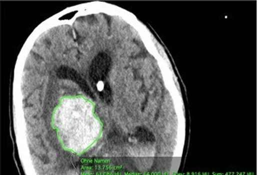
Akut parankimal kanamada BT okuması yalnız kan odağını saptamakla kalmaz; hematom hacmi, çevre ödemi ve beyin yapılarındaki bası etkisini de tanımlar. Kontrastsız BT kanın sınırlarını, intraventriküler yayılımı, ventrikül basısını, orta hat kaymasını ve sistern açıklığını gösterir. ABC/2 yatak başı için hızlı bir yaklaşık hacim hesabıdır; düzensiz veya çok loblu hematomda gerçek hacmi olduğundan farklı tahmin edebilir. Kitle etkisi veya herniasyonun eşlik ettiği büyük hematomlar kötü prognozla ilişkilidir; bu nedenle BT’de bu bulgular da değerlendirilmelidir.
Faz ve pencere
- Kontrastsız tanısal
- Hemorajik odağın çevresindeki perihematomal ödem, BT’de düşük dansiteli bir alan olarak görülebilir. En geniş kesitte iki dik çap ölçülür; kraniokaudal yayılım kesit sayısı ve kesit kalınlığıyla yaklaşık belirlenir. Aynı seride ventrikül içi kan, ventrikül sıkışması, septum pellucidum/pineal düzeyinde orta hat yer değiştirmesi ve bazal sisternlerin silinmesi değerlendirilir; bu hacim-kitle etkisi sorusu için kontrast gerekmez.
Önerilen pencereler: Beyin (G 80 / M 40), Subdural (G 200 / M 75).
BT bulguları
- Hematoma üç eksen ölçümü — en geniş aksiyel kesitte uzun çap A ve ona dik çap B, kraniokaudal yayılım C ile yaklaşık elipsoid hacmi hesaplamaya yarar.
- ABC/2 tahmini — A×B×C/2 yaklaşımı hızlıdır; düzenli elipsoid biçime daha uygundur ve düzensiz sınırlı kanamada hata artar.
- Perihematomal ödem, BT’de kanama odağını çevreleyen düşük dansiteli alan olarak görülebilir ve kitle etkisini artırabilir.
- Ventrikül içi yayılım — lateral ventrikül, üçüncü veya dördüncü ventrikülde hiperdens kan; eşlik eden ventrikül genişliği ve hidrosefaliyi de kaydet.
- Ventrikül basısı — hematom tarafındaki lateral ventrikül gövde veya frontal/oksipital boynuzlarında karşı tarafa göre daralma ya da silinme.
- Orta hat kayması — septum pellucidumun, aynı düzeyde belirlenen ideal orta hatta göre milimetre cinsinden sapmasını ölçün; falxı orta hat referansını belirlemede kullanın.
- Sisternal silinme — suprasellar, ambient veya perimezensefalik sisternlerde BOS aralığının daralması, aşağı yönlü kitle etkisi açısından önemlidir.
- Orta hat kaymasına eşlik edebilen subfalsin veya unkal herniasyonu ve bazal sisternlerin silinmesini tarayın.
Ölçütler ve sınıflamalar
- ABC/2 yaklaşık hematom hacmi
- A × B × C ÷ 2 cm³ (A ve B cm; C cm)
Normalde
Normal karşılaştırmada septum pellucidumun ideal orta hatta göre yer değiştirmesi değerlendirilir; ideal orta hat çoğunlukla falx cerebri'nin kafatasına tutunma hattıyla belirlenir. Hematom tarafındaki ventrikül ve sulkusları karşı tarafla kıyaslayın; orta hat yer değiştirmesini aynı düzeyde septum pellucidumdan, sisternal daralmayı suprasellar ve ambient sisternlerden değerlendirin.
Normal BT ile kıyasla
Aynı bölgenin, kaynak veri setinde patoloji içermediği belirtilen bir BT incelemesini kesit kesit kaydırın; organ sınırlarını açarak anatomiyi hasta görüntüsüyle karşılaştırın. Küçük rastlantısal bulgular tümüyle dışlanmamıştır.
Kaynak: TotalSegmentator v2.01 veri seti (Wasserthal ve ark., Radiology: Artificial Intelligence 2023), CC BY 4.0, vaka s0103; organ sınırları veri setinin otomatik segmentasyonundan, birleştirilerek sadeleştirildi. Görüntüler 2,5 mm kesit aralığına yeniden örneklendi.
Ayırıcı tanı
- Hemorajik venöz infarkt
- arteriyel dağılımı aşan hemorajik parankimal lezyonlar venöz tutulumu düşündürmelidir.
- Hemorajik tümör
- nodüler veya kitle biçimli doku, çevre ödemi ve kontrast sonrası solid bileşen şüphesi doğurabilir; akut tek faz BT karakterizasyonu sınırlıdır.
- Hemorajik kontüzyon
- özellikle frontal ve temporal kutuplarda kortikal yüzeye yakın birden çok yama tarzı kan odağı ve ödem görülebilir.
- Kalsifikasyon
- kronik, keskin sınırlı hiperdens odak yapabilir; çevresel ödem ve akut kitle etkisi çoğunlukla beklenmez, belirsizlikte önceki inceleme veya dual-energy BT yardımcı olabilir.
- Kontrastlanma/iyot boyanması
- yakın zamanda kontrastlı işlem varsa damar içi iyot kanı taklit edebilir; tek enerjili kontrastsız seri veya dual-energy materyal haritaları ayrımda kullanılır.
Tuzaklar
- ABC/2'de C için hematom bulunan kesitleri kesit kalınlığıyla çarp; arada boş kesit veya farklı rekonstrüksiyon kalınlığını hesaba katmamak hacmi saptırır.
- Düzensiz, bikonveks olmayan veya çok odacıklı hematomda elipsoid varsayımı geçersizleşebilir; yaklaşık hesabı kesin planimetrik hacim gibi sunma.
- Orta hat kayması, septum pellucidum veya falx cerebri gibi orta hat yapılarının yer değiştirmesi olarak ölçülür.
- Ventrikül içi kanı parankimal hematom boyutuna eklemek ya da hidrosefaliyi gözden kaçırmak toplam kitle etkisini yanlış tarif eder.
Kendini dene
En geniş aksiyel dilimde A=4 cm, dik çap B=3 cm; yayılım C=5 cm. ABC/2 ile yaklaşık hacim nedir?
Cevabı göster
30 cm³. Çarpım 4×3×5=60 cm³; yarıya bölündüğünde yaklaşık 30 cm³ olur. Bu elipsoid yaklaşımıdır ve düzensiz şekilli hematomda kesin hacim olarak alınmaz.
Hematoma komşu ventrikül sıkışmış, septum karşıya itilmiş ve suprasellar sistern silik. Bu bulgular neyi gösterir?
Cevabı göster
Belirgin kitle etkisi. Ventrikül basısı, orta hat yer değiştirmesi ve sisternal daralma birlikte hematomun kitle etkisini gösterir. Hidrosefali genellikle ventrikül genişlemesi yapar; atrofi sulkus ve ventrikülleri genişletir; kist bu yaygın bası örüntüsünü açıklamaz.
İlgili konular
Kaynaklar
Bu sayfadaki 41 cümle ve 2 quiz sorusu aşağıdaki kaynaklardan alıntılarla otomatik olarak karşılaştırıldı; 10 cümle bu karşılaştırmada düzeltildi (2026-10-09).
- Rapid spontaneous regression of a massive post-thrombolysis intracerebral hemorrhage: A case reportpmc.ncbi.nlm.nih.gov
- The Effect of Intensive Blood Pressure Reduction on Perihematomal Edema After Intracerebral Hemorrhage: A Systematic Review and Meta‐Analysispmc.ncbi.nlm.nih.gov
- Life-Threatening Spontaneous Retroperitoneal Hemorrhage Associated with Acenocoumarol Anticoagulation in a Young Patient with a Mechanical Aortic Valve: A Case Reportpmc.ncbi.nlm.nih.gov
- AI-Based Approaches for the Identification and Quantification of Traumatic Brain Injury in Computed Tomography Imaging: Systematic Reviewpmc.ncbi.nlm.nih.gov
- Correlation of Contusion Morphology and Volume With Functional Recovery and in Hospital Mortality in Traumatic Brain Injurypmc.ncbi.nlm.nih.gov
- Airway-Occluding Mucus Plugs and Mortality in Patients With Chronic Obstructive Pulmonary Disease.pubmed.ncbi.nlm.nih.gov
- Visceral adiposity and inflammatory bowel disease.pubmed.ncbi.nlm.nih.gov
- Testosterone Levels in Pre-Menopausal Women are Associated With Nonalcoholic Fatty Liver Disease in Midlife.pubmed.ncbi.nlm.nih.gov
- Natural course of perihemorrhagic edema after intracerebral hemorrhage.pubmed.ncbi.nlm.nih.gov
- Improvement in abdominal and flank contouring by a novel adipocyte-selective non-contact radiofrequency device.pubmed.ncbi.nlm.nih.gov
- The effects of 3-month atorvastatin therapy on arterial inflammation, calcification, abdominal adipose tissue and circulating biomarkers.pubmed.ncbi.nlm.nih.gov
- Maresin 1 Resolves Inflammation and Aids Bone Healing in Periapical Lesions.pubmed.ncbi.nlm.nih.gov
- Dosimetry of [ 177 Lu]Lu-DOTATATE in Patients with Advanced Midgut Neuroendocrine Tumors: Results from a Substudy of the Phase III NETTER-1 Trial.pubmed.ncbi.nlm.nih.gov
- Ten cases of intradiaphragmatic extralobar pulmonary sequestration: a single-center experience.pubmed.ncbi.nlm.nih.gov
- High Throughput Detection of 327 Drugs in Blood by LC-MS-MS with Automated Data Processing.pubmed.ncbi.nlm.nih.gov
- Sclerostin antibody inhibits skeletal deterioration due to reduced mechanical loading.pubmed.ncbi.nlm.nih.gov
- Early Diagnostic Features of Left-to-Right Shunt-Induced Pulmonary Arterial Hypertension in Piglets.pubmed.ncbi.nlm.nih.gov
- Alterations in Body Composition, Resting Metabolic Rate, Muscular Strength, and Eating Behavior in Response to Natural Bodybuilding Competition Preparation: A Case Study.pubmed.ncbi.nlm.nih.gov
- Reduction of Radiation Dose and Contrast Medium Volume in Photon-Counting Detector Computed Tomography Head and Neck Angiography: A Comparison to Energy-Integrating Detector Computed Tomography.pubmed.ncbi.nlm.nih.gov
- Spontaneous Rectus Sheath Hematoma: Factors Predictive of Conservative Management Failure.pubmed.ncbi.nlm.nih.gov
- Case Report: Cerebrospinal fluid cytology as a diagnostic clue in hemorrhagic glioblastoma mimicking hypertensive intracranial hemorrhage.pubmed.ncbi.nlm.nih.gov
- Multimodal Monitoring in Eclamptic Intracerebral Hemorrhage Complicated by Reversible Cerebral Vasoconstriction Syndrome (RCVS): A Case Report.pubmed.ncbi.nlm.nih.gov
- Rapid spontaneous regression of a massive post-thrombolysis intracerebral hemorrhage: A case report.pubmed.ncbi.nlm.nih.gov
- AI-Based Approaches for the Identification and Quantification of Traumatic Brain Injury in Computed Tomography Imaging: Systematic Review.pubmed.ncbi.nlm.nih.gov
- Decision-making for emergency craniotomy in traumatic intracranial hemorrhage with severe thrombocytopenia and coagulopathy in a patient with suspected acute leukemia: A case-based review.pubmed.ncbi.nlm.nih.gov
- Traumatic basal ganglia hemorrhage: illustrative cases.pubmed.ncbi.nlm.nih.gov
- Development and Internal Validation of the CLIMPOUT Score: A Multimodal Prediction Model for Neurological Outcome After Pediatric Intracerebral Hemorrhage.pubmed.ncbi.nlm.nih.gov
- Clinical and Radiological Evaluation of Warfarin-Associated Intracerebral Hemorrhage (ICH) in Patients With Mechanical Prosthetic Heart Valves: A Retrospective Observational Study.pubmed.ncbi.nlm.nih.gov
- Clinical, Laboratory, and CT Morphological Factors Associated with 3-Month Functional Outcomes in Spontaneous Intracerebral Hemorrhage: A Retrospective Cohort Study.pubmed.ncbi.nlm.nih.gov
- Delayed Expanding Intracerebral Pneumatocele Following Severe Craniofacial Trauma: A Case Report With Serial Imaging Correlation.pubmed.ncbi.nlm.nih.gov
- Early secondary brain abscess formation caused by <i>Bacteroides fragilis</i> at the intracerebral hemorrhage site: A case report.pubmed.ncbi.nlm.nih.gov
- Multifocal intracranial hemorrhage on emergency brain CT in a child with end-stage nephrotic syndrome.pubmed.ncbi.nlm.nih.gov
- Pigmented Peril: A fatal case of Primary intracranial melanoma in a paediatric patient.pubmed.ncbi.nlm.nih.gov
- Central Nervous System Involvement in Acute Myeloid Leukemia: From Pathophysiology to Neuroradiologic Features and the Emerging Role of Artificial Intelligence.pubmed.ncbi.nlm.nih.gov
- Tumor-like Lesions in Primary Angiitis of the Central Nervous System: The Role of Magnetic Resonance Imaging in Differential Diagnosis.pubmed.ncbi.nlm.nih.gov
- Role of Neuroimaging in COVID 19 Infection-A Retrospective Study.pubmed.ncbi.nlm.nih.gov
- Spontaneous resolution of cerebrospinal fluid obstruction due to intraventricular hemorrhage: A case report.pubmed.ncbi.nlm.nih.gov
- Jugular Venous Compression via Soft Cervical Collar in Shunt-Refractory Low-Pressure Hydrocephalus: A Report of Two Cases.pubmed.ncbi.nlm.nih.gov
- Progress in the diagnosis and treatment of post-traumatic hydrocephalus.pubmed.ncbi.nlm.nih.gov
- Eccentric edema pattern preceding postoperative porencephalic cyst formation in subcortical hemorrhage: A case series and literature review.pubmed.ncbi.nlm.nih.gov
- A Silent Vasculitis With a Loud Presentation: Takayasu Arteritis Causing Intracerebral Hemorrhage.pubmed.ncbi.nlm.nih.gov
- Individualized treatment for pediatric intracranial aneurysms in China: case series and literature review.pubmed.ncbi.nlm.nih.gov
- Predictive modeling of brain herniation risk factors and critical thresholds in spontaneous intracerebral hemorrhage: a pilot study.pubmed.ncbi.nlm.nih.gov
- Brain Shift Patterns: Upward, Lateral and Downward Herniation, Its Correlation with Clinical Patterns in Acute Intracranial Pathologies and Neurosurgical Management.pubmed.ncbi.nlm.nih.gov
- A decision tree model for hematoma expansion prediction in women after spontaneous intracerebral hemorrhage.pubmed.ncbi.nlm.nih.gov
- In-hospital unexpected cerebral herniation-New neurosurgical quality control standards: A case report.pubmed.ncbi.nlm.nih.gov
- Accuracy, Hemorrhagic Complications and CT Radiation Dose of Emergency External Ventricular Drain (EVD) Placement in Pediatric Patients: A 15-Year Retrospective Analysis.pubmed.ncbi.nlm.nih.gov
- Structural neuroimaging markers of normal pressure hydrocephalus versus Alzheimer's dementia and Parkinson's disease, and hydrocephalus versus atrophy in chronic TBI-a narrative review.pubmed.ncbi.nlm.nih.gov
- Venous-significant intracranial hemorrhage in neonatal HIE treated with therapeutic hypothermia: imaging phenotypes, diagnostic pitfalls, and prognostic implications.pubmed.ncbi.nlm.nih.gov
- Intracranial venous thrombosis imaging by anatomical location: an educational review with mimics and diagnostic pitfalls.pubmed.ncbi.nlm.nih.gov
- Coexistence of a Hemispheric Developmental Venous Anomaly and Two Ipsilateral Arteriovenous Malformations: A Rare Neurovascular Association.pubmed.ncbi.nlm.nih.gov
- Hemorrhagic brain abscess mimicking an intracranial neoplasm in invasive Klebsiella pneumoniae syndrome: illustrative case.pubmed.ncbi.nlm.nih.gov
- Delayed multiple hemorrhagic brain metastases from atrial myxoma: a case report and literature review.pubmed.ncbi.nlm.nih.gov
- Delayed intracranial lesions following cardiac myxoma resection in a patient with concurrent cerebral embolism: a diagnostic challenge in the absence of pathological evidence.pubmed.ncbi.nlm.nih.gov
- Correlation of Contusion Morphology and Volume With Functional Recovery and in Hospital Mortality in Traumatic Brain Injury.pubmed.ncbi.nlm.nih.gov
- Successful endoscopic evacuation of traumatic intracerebral hemorrhage: A report of two cases.pubmed.ncbi.nlm.nih.gov
- Dual Impact: Coup-Contrecoup Brain Injury in a Young Cyclist-A Case Report.pubmed.ncbi.nlm.nih.gov
- Diabetic striatopathy and concurrent peri-ictal cortical diffusion restriction in nonketotic hyperglycemia: A rare stroke mimic.pubmed.ncbi.nlm.nih.gov
- Diabetic striatopathy presenting as stroke-mimicking hemiparesis without involuntary movements: A case report.pubmed.ncbi.nlm.nih.gov
- PCR-Confirmed Convexity Subarachnoid Neurocysticercosis with Negative Serology: A Case Report.pubmed.ncbi.nlm.nih.gov
- Spectral Computed Tomography Angiography in Visceral Artery Aneurysms: Technical Principles and Clinical Applications.pubmed.ncbi.nlm.nih.gov
- Research progress on the application of dual energy CT in gastric diseases.pubmed.ncbi.nlm.nih.gov
- Dual-energy computed tomography in children: Technique and clinical applications.pubmed.ncbi.nlm.nih.gov
- Retrospective validation of 3D Slicer against CTA source images for accurate intracerebral hematoma volumetry: a comparative study with the Tada formula.pubmed.ncbi.nlm.nih.gov
- The prognostic value of systemic inflammatory indices in elderly patients with spontaneous intracerebral hemorrhage.pubmed.ncbi.nlm.nih.gov
- Perihematomal Edema Characteristics After Minimally Invasive Surgery in Intracerebral Hemorrhage.pubmed.ncbi.nlm.nih.gov
- Simple Ways to Estimate Meningioma Volume: Can ABC- and SH-Derived Methods Be Used in Clinical Practice Reliably?pubmed.ncbi.nlm.nih.gov
- Role of artificial intelligence and machine learning in the diagnosis of cerebrovascular disease.pubmed.ncbi.nlm.nih.gov
- Automation of CT-based haemorrhagic stroke assessment for improved clinical outcomes: study protocol and design.pubmed.ncbi.nlm.nih.gov
- Dynamic Changes in Optic Nerve Sheath Diameter on Computed Tomography Predict Decompressive Efficacy and Outcomes in Severe Traumatic Brain Injury.pubmed.ncbi.nlm.nih.gov
- Topographical Patterns of Intraventricular Hemorrhage as a Novel Prognostic Imaging Biomarker in Intracerebral Hemorrhage.pubmed.ncbi.nlm.nih.gov
- Size and location of intracranial hemorrhage in acute stroke presentations: correlation with baseline NIHSS and relevance to direct-to-angiography workflows.pubmed.ncbi.nlm.nih.gov
- The Effect of Intensive Blood Pressure Reduction on Perihematomal Edema After Intracerebral Hemorrhage: A Systematic Review and Meta-Analysis.pubmed.ncbi.nlm.nih.gov
- Advances in Stereotactic Hematoma Evacuation for Hypertensive Intracerebral Hemorrhage and the Value of Multimodal Monitoring.pubmed.ncbi.nlm.nih.gov
- Neutrophil Percentage-to-Albumin Ratio is Associated with Functional Outcome, Hematoma Clearance, and Peak Perihematomal Edema in Supratentorial Intracerebral Hemorrhage.pubmed.ncbi.nlm.nih.gov
- Tumor-like cortical venous thrombosis: Case report and literature review.pubmed.ncbi.nlm.nih.gov
- Magnetic resonance imaging findings of degenerating uterine fibroids: A pictorial review.pubmed.ncbi.nlm.nih.gov
- Adnexal Torsion of Normal Adnexa at 19 Weeks of Gestation: A Case Report.pubmed.ncbi.nlm.nih.gov
- Alobar Holoprosencephaly with Severe Craniofacial Anomalies and Congenital Diaphragmatic Hernia: First Reported Case from Somalia.pubmed.ncbi.nlm.nih.gov
- Cardiac arrest after severe traumatic brain injury can be survivable with good outcomes.pubmed.ncbi.nlm.nih.gov
- Imaging for the diagnosis and management of traumatic brain injury.pubmed.ncbi.nlm.nih.gov
- Technical nuances of endoscopic transcortical transventricular fenestration for management of giant suprasellar arachnoid cysts in adults: illustrative cases.pubmed.ncbi.nlm.nih.gov
- Beyond Imaging: A 12-Year Clinicopathological Analysis of Surgically Excised Neurocysticercosis.pubmed.ncbi.nlm.nih.gov
- Distribution and predictive performance of the temporal phase of dynamic spot sign appearance in acute intracerebral hemorrhage.pubmed.ncbi.nlm.nih.gov
- Giant Right Sphenoid Wing Meningioma as a Reversible Frontal Network Lesion: A Pseudo-bvFTD Case with Venous-Sparing Skull-Base Resection.pubmed.ncbi.nlm.nih.gov
- An Atlas of Neonatal Neurovascular Imaging Anatomy as Depicted with Microvascular Imaging: The Intracranial Arteries.pubmed.ncbi.nlm.nih.gov
Eğitim amaçlıdır; hasta başında tanı ve tedavi kararının yerine geçmez, tıbbi tavsiye değildir. Metinler yapay zekâ desteğiyle yazıldı ve otomatik bir adımda kaynak alıntılarıyla karşılaştırıldı; bu bir tıbbi doğrulama değildir ve içerik uzman radyolog incelemesinden geçmedi. Klinik kararlarda güncel kılavuzları, kurum protokollerini ve radyoloji raporunu esas alın.