Özet
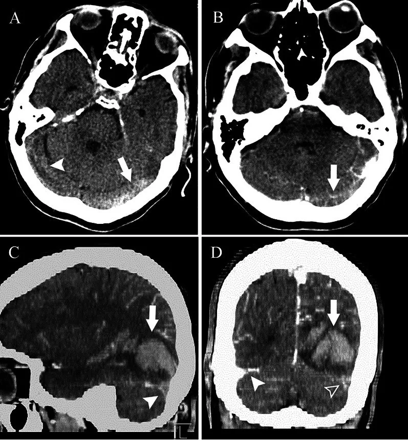
Baş ağrısı, nöbet veya fokal nörolojik bulguyla başvuran hastada serebral venöz tromboz, venöz basınç artışı ve hemorajik infarktla ortaya çıkabilir. Kontrastsız BT'de tromboze sinüs ya da kortikal ven hiperdens olabilir; eşlik eden ödem ve kanama çoğu zaman doğrudan trombüsü göstermeyen dolaylı işaretlerdir. Venöz fazda BT venografi (BT V) sinüs lümenindeki dolum kusurunu ve trombozun uzanımını gösterir. Tanı atlanırsa arteriyel inme gibi yorumlanan, arter sınırlarını aşan hemorajik lezyonun venöz nedeni gözden kaçar.
Faz ve pencere
- Kontrastsız tanısal
- Tromboze kortikal ven veya dural sinüslerde akut trombüse bağlı spontan hiperdensite (kord işareti) görülebilir. Parankimde kortikal şişme, beyaz cevher ödemi, gri-beyaz ayrımında silinme ve ödem içine serpiştirilmiş kan odakları görülebilir; kontrast gerekmez ancak normal görünüm trombozu dışlamaz.
- Venöz tanısal
- Venöz zamanlamalı BT venografide opaklaşması beklenen dural sinüs segmentinde intraluminal dolum kusuru, trombüs boyunca opaklaşmama veya kontrastla çevrili merkezî defekt aranır. Kortikal venlerin asimetrik görünümü ve kollateral drenaj, venöz fazda (cerebral ve juguler venlerde kontrast görünüp başlatılan) görüntülemede incelenir; arteriyel zamanlama bu değerlendirmeyi sağlamaz.
Önerilen pencereler: Beyin (G 80 / M 40), Anjiyo (G 600 / M 150).
BT bulguları
- Kord işareti — sulkus boyunca uzanan kortikal ven içinde çevre beyin parankiminden belirgin hiperdens lineer yapı.
- Yoğun sinüs işareti — superior sagittal veya transvers sinüs lümeninde beklenmeyen hiperdensite; hematokrit ve dehidratasyon gibi etkenlerle özgüllüğü azalır.
- BT venografide dolum kusuru — opaklaşan sinüs içinde trombüsle uyumlu opaklaşmayan bölüm ya da segment boyunca lümen opasifikasyonunun kaybı.
- Boş delta görünümü — kontrastlanan sinüs duvarları arasında dolmayan üçgensel merkez; tromboze superior sagittal sinüste görülebilir.
- Arter dışı dağılımlı ödem — kortikal-subkortikal hipodensite bir arter havzasına uymaz, birden fazla komşu lob veya iki taraflı talamusları etkileyebilir.
- Hemorajik venöz infarkt — ödemli dokunun içinde peteşiyal ya da birleşen kan odakları; venöz konjesyonla birlikte görülür.
- Kortikal venöz kollateraller — tıkanıklık yakınında genişlemiş veya belirginleşmiş venler, çıkış engelini destekler.
Normalde
Normal BT'de superior sagittal, düz, transvers ve sigmoid sinüslerin lümeni kontrastsız incelemede beyin dokusundan belirgin yoğun bir kord oluşturmaz; venöz zamanlı incelemede sinüs boyunca kesintisiz kontrast dolumu beklenir. Şüpheli segmenti karşı transvers sinüs ve devamındaki sigmoid sinüsle kıyaslayın; asimetriyi konjenital hipoplazi olasılığını da gözeterek değerlendirip parankim hipodensitesinin arteriyel sınıra uyup uymadığını kontrol edin.
Normal BT ile kıyasla
Aynı bölgenin, kaynak veri setinde patoloji içermediği belirtilen bir BT incelemesini kesit kesit kaydırın; organ sınırlarını açarak anatomiyi hasta görüntüsüyle karşılaştırın. Küçük rastlantısal bulgular tümüyle dışlanmamıştır.
Kaynak: TotalSegmentator v2.01 veri seti (Wasserthal ve ark., Radiology: Artificial Intelligence 2023), CC BY 4.0, vaka s0103; organ sınırları veri setinin otomatik segmentasyonundan, birleştirilerek sadeleştirildi. Görüntüler 2,5 mm kesit aralığına yeniden örneklendi.
Ayırıcı tanı
- Arteriyel infarkt
- çoğunlukla belirli arter havzasına uyan kama biçimli değişiklik yapar; venöz ödem sınırları aşabilir ve kanama daha sık eşlik eder.
- Hipoksik-iskemik hasar
- bazal ganglionlar, korteks veya sınır bölgelerinde yaygın simetrik tutulum olabilir; sinüs içi trombüs ve dolum defekti beklenmez.
- Ensefalit
- temporal veya limbik ağırlıklı ödem yapabilir; dural sinüs içinde lümen defekti oluşturmaz.
- Hiperdens sinüs taklidi
- yüksek hematokrit/dehidratasyonda sinüs yoğunluğu artabilir; karşı sinüs ve damarlarla kıyas ile BT venografi lümen açıklığını gösterir.
- Sinüs hipoplazisi
- özellikle transvers sinüste küçük kalibrasyon ve kemik oluğunun gelişimsel küçüklüğü görülebilir; ani kesilme ya da intraluminal trombüs görünümü yoktur.
Tuzaklar
- Kontrastsız BT'de normal sinüs dansitesi trombozu dışlamaz; parankimal venöz infarkt veya klinik şüphe varsa venöz zamanlı BT venografi gerekir.
- Tek transvers sinüsün küçük olması tromboz anlamına gelmez; karşı taraf, sigmoid devamlılık ve kemik oluğu kalibresiyle gelişimsel hipoplaziyi ayır.
- Hemorajik venöz infarktı primer lobar kanama olarak etiketleme; çevresindeki ödemin arteriyel havzaya uyup uymadığını ve venöz sinüsleri birlikte incele.
- BT venografide doğru venöz zamanlama (cerebral/juguler venlerde kontrast görünüp başlatılan) sağlanmazsa sinüsler yetersiz dolabilir; tromboz için yeterli venöz opaklaşma gerekir.
Kendini dene
Baş ağrısı ve nöbeti olan hastada parasagittal ödem arter havzasını aşıyor, içinde kan odakları var. En olası neden?
Cevabı göster
Serebral venöz tromboz. Arter dışı dağılım ve ödem içi kan venöz çıkış tıkanıklığıyla uyumludur. Arter infarktı damar havzasını izler; glioblastom kitle biçiminde gelişir; ensefalit bu venöz dağılımı açıklamaz.
Kontrastsız BT'de transvers sinüs yoğun; venöz zamanlı BT'de lümen kesintisiz opaklaşıyor. En olası açıklama nedir?
Cevabı göster
Yüksek hematokrit. Venöz lümenin devamlı kontrast alması tıkayıcı trombüsü desteklemez; yüksek kan yoğunluğu sinüs dansitesini artırabilir. Fistül anormal damar bağlantısı ve venöz erken doluşla, kortikal ven trombozu ise ayrı bir ven segmentindeki dolum kaybıyla değerlendirilir.
İlgili konular
Kaynaklar
Bu sayfadaki 40 cümle ve 2 quiz sorusu aşağıdaki kaynaklardan alıntılarla tek tek karşılaştırıldı; 3 cümle bu karşılaştırmada düzeltildi (2026-10-08).
- Imaging of Cerebral Venous Thrombosispmc.ncbi.nlm.nih.gov
- Isolated cortical vein thrombosis: case series.pubmed.ncbi.nlm.nih.gov
- Pathways for Neuroimaging of Neonatal Stroke.pubmed.ncbi.nlm.nih.gov
- [Autopsy case of dural thickening caused by widespread dural vein thrombosis associated with disseminated bone marrow carcinomatosis].pubmed.ncbi.nlm.nih.gov
- [Jugular vein reconstruction by longitudinal constriction suture venoplasty and microvascular anastomosis].pubmed.ncbi.nlm.nih.gov
- Venous injury in abusive head trauma.pubmed.ncbi.nlm.nih.gov
- [Cerebral venous thrombosis revealing neurosyphilis].pubmed.ncbi.nlm.nih.gov
- 36th International Symposium on Intensive Care and Emergency Medicine : Brussels, Belgium. 15-18 March 2016.pubmed.ncbi.nlm.nih.gov
- Cerebral venous thrombosis: state of the art diagnosis and management.pubmed.ncbi.nlm.nih.gov
- Assessment of Patients with Intracerebral Hemorrhage or Hemorrhagic Transformation in the VENOST Study.pubmed.ncbi.nlm.nih.gov
- Pathological confirmation of 4 cases with isolated cortical vein thrombosis previously misdiagnosed as brain tumor.pubmed.ncbi.nlm.nih.gov
- Cerebral Venous Sinus Thrombosis in an Immunocompetent HIV Patient.pubmed.ncbi.nlm.nih.gov
- Cerebral Venous Thrombosis Presenting as Simultaneous Intracerebral Hemorrhage.pubmed.ncbi.nlm.nih.gov
- Postpartum Superior Sagittal Sinus Thrombosis: A Case Report.pubmed.ncbi.nlm.nih.gov
- Whole-Brain Computed Tomographic Perfusion Imaging in Acute Cerebral Venous Sinus Thrombosis.pubmed.ncbi.nlm.nih.gov
- Bilateral Vein of Trolard Thrombosis Presenting with Seizure and Minimal Deficits: A Rare Case Report.pubmed.ncbi.nlm.nih.gov
- Efficacy and safety of anticoagulation for pediatric cerebral sinovenous thrombosis: systematic review and meta-analysis.pubmed.ncbi.nlm.nih.gov
- Clinical case of a pregnant woman with arteriovenous malformation, hemorrhagic venous infarcts and epileptic seizures: a case report.pubmed.ncbi.nlm.nih.gov
- Extensive cerebral venous sinus thrombosis with hemorrhagic venous infarction as the initial presentation of acute myeloid leukemia: A case report.pubmed.ncbi.nlm.nih.gov
- The Commonly Uncommon Presentation of Cerebral Venous Thrombosis: A Case Report.pubmed.ncbi.nlm.nih.gov
- Computed tomography perfusion stroke mimics on RAPID commercial software: A case-based review.pubmed.ncbi.nlm.nih.gov
- Intracranial venous thrombosis imaging by anatomical location: an educational review with mimics and diagnostic pitfalls.pubmed.ncbi.nlm.nih.gov
- Acute otitis media complicated by mastoiditis and otogenic transverse-sigmoid sinus thrombosis in an adult: a case report on infectious pathogenesis and multidisciplinary therapy.pubmed.ncbi.nlm.nih.gov
- Seizures as the initial presentation of cerebral venous sinus thrombosis in an elderly patient: a case report and literature review.pubmed.ncbi.nlm.nih.gov
- Tumor-like cortical venous thrombosis: Case report and literature review.pubmed.ncbi.nlm.nih.gov
- Dengue-Associated Encephalopathy With Multiple Cranial Neuropathies and Multisystem Involvement in a Patient With Epilepsy: A Comprehensive Case Report With Longitudinal Multimodality Imaging.pubmed.ncbi.nlm.nih.gov
- Emergency in pediatric rheumatology: a narrative review.pubmed.ncbi.nlm.nih.gov
- Computed Tomography Findings of Cerebral Venous Thrombosis: A Case Report.pubmed.ncbi.nlm.nih.gov
- Cerebral Venous Sinus Thrombosis Following Nitrous Oxide Abuse: A Clinical Reminder of a Growing Trend.pubmed.ncbi.nlm.nih.gov
- Explainable Artificial Intelligence in Non-Contrast Brain Computed Tomography Scan for Intracerebral Hemorrhage: A Scoping Review.pubmed.ncbi.nlm.nih.gov
- Developmental venous anomaly related to de novo cerebral cavernous malformation: Imaging findings from two cases.pubmed.ncbi.nlm.nih.gov
- Cerebral Venous Thrombosis: Pathophysiologic Insights, Clinical Evaluation Tools, and Novel Therapeutic Strategies.pubmed.ncbi.nlm.nih.gov
- Morphological variations of the dural venous sinuses: a cadaveric study.pubmed.ncbi.nlm.nih.gov
- Venous-significant intracranial hemorrhage in neonatal HIE treated with therapeutic hypothermia: imaging phenotypes, diagnostic pitfalls, and prognostic implications.pubmed.ncbi.nlm.nih.gov
- Polycythemia, a radiologic mimic in plain CT head: A case report.pubmed.ncbi.nlm.nih.gov
- Admission hyperdense sinus sign predicts poorer outcomes in cerebral venous sinus thrombosis.pubmed.ncbi.nlm.nih.gov
- Coexistence of cerebral venous thrombosis and dural arteriovenous fistula in an adolescent: A case report.pubmed.ncbi.nlm.nih.gov
- Case Report: Mechanical thrombectomy of cerebral venous sinuses using the NeVa stent retriever.pubmed.ncbi.nlm.nih.gov
- Fatal Cerebral Air Embolism Following Central Venous Catheter Mishandling in a Stroke Patient: A Case Report.pubmed.ncbi.nlm.nih.gov
- Tissue clock-guided prediction and intervention of futile recanalization: towards precision therapy in mechanical thrombectomy.pubmed.ncbi.nlm.nih.gov
- Lemierre-like syndrome presenting with extensive intracranial and central venous thrombosis without pulmonary embolism: illustrative case.pubmed.ncbi.nlm.nih.gov
- Obstructive Sleep Apnea as a Modifier of Systemic Thromboembolic Risk Following Retinal Vein Occlusion.pubmed.ncbi.nlm.nih.gov
- Extensive Cerebral Venous Sinus Thrombosis With Obstructive Hydrocephalus in a Young Female With Underlying Essential Thrombocythemia: A Case Report.pubmed.ncbi.nlm.nih.gov
- Cerebral venous disorders: clinical presentation, diagnostic strategy, and contemporary management.pubmed.ncbi.nlm.nih.gov
- Cerebral venous sinus thrombosis secondary to relapsed minimal change disease: a case managed with endovascular thrombolysis.pubmed.ncbi.nlm.nih.gov
- Congenital and Breed-Associated Vertebral Anomalies in Dogs and Cats: Developmental Anatomy, Genetic Determinants, and Biological Significance.pubmed.ncbi.nlm.nih.gov
- Transverse Sinus Hypoplasia as a Differential Diagnosis for Cerebral Vein Thrombosis: A Case Report.pubmed.ncbi.nlm.nih.gov
- Natural History and Factors Influencing 90-Day Clinical Outcomes and 1-Year Recurrence of Thalamic Stroke.pubmed.ncbi.nlm.nih.gov
- Pediatric Cardiac Arrest From Tumor-Associated Pulmonary Embolism: Diagnostic Insights From Imaging After Extracorporeal Cardiopulmonary Resuscitation.pubmed.ncbi.nlm.nih.gov
- Neonatal Thromboembolic Disease: Clinical Spectrum and Emerging Evidence for Diagnostic and Therapeutic Strategies Addressing Unmet Needs.pubmed.ncbi.nlm.nih.gov
- When headache meets visual disturbance: An evidence-based review of common and critical entities.pubmed.ncbi.nlm.nih.gov
- Imaging in scrub typhus - a pictorial review.pubmed.ncbi.nlm.nih.gov
- Neurological and Neuropsychiatric Manifestations of Pediatric Inflammatory Multisystem Syndrome (PIMS/MIS-C): A Narrative Review.pubmed.ncbi.nlm.nih.gov
- PHACE syndrome: a systematic literature review and illustrative case report of a patient with severe cerebrovascular and neurodevelopmental sequelae.pubmed.ncbi.nlm.nih.gov
- Morphological and Epidemiological Analysis of the Sphenoid Sinus Based on Computed Tomography and Magnetic Resonance Imaging: A Narrative Review.pubmed.ncbi.nlm.nih.gov
- A Practical Approach to Intracranial Dural Arteriovenous Fistulas: Pathogenesis, Classification and Management.pubmed.ncbi.nlm.nih.gov
- Stepwise clinical and diagnostic strategy for coma of unknown origin.pubmed.ncbi.nlm.nih.gov
- Spinal cord reversible cerebral vasoconstriction syndrome.pubmed.ncbi.nlm.nih.gov
- Value of pre-intervention CT perfusion imaging in acute ischemic stroke prognosis.pubmed.ncbi.nlm.nih.gov
- Mastoid emissary vein, mastoid emissary foramen, and mastoid emissary canal: anatomy, variability, imaging, and clinical implications.pubmed.ncbi.nlm.nih.gov
- Reversible cerebral vasoconstriction syndrome: A narrative review.pubmed.ncbi.nlm.nih.gov
- The pathogenesis of cerebral small vessel disease and vascular cognitive impairment.pubmed.ncbi.nlm.nih.gov
- An Integrated Approach on the Diagnosis of Cerebral Veins and Dural Sinuses Thrombosis (a Narrative Review).pubmed.ncbi.nlm.nih.gov
- Balloon-assisted retrograde cerebral venography for enhanced visualization of the posterior and lateral dural venous sinuses: a feasibility study.pubmed.ncbi.nlm.nih.gov
- Acute Innominate Vein Dissection During Pacemaker Revision Procedure: A Case Report and Conservative Management.pubmed.ncbi.nlm.nih.gov
- Imaging features, diagnosis, and clinical management of hepatic involvement in hereditary hemorrhagic telangiectasia: a case series report and literature review.pubmed.ncbi.nlm.nih.gov
- Interesting neuroimaging findings in a child with hyperlipidemia: a case report.pubmed.ncbi.nlm.nih.gov
- Clinical and Angiographic Imaging Features of Isolated Cortical Venous Thrombosis.pubmed.ncbi.nlm.nih.gov
- Isolated cortical venous thrombosis after fetal demise.pubmed.ncbi.nlm.nih.gov
- Venous-phase Tan-based collateral assessment on CTP-derived CTA for outcome stratification after reperfusion therapy in acute ischemic stroke.pubmed.ncbi.nlm.nih.gov
- Where Does the Signal Go? Technical Challenges of CT Perfusion in Lacunar and Infratentorial Stroke.pubmed.ncbi.nlm.nih.gov
- Perforator-Sparing Microsurgical Clipping of Tandem Dominant-Hemisphere Middle Cerebral Artery Aneurysms: Geometry-Guided Reconstruction of a Wide-Neck Bifurcation and Dorsal M1 Fusiform Lesion.pubmed.ncbi.nlm.nih.gov
- Aquaporin-4 in Stroke and Brain Edema-Friend or Foe?pubmed.ncbi.nlm.nih.gov
- Neutrophil extracellular traps and microglia/macrophages interactions in stroke: from thromboinflammation to immunotherapy.pubmed.ncbi.nlm.nih.gov
- Penetrating injuries of the posterior dural venous sinuses: a systematic review of injury mechanisms and repair techniques.pubmed.ncbi.nlm.nih.gov
- Supratentorial Dural Venous Channel (SDVC): Imaging Anatomy Evaluated on CT Venography.pubmed.ncbi.nlm.nih.gov
- Radiological Findings of Retrograde Venous Cerebral Air Embolism Infarcts: A Case Report and Literature Review.pubmed.ncbi.nlm.nih.gov
- Transcirculation repair of a direct carotid-cavernous fistula in a patient who presented with hydrocephalus: illustrative case.pubmed.ncbi.nlm.nih.gov
- Spontaneous Intracranial Hypotension and CSF-Venous Fistulae: from Diagnostic Imaging to Interventional Management.pubmed.ncbi.nlm.nih.gov
- Complex Management of Extensive Stanford Type B Aortic Dissection With Multiorgan Failure and Acute Limb Ischemia Treated by Femoro-Femoral Bypass and Complicated by Iatrogenic Brachial Arteriovenous Fistula: A Case Report.pubmed.ncbi.nlm.nih.gov
- Spontaneous Non-Aneurysmal Convexity Subarachnoid Hemorrhage: A Scoping Review of Different Etiologies beyond Cerebral Amyloid Angiopathy.pubmed.ncbi.nlm.nih.gov
- Rare cause of cerebral venous sinus thrombosis: Spontaneous intracranial hypotension syndrome: A case report.pubmed.ncbi.nlm.nih.gov
- Non-Thrombotic Filling Defects in Cerebral Veins and Sinuses: When Normal Structures Mimic a Disease.pubmed.ncbi.nlm.nih.gov
- The Anticoagulation Dilemma: Concurrent Traumatic Brain Hemorrhage and Cerebral Venous Thrombosis in a Blast Injury Patient.pubmed.ncbi.nlm.nih.gov
Eğitim amaçlıdır; hasta başında tanı ve tedavi kararının yerine geçmez. İçerik yapay zekâ destekli bir süreçle güncel literatüre göre cümle cümle kaynaklarla karşılaştırıldı, ancak uzman radyolog incelemesinden geçmedi. Klinik kararlarda güncel kılavuzları, kurum protokollerini ve radyoloji raporunu esas alın.