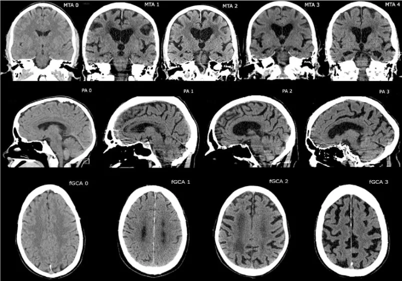
,_posterior_atrophy_(PA)_and_frontal_cortical_atrophy_(fGCA).png)
Özet
Serebral hacim kaybında kortikal sulkuslar ve subaraknoid BOS aralıkları yaygın genişler; bu görünüm subdural koleksiyonla karışabilir. Kontrastsız BT’de sulkusların belirgin genişlemesi ve parankim hacim kaybı, atrofi lehine yorumlanır; kitle etkisi bulunmaması bu görünümü destekler. Kortikal ven ve antikortikal ven işaretleri MR’da, kortikal atrofi zeminindeki subdural higromanın ayırıcı tanısı bağlamında bildirilmiştir. Atrofiye bağlı genişlemiş BOS aralığının subdural koleksiyon olarak yanlış yorumlanması yanlış pozitife; gerçek bir subdural koleksiyonu yalnızca atrofi sanmak ise kitle etkisinin ve tedavi ihtiyacının kaçırılmasına yol açar.
Faz ve pencere
- Kontrastsız tanısal
- Kontrastsız BT’de geniş sulkuslar ve konveksite boyunca BOS aralıkları değerlendirilir; yaygın parankim hacim kaybının varlığı ile fokal kitle basısının bulunmaması atrofi lehine yorumlanır. Kortikal ve antikortikal ven işaretleri MR’da bildirilmiştir. Kontrastsız BT yeterli olup; parankim hacmi, sulkuslar ve ventrikül boyutları birlikte değerlendirilebilir.
Önerilen pencereler: Beyin (G 80 / M 40), Subdural (G 200 / M 75).
BT bulguları
- Sulkus genişlemesi — frontal, parietal veya temporal korteks olukları kalvaryuma doğru belirginleşir ve birbirinden ayrılır.
- Subaraknoid BOS devamlılığı — konveksitedeki sıvı aralığı kortikal sulkusların içine kesintisiz uzanır.
- Kortikal ven işareti (MR) — kortikal ven bulgusu, atrofi ile subdural koleksiyon ayrımında MR’da bildirilmiştir.
- Parankim hacim azalması — giruslar incelmiş ve aralarındaki sulkal boşluklar yaygın genişlemiştir.
- Lateral ve üçüncü ventriküllerin genişlemesi, parankim hacim kaybına (atrofi) bağlı ex vacuo değişimi yansıtabilir; tek başına hidrosefali veya BOS akış bozukluğu anlamına gelmez.
- Kitle etkisinin olmaması — korteks içe itilmez, sulkuslar koleksiyon altında silinmez ve orta hat yapıları basıyla yer değiştirmez.
- Yaygın veya bölgesel dağılım — geniş BOS aralıkları birden çok lobda hacim kaybıyla uyumlu örüntü gösterir, tek hilal koleksiyona sınırlı kalmaz.
Normalde
Normal yaş ve anatomiye uygun BT’de konveksite sulkusları ince BOS çizgileri şeklindedir ve giruslar kalvaryuma daha yakın durur. Kıyaslarken yaygın sulkus genişliğini, parankim hacmini ve ventrikül genişlemesinin sulkuslarla uyumunu değerlendirin; ventriküler boyutlardaki artışın atrofya mı yoksa BOS akış dinamiklerindeki değişikliğe mi dayandığı birlikte incelenir.
Normal BT ile kıyasla
Aynı bölgenin, kaynak veri setinde patoloji içermediği belirtilen bir BT incelemesini kesit kesit kaydırın; organ sınırlarını açarak anatomiyi hasta görüntüsüyle karşılaştırın. Küçük rastlantısal bulgular tümüyle dışlanmamıştır.
Kaynak: TotalSegmentator v2.01 veri seti (Wasserthal ve ark., Radiology: Artificial Intelligence 2023), CC BY 4.0, vaka s0103; organ sınırları veri setinin otomatik segmentasyonundan, birleştirilerek sadeleştirildi. Görüntüler 2,5 mm kesit aralığına yeniden örneklendi.
Ayırıcı tanı
- Subdural higroma
- fokal koleksiyon oluşturarak korteksi içe itebilir ve alttaki sulkusları daraltabilir; kitle etkisi ve orta hat kayması ayırıcı tanıda önemlidir.
- Kronik subdural hematom
- iç yapı heterojen veya bölmeli olabilir; koleksiyon beyin yüzeyini basıyla yer değiştirebilir.
- Beyin atrofisi olmayan hidrosefali
- ventriküller orantısız genişlerken konveksite sulkusları silikleşebilir; sulkuslar yaygın genişlemiş olmaz.
- Genişlemiş subaraknoid alan
- BOS aralığı ve damarlar kortikal yüzeyin dış konturunu izler; beyin hacminin genel görünümü ile birlikte yorumlanır.
Tuzaklar
- BOS yoğunluklu aralığın hilal biçimli görünmesi tek başına subdural yerleşimi kanıtlamaz; sulkuslarla devamlılığı ve içindeki damarları izleyin.
- Ventrikül genişliğini tek başına hidrosefali kabul etmeyin; kortikal sulkusların da geniş olup olmadığını ve parankim hacmini karşılaştırın.
- Kortikal ven işareti MR’da bildirilmiştir; kortikal ve antikortikal ven bulgularının birlikte görülmesi atrofi ile subdural koleksiyon ayrımını güçleştirebilir.
Kendini dene
Konveksitede BOS aralıkları yaygın geniş, kortikal sulkuslar bu aralıklarla devamlı ve belirgin fokal bası yok.
Cevabı göster
Kortikal atrofi. Yaygın sulkus genişlemesi ve fokal kortikal bası olmaması, BOS aralıklarının hacim kaybına (atrofi) bağlı olduğunu destekler; kitle etkisi veya altta yatan sulkusların basılması yoktur.
Genişlemiş lateral ventriküller ve yaygın sulkus genişlemesi birlikte görüldüğünde bu bulgu en çok aşağıdakilerden hangisiyle ilişkilidir?
Cevabı göster
Beyin atrofisi (ex vacuo ventrikülomejali). Sulkusların da geniş olması ve parankim hacim kaybı ventrikül genişlemesinin ex vacuo olabileceğini destekler. Hidrosefalide ventrikül genişlemesine sulkus basısı eşlik edebilir; kanama ve kitle için BT'de ayrı intraventriküler veya fokal bulgu aranır.
Bu konunun yer aldığı turlar
Kaynaklar
Bu sayfadaki 33 cümle ve 2 quiz sorusu aşağıdaki kaynaklardan alıntılarla tek tek karşılaştırıldı; 12 cümle bu karşılaştırmada düzeltildi (2026-10-09).
- Assessing CT-based volumetric analysis via deep learning for idiopathic normal pressure hydrocephaluspmc.ncbi.nlm.nih.gov
- Probable Sporadic Creutzfeldt-Jakob Disease Presenting as Rapidly Progressive Dementia: A Case Report From Georgiapmc.ncbi.nlm.nih.gov
- Cerebral small vessel disease: pearls and pitfalls from unexplored risk factors to treatmentpmc.ncbi.nlm.nih.gov
- Copine-5-IgG-related autoimmune encephalitis: a novel paraneoplastic neurological syndrome with a strong melanoma associationpmc.ncbi.nlm.nih.gov
- Dystonic Head Tremor as a Presenting Feature of FMR1 Premutation: A Case Reportpmc.ncbi.nlm.nih.gov
- Review of imaging modalities and radiological findings of calvarial lesions.pubmed.ncbi.nlm.nih.gov
- Cerebral Superficial Siderosis : Etiology, Neuroradiological Features and Clinical Findings.pubmed.ncbi.nlm.nih.gov
- Cerebral Venous Outflow Implications in Idiopathic Intracranial Hypertension-From Physiopathology to Treatment.pubmed.ncbi.nlm.nih.gov
- Granulomatous Diseases of the CNS: Aetiology, Neuroradiological Features and Clinical Findings.pubmed.ncbi.nlm.nih.gov
- MRI Correlates of Depression and Anxiety in Multiple Sclerosis.pubmed.ncbi.nlm.nih.gov
- Evaluation of disproportionately enlarged subarachnoid-space hydrocephalus in progressive supranuclear palsy.pubmed.ncbi.nlm.nih.gov
- Role of neuroimaging in multidisciplinary approach towards Non-Alzheimer's dementia.pubmed.ncbi.nlm.nih.gov
- Automated brain computed tomographic densitometry of early ischemic changes in acute stroke.pubmed.ncbi.nlm.nih.gov
- Beta-Thalassemia Presenting as Moyamoya Syndrome With a Review of Skeletal Manifestations.pubmed.ncbi.nlm.nih.gov
- A Homozygous MAN2B1 Missense Mutation in a Doberman Pinscher Dog with Neurodegeneration, Cytoplasmic Vacuoles, Autofluorescent Storage Granules, and an α-Mannosidase Deficiency.pubmed.ncbi.nlm.nih.gov
- Differential diagnoses of cerebral hemiatrophy in childhood: a review of literature with illustrative report of two cases.pubmed.ncbi.nlm.nih.gov
- A comparative assessment of age- and gender-related variations in brain morphometry using linear and volumetric MRI analysis.pubmed.ncbi.nlm.nih.gov
- Perivascular spaces and basilar artery remodeling in Fabry disease-a dual vascular pathology.pubmed.ncbi.nlm.nih.gov
- Caution: shortcomings of traditional segmentation methods from magnetic resonance imaging brain scans intended for 3-dimensional surface modelling in children with pathology.pubmed.ncbi.nlm.nih.gov
- Radiological Correlates of Headache and Papilledema in Patients Undergoing Cranial MRI: Clinical Relevance of the Evans Index and Fazekas Score.pubmed.ncbi.nlm.nih.gov
- Surgery for post-traumatic hydrocephalus: lessons, challenges and future directions.pubmed.ncbi.nlm.nih.gov
- Reversal of cerebral pseudoatrophy in normal pressure hydrocephalus after ventriculoatrial shunt placement.pubmed.ncbi.nlm.nih.gov
- Paediatric cranial ultrasound: abnormalities of the brain in term neonates and young infants.pubmed.ncbi.nlm.nih.gov
- Preoperative CT markers and poor discharge functional status after burr-hole drainage for chronic subdural hematoma: a retrospective cohort study.pubmed.ncbi.nlm.nih.gov
- Risk Factors of Chronic Subdural Hematoma Recurrence: A Systematic Review.pubmed.ncbi.nlm.nih.gov
- Can Low-Dose Radiation Therapy Aid in Chronic Subdural Hematoma Management? A Historical Review.pubmed.ncbi.nlm.nih.gov
- Chronic Subdural Hematoma: Pathophysiology, Diagnosis, and the Emerging Role of Middle Meningeal Artery Embolization.pubmed.ncbi.nlm.nih.gov
- Continuum of Irreversible and Potentially Reversible Traumatic Brain Injury Sequelae: Axonal Injury and Hydrocephalus.pubmed.ncbi.nlm.nih.gov
- Hydrocephalic Dementia: Revisited with Multimodality Imaging and toward a Unified Imaging Approach.pubmed.ncbi.nlm.nih.gov
- Isolated leptomeningeal carcinomatosis and possible fungal meningitis as late sequelae of oesophageal adenocarcinoma.pubmed.ncbi.nlm.nih.gov
- Overview of the Current Knowledge and Conventional MRI Characteristics of Peri- and Para-Vascular Spaces.pubmed.ncbi.nlm.nih.gov
- Choroid Plexus Fibroblast-ILC2 Niche Promotes Adult Hippocampal Neurogenesis after Traumatic Brain Injury.pubmed.ncbi.nlm.nih.gov
- Structural neuroimaging markers of normal pressure hydrocephalus versus Alzheimer's dementia and Parkinson's disease, and hydrocephalus versus atrophy in chronic TBI-a narrative review.pubmed.ncbi.nlm.nih.gov
- Fetal brain MRI: neurometrics, typical diagnoses, and resolving common dilemmas.pubmed.ncbi.nlm.nih.gov
- Radiological assessment of dementia: the Italian inter-society consensus for a practical and clinically oriented guide to image acquisition, evaluation, and reporting.pubmed.ncbi.nlm.nih.gov
- A Deep Learning-Based Multimodal Fusion Model for Predicting Bone Cement Leakage in Percutaneous Kyphoplasty: Development and Validation.pubmed.ncbi.nlm.nih.gov
- Diffusion-Weighted Imaging in the Musculoskeletal System: Evolving Role in Modern Imaging Practice.pubmed.ncbi.nlm.nih.gov
- Admission hyperdense sinus sign predicts poorer outcomes in cerebral venous sinus thrombosis.pubmed.ncbi.nlm.nih.gov
- Systemic endothelial glycocalyx shedding mediates vascular hyperpermeability after traumatic brain injury.pubmed.ncbi.nlm.nih.gov
- Anatomy-Guided Microsurgical Resection of a Dominant Frontal Lobe Tumor Without Intraoperative Adjuncts: A Case Report from a Resource-Limited Context.pubmed.ncbi.nlm.nih.gov
- Fetal intracranial hemorrhage and infarct: Main sonographic and MRI characteristics: A review article.pubmed.ncbi.nlm.nih.gov
- Canine Cognitive Dysfunction and Alzheimer's Disease: Pathophysiological Relationships and the Impact of Glymphatic System Impairment on Neurodegeneration.pubmed.ncbi.nlm.nih.gov
- Prenatal diagnosis of infratentorial anomalies: ultrasound, fetal MRI, and implications for counseling.pubmed.ncbi.nlm.nih.gov
- The Effects of Endoscopic Third Ventriculostomy Versus Ventriculoperitoneal Shunt on Neuropsychological and Motor Performance in Patients with Idiopathic Normal Pressure Hydrocephalus-ENVENTOR-iNPH: Study Protocol.pubmed.ncbi.nlm.nih.gov
- Lightweight Deep Learning Models with Explainable AI for Early Alzheimer's Detection from Standard MRI Scans.pubmed.ncbi.nlm.nih.gov
- Cognitive impairment, neuroimaging abnormalities, and their correlations in myotonic dystrophy: a comprehensive review.pubmed.ncbi.nlm.nih.gov
- What's that smell? A pictorial review of the olfactory pathways and imaging assessment of the myriad pathologies that can affect them.pubmed.ncbi.nlm.nih.gov
- Neonatal cranial sonography: ultrasound findings in neonatal meningitis-a pictorial review.pubmed.ncbi.nlm.nih.gov
- Comparing the Efficacy of Two Cognitive Screening Tools in Identifying Gray and White Matter Brain Damage among Older Adults.pubmed.ncbi.nlm.nih.gov
- A pilot study of quantitative MRI measurements of ventricular volume and cortical atrophy for the differential diagnosis of normal pressure hydrocephalus.pubmed.ncbi.nlm.nih.gov
- Navigating Diagnostic Ambiguity in Palliative Pediatric Neuro-Oncology: A Conceptual Framework for Suspected Shunt Malfunction and Refractory Hyponatremia.pubmed.ncbi.nlm.nih.gov
- Redefining Idiopathic Normal Pressure Hydrocephalus Using AI-Driven Brain Volumetry.pubmed.ncbi.nlm.nih.gov
- A Framework for MRI Characterization of Intracranial Metastatic Disease.pubmed.ncbi.nlm.nih.gov
- Anatomically Precise Microsurgical Resection of a Posterior Fossa Cerebellar Metastasis in an Elderly Patient with Preservation of Venous Outflow, Dentate Nucleus, and Cerebrospinal Fluid Pathways.pubmed.ncbi.nlm.nih.gov
- Neurological manifestations and neuroimaging findings in patients with SARS-CoV2-a systematic review.pubmed.ncbi.nlm.nih.gov
- Encephalitis in Patients with COVID-19: A Systematic Evidence-Based Analysis.pubmed.ncbi.nlm.nih.gov
- Cerebrospinal fluid dysregulation and post-traumatic hydrocephalus after paediatric traumatic brain injury.pubmed.ncbi.nlm.nih.gov
- Assessing CT-based volumetric analysis via deep learning for idiopathic normal pressure hydrocephalus.pubmed.ncbi.nlm.nih.gov
- Copine-5-IgG-related autoimmune encephalitis: a novel paraneoplastic neurological syndrome with a strong melanoma association.pubmed.ncbi.nlm.nih.gov
- Surgery of the ear and the lateral skull base: pitfalls and complications.pubmed.ncbi.nlm.nih.gov
- Unraveling the Role of WDR91: Case Report of a Previously Unrecognized Clinical Entity.pubmed.ncbi.nlm.nih.gov
- Bilateral post-traumatic hygromas in patient with frontotemporal dementia.pubmed.ncbi.nlm.nih.gov
- Cerebellar mutism syndrome of non-tumour surgical aetiology-a case report and literature review.pubmed.ncbi.nlm.nih.gov
- Intracranial venous thrombosis imaging by anatomical location: an educational review with mimics and diagnostic pitfalls.pubmed.ncbi.nlm.nih.gov
- An analysis of the imaging features and diagnostic value of osteoid osteoma on DR, CT, and MRI.pubmed.ncbi.nlm.nih.gov
- Diagnostic Approach to Macrocephaly in Children.pubmed.ncbi.nlm.nih.gov
- Fetal Brain Development: Regulating Processes and Related Malformations.pubmed.ncbi.nlm.nih.gov
- Benign external hydrocephalus: a review, with emphasis on management.pubmed.ncbi.nlm.nih.gov
- Triple-modality management of complex septated chronic subdural hematoma: a preliminary technical note on feasibility and safety.pubmed.ncbi.nlm.nih.gov
- Intraoperative Ultrasound in the Management of Rare Lesions Involving the Intradural Extramedullary Spinal Compartment: A Quick, but Effective Helping Hand to Define the Optimal Surgical Strategy.pubmed.ncbi.nlm.nih.gov
- Differentiating Tuberculous and Pyogenic Spondylodiscitis: Part II: MRI/CT Features, Prediction Models, and the Role of PET/CT-A Narrative Review.pubmed.ncbi.nlm.nih.gov
- Spatial organization of AQP4 water channel expression in the human brain.pubmed.ncbi.nlm.nih.gov
- Expert Panel Consensus Guidelines of the German Society of Neuroradiology on the Use of Magnetic Resonance Imaging in the Diagnosis and Monitoring of Multiple Sclerosis.pubmed.ncbi.nlm.nih.gov
- Spinal cord imaging in multiple sclerosis: from diagnosis to disease progression.pubmed.ncbi.nlm.nih.gov
- Adrenohepatic Adhesion/Fusion Mimicking Hepatic Invasion: Two Cases of Benign Right Adrenal Lesions and a Review of the Literature.pubmed.ncbi.nlm.nih.gov
- Prevalence of venous extension in malignant adrenal neoplasia: beyond primary tumors and identification of a novel imaging sign.pubmed.ncbi.nlm.nih.gov
- Correlation between clinical and brain computed tomography findings of stroke patients: A cross-sectional study.pubmed.ncbi.nlm.nih.gov
- Case Report: Mechanical thrombectomy of cerebral venous sinuses using the NeVa stent retriever.pubmed.ncbi.nlm.nih.gov
- Reversible hemi-parkinsonism syndrome secondary to bilateral subdural hematomas: case report and literature review.pubmed.ncbi.nlm.nih.gov
- Excessive subgaleal suction from a variable vacuum drainage system used for managing chronic subdural hematomas: a cautionary note. Illustrative case.pubmed.ncbi.nlm.nih.gov
- Stroke-Like Migraine Attacks After Radiation Therapy (SMART) Syndrome: A Case Report and Review of a Rare Delayed Complication of Cranial Irradiation.pubmed.ncbi.nlm.nih.gov
- Dyke-Davidoff-Masson Syndrome: A Case of Unilateral Cerebral Atrophy and Seizure Disorder.pubmed.ncbi.nlm.nih.gov
- <i>Cryptococcus gattii</i> CNS infection presenting as a periaqueductal "soap-bubble" lesion causing obstructive hydrocephalus with refractory CSF diversion.pubmed.ncbi.nlm.nih.gov
- Otogenic Sigmoid Sinus Thrombosis in a 34-Year-Old Woman: A Rare Intracranial Complication.pubmed.ncbi.nlm.nih.gov
- Disproportionately elevated sulcal index (DESI): An automatically driven index representing disproportionate subarachnoid space enlargement in brain MRI scans.pubmed.ncbi.nlm.nih.gov
- Clinical-Radiological Spectrum of Cerebral Amyloid Angiopathy-Related Inflammation.pubmed.ncbi.nlm.nih.gov
- Clinical Image Quality and Reader Variability in 3D Synthetic Brain MRI Compared with Conventional MRI.pubmed.ncbi.nlm.nih.gov
- Brain Magnetic Resonance Imaging of Children With Molybdenum Cofactor Deficiency.pubmed.ncbi.nlm.nih.gov
- Delayed diagnosis of bilateral subdural effusions complicating intracranial hypotension in a patient presenting with post lumbar puncture headache.pubmed.ncbi.nlm.nih.gov
- Structural Brain Magnetic Resonance Imaging to Rule Out Comorbid Pathology in the Assessment of Alzheimer's Disease Dementia: Findings from the Ontario Neurodegenerative Disease Research Initiative (ONDRI) Study and Clinical Trials Over the Past 10 Years.pubmed.ncbi.nlm.nih.gov
- The Dystonias.pubmed.ncbi.nlm.nih.gov
- Neuroimaging findings in children with retinopathy-confirmed cerebral malaria.pubmed.ncbi.nlm.nih.gov
- Bacterial Meningoencephalitis in Newborns.pubmed.ncbi.nlm.nih.gov
- Congenital central nervous system malformations in piedmontese calves-part 1. structural brain disorders.pubmed.ncbi.nlm.nih.gov
- Clinical validation pipeline of a deep learning model for segmenting and quantifying intracranial and ventricular volumes on computed tomography.pubmed.ncbi.nlm.nih.gov
- Differential diagnosis of non-hydrocephalus ventricular dilation and hydrocephalus.pubmed.ncbi.nlm.nih.gov
- Dyke-Davidoff-Masson Syndrome Underlying Hemiplegic Cerebral Palsy in an Adolescent: A Case Report.pubmed.ncbi.nlm.nih.gov
- Clinical and literature insights into the frontotemporal dementia and motor neuron disease spectrum.pubmed.ncbi.nlm.nih.gov
- Ischemic stroke can have a T1w hyperintense appearance in absence of intralesional hemorrhage.pubmed.ncbi.nlm.nih.gov
- A Comprehensive Review of the Role of the Latest Minimally Invasive Neurosurgery Techniques and Outcomes for Brain and Spinal Surgeries.pubmed.ncbi.nlm.nih.gov
- Comparative CT Ventricular Morphometrics in Hydrocephalus, Stroke, and Traumatic Brain Injury: A Distortion-Controlled Analysis.pubmed.ncbi.nlm.nih.gov
- Consensus Statement on Middle Meningeal Artery Embolization in Chronic Subdural Hematoma Treatment: A Guideline from the Society of Vascular and Interventional Neurology Guidelines and Practice Standards Committee.pubmed.ncbi.nlm.nih.gov
- Chronic subdural hematoma.pubmed.ncbi.nlm.nih.gov
- The clinicopathologic spectrum of focal cortical dysplasias: a consensus classification proposed by an ad hoc Task Force of the ILAE Diagnostic Methods Commission.pubmed.ncbi.nlm.nih.gov
- Long-term safety and effectiveness of the "OptEase" vena cava filter.pubmed.ncbi.nlm.nih.gov
- Determination of cystathionine beta-synthase activity in human plasma by LC-MS/MS: potential use in diagnosis of CBS deficiency.pubmed.ncbi.nlm.nih.gov
- Secondary thalamic neuroinflammation associates with disturbed corticothalamic connectivity in a model of severe traumatic brain injury in male rats-a longitudinal study.pubmed.ncbi.nlm.nih.gov
- Hydrocephalus: Molecular and Neuroimaging Biomarkers in Diagnosis and Management.pubmed.ncbi.nlm.nih.gov
- Distinct cerebral cortical microstructural changes in idiopathic normal-pressure hydrocephalus.pubmed.ncbi.nlm.nih.gov
- Parasagittal subdural space: a novel quantitative marker of spontaneous intracranial hypotension syndrome-induced chronic subdural hematoma.pubmed.ncbi.nlm.nih.gov
- A deep learning approach to predict temporal changes of subdural hemorrhage on computed tomography.pubmed.ncbi.nlm.nih.gov
- An Imaging Overview of COVID-19 ARDS in ICU Patients and Its Complications: A Pictorial Review.pubmed.ncbi.nlm.nih.gov
Eğitim amaçlıdır; hasta başında tanı ve tedavi kararının yerine geçmez. İçerik yapay zekâ destekli bir süreçle güncel literatüre göre cümle cümle kaynaklarla karşılaştırıldı, ancak uzman radyolog incelemesinden geçmedi. Klinik kararlarda güncel kılavuzları, kurum protokollerini ve radyoloji raporunu esas alın.