Özet
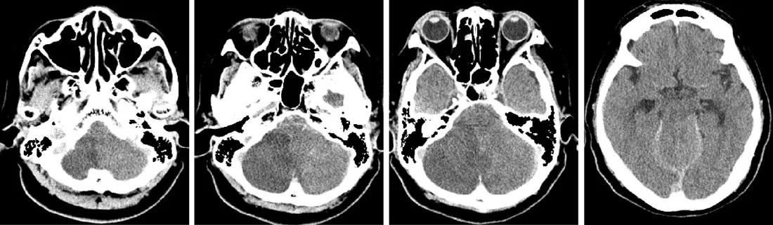
Posterior fossa hematomu, tümörü, infarktı veya ödemi serebellum ile vermisi tentoryal açıklıktan yukarı iter. Obstrüktif hidrosefali arka çukur lezyonlarına eşlik edebilir; supratentoryal BOS diversiyonu ise basınç gradyanını değiştirerek yukarı herniasyonu tetikleyebilir. Kontrastsız BT'de quadrigeminal ve superior serebellar sisternlerin silinmesi, orta beyin şekil değişikliği ve akuadukt basısı aranır. Akuadukt basısı üçüncü ve lateral ventriküllerde genişlemeye yol açabilir. Yukarı yer değiştirme fark edilmezse mezensefalon ve diensefalon basısı ilerleyebilir.
Faz ve pencere
- Kontrastsız tanısal
- Tentoryal açıklık çevresinde serebellar vermisin yukarı bombeleşmesi, quadrigeminal/superior serebellar sisternlerde BOS yoğunluğunun silinmesi, orta beyin konturunun basılması ve akuadukt/üçüncü ventrikül deformasyonu aranır. Kontrastsız faz akut kanama ve hidrosefaliyi birlikte gösterir; kontrastlı faz herniasyonun tanısı için gerekli değildir.
Önerilen pencereler: Beyin (G 80 / M 40).
BT bulguları
- Quadrigeminal sistern effasmanı — orta beyin arkasındaki BOS boşluğu daralır veya tamamen silinir.
- Superior serebellar sistern kaybı — serebellar vermisin üst yüzü ile tentoryum arasındaki BOS aralığı basıyla daralır veya silinir.
- Vermis ve serebellumun yukarı yer değiştirmesi — posterior fossa içeriği tentoryal çentik düzeyinde yukarı bombeleşir.
- Orta beyin deformasyonu — mezensefalon tentoryal kenara temasla basık veya deforme olabilir; akuadukt lümeni daralması ve posterior üçüncü ventrikül düzleşmesi eşlik eder.
- Posterior üçüncü ventrikül basısı — üçüncü ventrikül arka bölümü düzleşir veya yukarı kitle etkisiyle biçim değiştirir.
- Akuadukt daralması — serebral akuadukt lümeni seçilemez ya da bası altında incelir.
- Obstrüktif hidrosefali — lateral ve üçüncü ventriküller genişlerken dördüncü ventrikül basının seviyesine göre küçük ya da deforme kalabilir.
- Altta yatan arka çukur lezyonu — hematom, infarkt-ödem veya kitle tentoryal açıklığa yönelen basının kaynağıdır.
Normalde
Normalde quadrigeminal sistern mezensefalonun arkasında, superior serebellar sistern tentoryal açıklığın altında açık BOS alanlarıdır; serebellar vermis tentoryal çentik altında kalır. Orta beyin simetrik konturunu, akuadukt lümenini ve üçüncü ventrikül arka duvarını aynı seviyedeki normal anatomiyle kıyaslayın.
Normal BT ile kıyasla
Aynı bölgenin, kaynak veri setinde patoloji içermediği belirtilen bir BT incelemesini kesit kesit kaydırın; organ sınırlarını açarak anatomiyi hasta görüntüsüyle karşılaştırın. Küçük rastlantısal bulgular tümüyle dışlanmamıştır.
Kaynak: TotalSegmentator v2.01 veri seti (Wasserthal ve ark., Radiology: Artificial Intelligence 2023), CC BY 4.0, vaka s0103; organ sınırları veri setinin otomatik segmentasyonundan, birleştirilerek sadeleştirildi. Görüntüler 2,5 mm kesit aralığına yeniden örneklendi.
Ayırıcı tanı
- Orta beyin kitle lezyonu
- sisterna daralması yerel mezensefalon genişlemesi veya odak içi dansite değişikliğiyle ilişkilidir; yukarı itilen vermis paterni olmayabilir.
- Aşağı transtentorial/uncal herniasyon
- medial temporal lob tentoryal kenardan aşağı kayar, ambient sistern ve ipsilateral mezensefalon basısı öne çıkar.
- Tonsiller herniasyon
- tonsiller foramen magnum düzeyine veya ötesine inerken aşağıya doğru yer değiştirir; yukarı herniasyonda ise ana yer değiştirme posterior fossa içeriğinin tentoryal açıklığa doğru yükselmesidir.
- Hidrosefaliye bağlı sisternal bası
- ventriküler genişleme mevcut olsa da arka çukur kitle etkisi ve vermisin yukarı yer değiştirmesi aranarak ayrılır.
Tuzaklar
- Quadrigeminal sistern silinmesini tek başına herniasyon diye yorumlama; arka çukur kaynaklı bası, vermis seviyesi ve orta beyin şekli birlikte değişmelidir.
- Posterior fossa kitleli hastada ventriküllerin küçük olması herniasyonu dışlamaz; akuadukt tıkanması ve sisternal effasman daha erken olabilir.
- Posterior üçüncü ventrikül basısı ve orta beyin deformasyonu değerlendirilirken yalnızca bir inceleme düzlemine güvenilmemeli; aksiyel ve sagittal reformasyonlar arasında korrelasyon kurularak tentoryal açıklık ilişkisi doğrulanmalıdır.
Kendini dene
Posterior fossa hematomu sonrası quadrigeminal sistern silinmiş, vermis yukarı itilmiş ve akuadukt seçilemiyor. Tanı hangisidir?
Cevabı göster
Yukarı transtentorial herniasyon. Vermisin tentoryal açıklığa doğru yükselmesi, quadrigeminal sistern effasmanı ve akuadukt basısı yukarı transtentorial herniasyon paternidir. Tonsiller ve unkal herniasyonun yer değiştirme yönü aşağıdır; normal varyant orta beyin basısı oluşturmaz.
Aşağıdakilerden hangisi yukarı transtentorial herniasyonda beklenen BOS yolu etkisidir?
Cevabı göster
Akuadukt basısı. Tentoryal açıklık çevresindeki yukarı bası akuadukt lümenini kapatarak obstrüktif hidrosefaliye yol açabilir. Diğer bulgular özgül akuadukt tıkanma paterni değildir ve tek başlarına bu herniasyonu tanımlamaz.
İlgili konular
Kaynaklar
Bu sayfadaki 36 cümle ve 2 quiz sorusu aşağıdaki kaynaklardan alıntılarla otomatik olarak karşılaştırıldı; 3 cümle bu karşılaştırmada düzeltildi (2026-10-09).
- Posterior fossa hematoma: CT perfusion as a tool to reveal occult intracranial hypertension and support surgical decision-making – a case reportpmc.ncbi.nlm.nih.gov
- Successfully treated delayed upward trans-tentorial herniation (UTTH) following ventriculoperitoneal shunt placement for an acute obstructive hydrocephalus secondary to midline posterior fossa mass in a low-resource setting: case reportpmc.ncbi.nlm.nih.gov
- Isolated brainstem death: case presentation demonstrating preserved supratentorial perfusion on 99m Tc-bicisate brain scintigraphypmc.ncbi.nlm.nih.gov
- External Ventricular Drainage for Hydrocephalus Following Cerebellar Infarction: A Scoping Reviewpmc.ncbi.nlm.nih.gov
- Brain Shift Patterns: Upward, Lateral and Downward Herniation, Its Correlation with Clinical Patterns in Acute Intracranial Pathologies and Neurosurgical Managementpmc.ncbi.nlm.nih.gov
- Brain Shift Patterns: Upward, Lateral and Downward Herniation, Its Correlation with Clinical Patterns in Acute Intracranial Pathologies and Neurosurgical Management.pubmed.ncbi.nlm.nih.gov
- Posterior fossa hematoma: CT perfusion as a tool to reveal occult intracranial hypertension and support surgical decision-making - a case report.pubmed.ncbi.nlm.nih.gov
- A Review of the Clinical Presentation, Causes, and Diagnostic Evaluation of Increased Intracranial Pressure in the Emergency Department.pubmed.ncbi.nlm.nih.gov
- Management of Posterior Fossa Tumors in Adults Based on the Predictors of Postoperative Hydrocephalus.pubmed.ncbi.nlm.nih.gov
- Surgical treatment of cerebellar metastases in elderly patients: A threshold that moves forward?pubmed.ncbi.nlm.nih.gov
- Ascending Transtentorial Herniation: A Rare Complication of Posterior Cranial Fossa Tumors and Review of Literature.pubmed.ncbi.nlm.nih.gov
- Cerebral venous disorders: clinical presentation, diagnostic strategy, and contemporary management.pubmed.ncbi.nlm.nih.gov
- Hidden hazard of remote cerebellar hemorrhage after cervical spine surgery: Case report.pubmed.ncbi.nlm.nih.gov
- Oculomotor Cranial Neuropathies: Diagnosis and Management.pubmed.ncbi.nlm.nih.gov
- Delayed posterior tilting of a giant basilar tip aneurysm following stent-assisted coiling with therapeutic P1 occlusion: A case report.pubmed.ncbi.nlm.nih.gov
- Comprehensive review of the cerebral aqueduct: anatomy, embryology, imaging, genetics, molecular, and pathology.pubmed.ncbi.nlm.nih.gov
- From shunt dependency to shunt freedom: Endoscopic third ventriculostomy in ventriculoperitoneal shunt-dependent communicating tetraventricular hydrocephalus: A case report.pubmed.ncbi.nlm.nih.gov
- Lumbar Puncture and Brain Herniation in Acute Bacterial Meningitis: An Updated Narrative Review.pubmed.ncbi.nlm.nih.gov
- Clival and Paraclival Lesions: A Pictorial Review.pubmed.ncbi.nlm.nih.gov
- Acute cerebellitis following SARS-CoV-2 infection: A case report and review of the literature.pubmed.ncbi.nlm.nih.gov
- External Ventricular Drainage for Hydrocephalus Following Cerebellar Infarction: A Scoping Review.pubmed.ncbi.nlm.nih.gov
- Supratentorial cerebrospinal fluid diversion using image-guided trigonal ventriculostomy during retrosigmoid craniotomy for cerebellopontine angle tumors.pubmed.ncbi.nlm.nih.gov
- Imaging of cranial nerves: a pictorial overview.pubmed.ncbi.nlm.nih.gov
- Remote cerebellar hemorrhage complicated after supratentorial surgery: retrospective study with review of articles.pubmed.ncbi.nlm.nih.gov
- Acute Epidural Hematoma: From Injury to Death.pubmed.ncbi.nlm.nih.gov
- Lymphatic Vessel Network Structure and Physiology.pubmed.ncbi.nlm.nih.gov
- Commentary: an eye on PET quantification.pubmed.ncbi.nlm.nih.gov
- Hemodynamic changes and their relationship with white matter hyperintensities in CSVD patients with cognitive impairment: a 4D flow study.pubmed.ncbi.nlm.nih.gov
- Pulsatile Tinnitus: A Comprehensive Clinical Approach to Diagnosis and Management.pubmed.ncbi.nlm.nih.gov
- Clinical and imaging findings after antiviral treatment in cats with presumptive neurologic feline infectious peritonitis: 9 cases (2020-2024).pubmed.ncbi.nlm.nih.gov
- Case Report: Primary Fourth Ventricle Outlet Obstruction in Childhood - New Clinical Insights and Long-Term Outcome.pubmed.ncbi.nlm.nih.gov
- Clinical Efficacy of Endoscopic Infratentorial Supracerebellar Approach for Pineal Region Tumors: A Retrospective Case-Control Study.pubmed.ncbi.nlm.nih.gov
- Imaging Evaluation of Acute Traumatic Brain Injury.pubmed.ncbi.nlm.nih.gov
- Chiari Malformation and Hindbrain Descent: Characterization and New Classification Based on Mechanism and Pathogenesis, and Surgical Management.pubmed.ncbi.nlm.nih.gov
- Functional and morphological changes in hypoplasic posterior fossa.pubmed.ncbi.nlm.nih.gov
- Fourth Ventricle Epidermoid Cyst: Case Report of Precision Telovelar Microsurgery, Functional Preservation, and Lifelong Surveillance.pubmed.ncbi.nlm.nih.gov
- Blake's pouch cyst.pubmed.ncbi.nlm.nih.gov
- Hydrocephalus in pediatric posterior fossa tumors: predictors and outcomes from a single center in Latin America.pubmed.ncbi.nlm.nih.gov
- Tumors of the posterior third ventricular region in pediatric patients: The Indian perspective and a review of literature.pubmed.ncbi.nlm.nih.gov
- Beyond Aqueductal Stenosis: A Case of an Atypical Teratoid/Rhabdoid Tumor Presenting With Neonatal Hydrocephalus.pubmed.ncbi.nlm.nih.gov
- Delayed mass effect following endovascular coiling of posterior inferior cerebellar artery aneurysms: Mechanism and treatment strategy: A case report.pubmed.ncbi.nlm.nih.gov
- Vestibular schwannomas and papilledema without hydrocephalus: a case report.pubmed.ncbi.nlm.nih.gov
- Successfully treated delayed upward trans-tentorial herniation (UTTH) following ventriculoperitoneal shunt placement for an acute obstructive hydrocephalus secondary to midline posterior fossa mass in a low-resource setting: case report.pubmed.ncbi.nlm.nih.gov
- The Tentorium Cerebelli: A Comprehensive Review Including Its Anatomy, Embryology, and Surgical Techniques.pubmed.ncbi.nlm.nih.gov
- Trapped Fourth Ventricle in a Pediatric Patient With a History of Post-Traumatic Hydrocephalus and Ventriculoperitoneal Shunt: A Case Report and Literature Review.pubmed.ncbi.nlm.nih.gov
- Isolated brainstem death: case presentation demonstrating preserved supratentorial perfusion on <sup>99m</sup>Tc-bicisate brain scintigraphy.pubmed.ncbi.nlm.nih.gov
- Is foramen magnum decompression for acquired Chiari I malformation like putting a finger in the dyke? - A simplistic overview of artificial intelligence in assessing critical upstream and downstream etiologies.pubmed.ncbi.nlm.nih.gov
- Management of severe acute encephalopathy in the ICU: an expert consensus statement from the french society of intensive care medicine.pubmed.ncbi.nlm.nih.gov
- A rare case of near-complete resolution of extensive cervicothoracic syrinx following bilateral cerebellar tonsillar resection in Chiari malformation type I.pubmed.ncbi.nlm.nih.gov
- Subarachnoid hemorrhage secondary to Chiari 1 malformation in a patient presenting with recurrent severe headache: A case report, imaging, and anatomic considerations.pubmed.ncbi.nlm.nih.gov
- Clinical and genetic features of syndromic craniosynostosis in 18 Chinese probands: novel candidate genes and phenotypes of known pathogenic genes.pubmed.ncbi.nlm.nih.gov
- Utility of Biometric Measurements from Fetal Magnetic Resonance Imaging for Improved Antenatal Diagnosis of Dandy-Walker Spectrum Posterior Fossa Lesions.pubmed.ncbi.nlm.nih.gov
- Loss of the maternal effect gene NLRP2 impairs embryonic and extra-embryonic development, revealing a novel genetic cause of congenital anomalies†.pubmed.ncbi.nlm.nih.gov
- Evaluation of ballistics euthanasia applied to stranded cetaceans using ethological and post-mortem computed tomography assessment.pubmed.ncbi.nlm.nih.gov
- Imaging of Skull Base Tumors.pubmed.ncbi.nlm.nih.gov
- 선천성 뇌 질환의 영상 소견: 임상 화보.pubmed.ncbi.nlm.nih.gov
- Evaluation and Treatment of Patients with Small Posterior Cranial Fossa and Chiari Malformation, Types 0 and 1.pubmed.ncbi.nlm.nih.gov
- The genetic basis of hydrocephalus: genes, pathways, mechanisms, and global impact.pubmed.ncbi.nlm.nih.gov
Eğitim amaçlıdır; hasta başında tanı ve tedavi kararının yerine geçmez, tıbbi tavsiye değildir. Metinler yapay zekâ desteğiyle yazıldı ve otomatik bir adımda kaynak alıntılarıyla karşılaştırıldı; bu bir tıbbi doğrulama değildir ve içerik uzman radyolog incelemesinden geçmedi. Klinik kararlarda güncel kılavuzları, kurum protokollerini ve radyoloji raporunu esas alın.