Özet
Glioblastom çoğunlukla erişkinlerde gelişen, hızla ilerleyebilen infiltratif bir intraaksiyel tümördür. Acilde BT, tümörün kendisini kesin sınıflandırmaktan çok kanamayı, ödemi ve yaşamı tehdit eden kitle etkisini hızla gösterir. Kontrastlı MRG tümörün yayılımını ve nekrotik kontrastlanan bileşenini daha iyi karakterize eder; BT bulguları histolojik ya da moleküler tanı yerine geçmez. Orta hat basısı, hidrosefali veya herniasyonun gözden kaçması acil değerlendirmeyi geciktirip nörolojik kötüleşmeye yol açabilir.
Faz ve pencere
- Kontrastsız tanısal
- Kontrastsız BT’de kitle genellikle hiperdens veya heterojen görünebilir; çevresinde vazojenik ödem kaynaklı hipodensite, ventrikül basısı ve intralezyonel kanama aranır. Kontrastsız BT nekrotik kontrastlanan halkayı göstermez.
- Kontrastlı tanısal
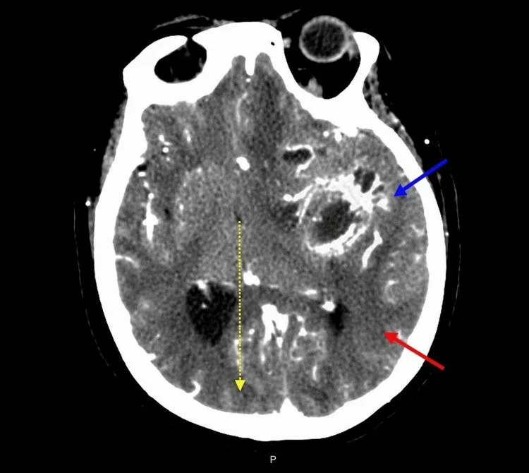
- Düzensiz, kalın ya da nodüler periferik/solid kontrastlanma ile çevrelenen santral kontrastlanmayan nekroz görülebilir; çevresel ödem eşlik edebilir. Bu örüntü yüksek dereceli nekrotik gliomu destekler ancak özgül değildir; metastaz ve apse gibi ayırıcı tanılar sürer. BT yumuşak doku yayılımını sınırlı gösterir; MRG daha ayrıntılıdır.
Önerilen pencereler: Beyin (G 80 / M 40), Kemik (G 1800 / M 400).
BT bulguları
- Yüksek dereceli astrositomlar tipik olarak intraaksiyel, diffüz infiltratif beyin tümörleridir.
- Santral kontrastlanmayan hipodens nekroz — kontrastlı incelemede düzensiz periferik/solid canlı tümör dokusuyla çevrelenebilir.
- Peritümöral hipodensite — glioblastomda çevresel vazojenik ödem görülebilir.
- Korpus kallozum boyunca uzanım — karşı hemisfere geçen infiltratif tümör kelebek biçimli kitle oluşturabilir.
- İntralezyonel hiperdens odak — akut kanama olabilir; kalsifikasyon daha seyrektir.
- Kitle etkisi — lateral ventrikül kompresyonu, orta hat kayması ve sisternlerde silinme şeklinde görülebilir.
Ölçütler ve sınıflamalar
- WHO CNS5 ve güncel terminoloji
- WHO CNS5 (2021) kapsamında glioblastom, IDH-yabanıl tip, CNS WHO derece 4 olarak adlandırılır. IDH-mutant derece-4 diffüz gliom “Astrositom, IDH-mutant, CNS WHO derece 4” olarak sınıflanır. Bu histomoleküler tanı yalnız görüntüleme ile kurulamaz. cIMPACT-NOW Update 11 (2025) tanı ölçütleri için öneriler sunar; WHO CNS5’i resmen değiştirmez.
Normalde
Aynı aksiyel düzeyde normal frontal ve parietal korteks simetrik gri cevher yoğunluğunda, aradaki beyaz cevher ise homojen ve daha düşük atenüasyondadır. Kontrastsız BT’de orta hat kayması, ventrikül basısı ve sisternlerde silinme gibi kitle etkisi bulguları dikkatlice değerlendirilir.
Normal BT ile kıyasla
Aynı bölgenin, kaynak veri setinde patoloji içermediği belirtilen bir BT incelemesini kesit kesit kaydırın; organ sınırlarını açarak anatomiyi hasta görüntüsüyle karşılaştırın. Küçük rastlantısal bulgular tümüyle dışlanmamıştır.
Kaynak: TotalSegmentator v2.01 veri seti (Wasserthal ve ark., Radiology: Artificial Intelligence 2023), CC BY 4.0, vaka s0103; organ sınırları veri setinin otomatik segmentasyonundan, birleştirilerek sadeleştirildi. Görüntüler 2,5 mm kesit aralığına yeniden örneklendi.
Ayırıcı tanı
- Beyin metastazlarında çevresel vazojenik ödem eşlik edebilir.
- Primer santral sinir sistemi lenfoması
- derin/periventriküler yerleşim ve kontrastlı incelemede daha homojen solid kontrastlanma düşündürür.
- Beyin apsesi
- halka biçimli lezyon yapabilir; merkezî difüzyon kısıtlanması MRG’de ayırıcıdır.
- Subakut enfarkt
- vasküler alana uyan kortikomedüller hipodensite ve zamanla gelişen kitle etkisi görülebilir.
Tuzaklar
- Ödem sınırını tümör sınırı sayma: infiltratif gliom kontrastlanmayan ödemli görünen beyaz cevherin içine uzanabilir; BT bu ayrımı kuramaz.
- Tek, halka biçimli lezyonu glioblastom kabul etme: soliter metastaz ve apse de benzer görünebilir; kontrastlı MRG ve klinik bağlam gerekir.
- Kontrastsız BT’de belirgin halka görmeyi bekleme: nekrozun kontrastlanan kenarı ancak kontrast sonrası incelemede değerlendirilir.
Kendini dene
Erişkinde korpus kallozum tutulumu, santral nekroz ve periferik kontrastlanma yüksek dereceli gliomla uyumlu olabilir. En olası görüntüleme tanısı nedir?
Cevabı göster
Glioblastom fenotipinde yüksek dereceli gliom. Kallozumu aşan infiltratif kitle, düzensiz kontrastlanan canlı periferik bölüm ve santral nekroz yüksek dereceli gliom, özellikle glioblastom fenotipiyle uyumludur. Halka kontrastlanan gliom benzeri lezyonlarda metastaz ve apse ayırıcı tanıda kalabilir; görüntüleme tek başına moleküler tümör sınıfını belirlemez.
Korpus kallozumu geçen frontal kitle ve orta hat basısı saptanıyor. Hangi bulgu acil kitle etkisini gösterir?
Cevabı göster
Bazal sistern silinmesi. Bazal sisternlerin silinmesi artmış basınç ve olası transtentoryal herniasyon açısından kritik kitle etkisidir. Dural kuyruk ekstraaksiyel menenjiyomu düşündürür; kalsifikasyon tek başına acil bası bulgusu değildir; sulkus genişlemesi bası değil hacim kaybını destekler.
İlgili konular
Kaynaklar
Bu sayfadaki 41 cümle ve 2 quiz sorusu aşağıdaki kaynaklardan alıntılarla tek tek karşılaştırıldı; 10 cümle bu karşılaştırmada düzeltildi (2026-10-09).
- Laser Interstitial Thermal Therapy for High-Grade Gliomas: Current Evidence, Clinical Applications and Emerging Role of Artificial Intelligencepmc.ncbi.nlm.nih.gov
- Snake venom bioprospecting as an approach to finding potential anti-glioblastoma moleculespmc.ncbi.nlm.nih.gov
- A phase 1/2a dose-finding study and biomarker assessment of oral lisavanbulin in patients with high-grade glioma or glioblastomapmc.ncbi.nlm.nih.gov
- Structural gray matter alterations in glioblastoma and high-grade glioma—A potential biomarker of survivalpmc.ncbi.nlm.nih.gov
- The CCL2-CCR4 axis promotes Regulatory T cell trafficking to canine glioma tissuespmc.ncbi.nlm.nih.gov
- Structural gray matter alterations in glioblastoma and high-grade glioma-A potential biomarker of survival.pubmed.ncbi.nlm.nih.gov
- Glioblastoma: Epidemiology and Imaging-Based Review.pubmed.ncbi.nlm.nih.gov
- Lomustine and Bevacizumab in Progressive Glioblastoma.pubmed.ncbi.nlm.nih.gov
- Circulating Liquid Biopsy Biomarkers in Glioblastoma: Advances and Challenges.pubmed.ncbi.nlm.nih.gov
- Targeting VPAC1 Receptors for Imaging Glioblastoma.pubmed.ncbi.nlm.nih.gov
- Glioblastoma: Radiation treatment margins, how small is large enough?pubmed.ncbi.nlm.nih.gov
- Immunocompetent murine models for the study of glioblastoma immunotherapy.pubmed.ncbi.nlm.nih.gov
- Oncolytic herpes simplex virus expressing IL-2 controls glioblastoma growth and improves survival.pubmed.ncbi.nlm.nih.gov
- Clinical implication of cellular vaccine in glioma: current advances and future prospects.pubmed.ncbi.nlm.nih.gov
- ImmunoPET-informed sequence for focused ultrasound-targeted mCD47 blockade controls glioma.pubmed.ncbi.nlm.nih.gov
- Molecular heterogeneity of glioblastomas: does location matter?pubmed.ncbi.nlm.nih.gov
- [Epidemiology of glioblastoma].pubmed.ncbi.nlm.nih.gov
- Visualization of Diagnostic and Therapeutic Targets in Glioma With Molecular Imaging.pubmed.ncbi.nlm.nih.gov
- Repeat-surgery at Glioblastoma recurrence, when and why to operate?pubmed.ncbi.nlm.nih.gov
- Imaging biomarkers guided anti-angiogenic therapy for malignant gliomas.pubmed.ncbi.nlm.nih.gov
- Glioblastoma Masquerading as a Presumed Ischemic Stroke: A Case of Delusional Jealousy and Fluctuating Psychosis.pubmed.ncbi.nlm.nih.gov
- Case Report: Rare presentations of glioblastoma: three multifocal and one cerebellar cases highlighting the value of shared clinical experience.pubmed.ncbi.nlm.nih.gov
- The role of dordaviprone in the multidisciplinary management of diffuse midline glioma.pubmed.ncbi.nlm.nih.gov
- Acute Truncal Ataxia After a Minor Head Trauma Revealing a Pediatric Cerebellar Pilocytic Astrocytoma.pubmed.ncbi.nlm.nih.gov
- Multidisciplinary Management of Emergency Neurosurgery for Intracerebral Hemorrhage During Pregnancy: A Case Report.pubmed.ncbi.nlm.nih.gov
- Von Hippel-Lindau syndrome presenting as a lateral ventricular space-occupying lesion: a case report.pubmed.ncbi.nlm.nih.gov
- Quantitative magnetic resonance imaging for accurate delineation of radiotherapy: a narrative review.pubmed.ncbi.nlm.nih.gov
- Research progress in imaging detection of brain metastases.pubmed.ncbi.nlm.nih.gov
- Prostate-specific membrane antigen in malignant brain tumors: vascular heterogeneity, imaging promise, and therapeutic challenges.pubmed.ncbi.nlm.nih.gov
- Case Report: Cerebrospinal fluid cytology as a diagnostic clue in hemorrhagic glioblastoma mimicking hypertensive intracranial hemorrhage.pubmed.ncbi.nlm.nih.gov
- Neuroimaging outcomes in suspected papilledema.pubmed.ncbi.nlm.nih.gov
- Primary cerebral melanoma of the frontal lobe: a case report and review of the literature.pubmed.ncbi.nlm.nih.gov
- Neuroradiology of acute pathologies in adults with hematologic malignancies: a pictorial review.pubmed.ncbi.nlm.nih.gov
- Neuroinflammation in Central Nervous System Tumors.pubmed.ncbi.nlm.nih.gov
- Predictors of pre-resection hydrocephalus in posterior cranial fossa tumors: development of a predictive scoring model.pubmed.ncbi.nlm.nih.gov
- Post-therapy imaging of adult brain tumours: patterns, pitfalls, and practical framework.pubmed.ncbi.nlm.nih.gov
- Predominantly cystic glioblastoma mimicking benign cystic lesions: A radiological diagnostic challenge.pubmed.ncbi.nlm.nih.gov
- Diagnostic imaging and emerging image-guided neurointervention in brain tumors: a narrative review.pubmed.ncbi.nlm.nih.gov
- Stiffness and Tissue Viscosity in a Cerebral Neoplasm Measured by Preoperative Multifrequency Magnetic Resonance Elastography (MRE) Guide the Differential Diagnosis of Brain Tumors by Ruling Out Glioma.pubmed.ncbi.nlm.nih.gov
- Spectrum of Neuroimaging Findings in CNS Lymphoproliferative Disorders.pubmed.ncbi.nlm.nih.gov
- Imaging diagnosis of cystic intraparenchymal brain neoplasms.pubmed.ncbi.nlm.nih.gov
- Prognostic Impact of Calcification, Corpus Callosum Invasion, and CDKN2A/B Hemizygous Deletion in Oligodendroglioma: A Single-center Retrospective Study.pubmed.ncbi.nlm.nih.gov
- Surgical decision making in the era of supramarginal glioma resections: a current perspective and narrative review.pubmed.ncbi.nlm.nih.gov
- Radiological, clinical, and molecular analyses reveal distinct subtypes of butterfly glioblastomas affecting the prognosis.pubmed.ncbi.nlm.nih.gov
- Low-dose temozolomide combined with valproic acid in the treatment of an elderly patient with MGMT-methylated glioblastoma and multiple comorbidities: a case report.pubmed.ncbi.nlm.nih.gov
- How I treat acute venous thromboembolism in patients with brain tumors.pubmed.ncbi.nlm.nih.gov
- Let Us Not Forget About Bleeding: A Case Report and Brief Literature Review on Hemorrhagic Vestibular Schwannoma.pubmed.ncbi.nlm.nih.gov
- Preliminary Evaluation of Preoperative Optic Nerve Sheath Diameter and CT Mass Effect in Relation to Pre-Excision Invasive Intracranial Pressure During Intracranial Tumor Surgery.pubmed.ncbi.nlm.nih.gov
- Anatomy-Guided Microsurgical Resection of a Dominant Frontal Lobe Tumor Without Intraoperative Adjuncts: A Case Report from a Resource-Limited Context.pubmed.ncbi.nlm.nih.gov
- Case Report: A case of Lynch syndrome-related glioblastoma with coexisting <i>MSH2</i> splicing defect and <i>MSH6</i> frameshift mutation.pubmed.ncbi.nlm.nih.gov
- Anterior Superior Longitudinal Fasciculus Microstructure Correlates With Bimanual Visuomotor Performance in Healthy Younger but Not Older Adults.pubmed.ncbi.nlm.nih.gov
- Electroencephalographic Aspects in a Sheep with Coenurosis.pubmed.ncbi.nlm.nih.gov
- Dopaminergic Radiopharmaceutical Imaging in Parkinsonian Syndromes: From Molecular Targets to Clinical Decision-Making.pubmed.ncbi.nlm.nih.gov
- Epidermal Growth Factor Receptor (EGFR) Amplification and Survival in Patients With Glioblastoma: A Systematic Review and Meta-Analysis.pubmed.ncbi.nlm.nih.gov
- Characterization of High-Grade Glial and Related Neuroepithelial Tumors Using the AOSNP-ADAPTR Framework: A Case Series With Review of Literature.pubmed.ncbi.nlm.nih.gov
- Clinical evaluation of the CrossNN DNA methylation classifier for central nervous system tumors.pubmed.ncbi.nlm.nih.gov
- Diagnostic Pitfalls of Oligodendroglioma-like Morphology: Integrated Reappraisal of 23 Non-Oligodendroglial Central Nervous System Tumors.pubmed.ncbi.nlm.nih.gov
- "Updates on diagnostic and prognostic molecular biomarkers of CNS tumors".pubmed.ncbi.nlm.nih.gov
- IDH-Mutant Diffuse Glioma: From Metabolic Origins to Targeted Therapy.pubmed.ncbi.nlm.nih.gov
- Comparison between molecular and histological IDH-wild-type glioblastoma and extensive subgroup analysis of IDH-wild-type astrocytic tumors without genomic glioblastoma-defining alterations.pubmed.ncbi.nlm.nih.gov
- Molecularly diagnosed glioblastoma: clinical presentation and outcomes.pubmed.ncbi.nlm.nih.gov
- Isocitrate Dehydrogenase (IDH)-Wildtype Glioblastoma With an Unusual EWSR1::RAD51B Gene Fusion: A Case Report and Review of the Literature.pubmed.ncbi.nlm.nih.gov
- Solitary neurocysticercosis mimicking tuberculoma and glioblastoma: A diagnostic challenge in a low-resource setting.pubmed.ncbi.nlm.nih.gov
- Butterfly Glioma of the Corpus Callosum Mimicking Rapidly Progressive Dementia.pubmed.ncbi.nlm.nih.gov
- Glioblastoma with pseudo-arteriovenous malformation features: Combined arterial spin-labeling assessment and preoperative embolization.pubmed.ncbi.nlm.nih.gov
- Masked autoencoding, generalizable pretraining, and integrated experts for enhanced glioma segmentation.pubmed.ncbi.nlm.nih.gov
- The role of diffusion-weighted MRI in biological image-guided radiation therapy: a roadmap.pubmed.ncbi.nlm.nih.gov
- Brain tumor classification using the diffusion tensor image segmentation (D-SEG) technique.pubmed.ncbi.nlm.nih.gov
- Solitary Brain Metastasis From Hepatocellular Carcinoma Masquerading as Acute Ischaemic Stroke: Radiological Diagnosis and Clinical Implications.pubmed.ncbi.nlm.nih.gov
- Primary glioblastoma mimicking brain metastasis in ALK-positive lung adenocarcinoma: a case report and literature review.pubmed.ncbi.nlm.nih.gov
- Clinical characteristics and prognostic analysis of calcified glioblastoma.pubmed.ncbi.nlm.nih.gov
- Anatomical and surgical technical aspects of intraparietal sulcus approach for posterolateral thalamic glioma: illustrative case.pubmed.ncbi.nlm.nih.gov
- Association of TERT promoter mutation and MGMT methylation with MRI features and overall survival in glioblastoma, IDH-wildtype.pubmed.ncbi.nlm.nih.gov
- The 2021 World Health Organization Central Nervous System Tumor Classification: The Spectrum of Diffuse Gliomas.pubmed.ncbi.nlm.nih.gov
- Imaging Spectrum of the Developing Glioblastoma: A Cross-Sectional Observation Study.pubmed.ncbi.nlm.nih.gov
- Association of Schimmelpenning Syndrome with Astrocytoma (WHO Grade 3): Case Report.pubmed.ncbi.nlm.nih.gov
- Intracranial ependymomas: The role of advanced neuroimaging in diagnosis and management.pubmed.ncbi.nlm.nih.gov
- Clinical application of 3D Slicer combined with Sina/MosoCam multimodal system in preoperative planning of brain lesions surgery.pubmed.ncbi.nlm.nih.gov
- Meningeal tuberculoma presenting as a space-occupying lesion in an immuno-competent young adult patient: A case report and literature review.pubmed.ncbi.nlm.nih.gov
- CNS Non-Hodgkin's Lymphoma With Atypical T2 Hyperintensity and Necrosis Mimicking Glioblastoma Multiforme: A Radiological Enigma.pubmed.ncbi.nlm.nih.gov
- Neuromelioidosis presenting with the 'tunnel sign': A case report.pubmed.ncbi.nlm.nih.gov
- Imaging features of a frontal neuroglial cyst in an adult patient: A rare case report.pubmed.ncbi.nlm.nih.gov
- Neonatal stress hyperglycemia mimicking diabetes mellitus secondary to massive intraparenchymal brain abscess due to escherichia coli sepsis: A case report.pubmed.ncbi.nlm.nih.gov
- Identification and characterisation of calcitonin receptor isoforms expressed in glioblastoma derived glioma stem and U-87 MG cells.pubmed.ncbi.nlm.nih.gov
- Integrated Transcriptomic Analysis Reveals Coordinated Gliovascular Remodeling Across Diffuse Glioma Grades.pubmed.ncbi.nlm.nih.gov
- Brain Midline Shift Measurement and Its Automation: A Review of Techniques and Algorithms.pubmed.ncbi.nlm.nih.gov
- Central neurocytomas: Decision-making process of postoperative treatment. Lesson learned.pubmed.ncbi.nlm.nih.gov
- Magnetic resonance imaging of primary central nervous system neoplasms of dogs and cats: Basic concepts for diagnostic pathologists.pubmed.ncbi.nlm.nih.gov
- Giant Right Sphenoid Wing Meningioma as a Reversible Frontal Network Lesion: A Pseudo-bvFTD Case with Venous-Sparing Skull-Base Resection.pubmed.ncbi.nlm.nih.gov
- Imaging in Neuro-oncology.pubmed.ncbi.nlm.nih.gov
- Successfully treated delayed upward trans-tentorial herniation (UTTH) following ventriculoperitoneal shunt placement for an acute obstructive hydrocephalus secondary to midline posterior fossa mass in a low-resource setting: case report.pubmed.ncbi.nlm.nih.gov
- In-hospital unexpected cerebral herniation-New neurosurgical quality control standards: A case report.pubmed.ncbi.nlm.nih.gov
- Transdural Skull Base Infiltration by Glioblastoma: Case Report and Review of the Literature.pubmed.ncbi.nlm.nih.gov
- Temporoinsular Extraventricular Neurocytoma Initially Diagnosed as Glioblastoma in a 57-Year-Old Woman: A Case Report and Literature Review.pubmed.ncbi.nlm.nih.gov
- Right Frontal Isocitrate Dehydrogenase (IDH)-Mutant WHO Grade 3 Oligodendroglioma Presenting With Morning Headache and Gait Imbalance.pubmed.ncbi.nlm.nih.gov
- Diagnostic challenges of gliosarcoma: case report of a rare glioblastoma histopathological variant.pubmed.ncbi.nlm.nih.gov
- Purely cystic meningioma: extremely rare clinical and radiographic mimic of intra-axial glioblastoma multiforme with systematic review of the literature. Illustrative case.pubmed.ncbi.nlm.nih.gov
- Follicular carcinoma of the thyroid presenting as metastasis in the wall of an arachnoid cyst.pubmed.ncbi.nlm.nih.gov
- Quantitative evaluation of neuroradiological and morphometric alteration of inferior Fronto-Occipital Fascicle across different brain tumor histotype: an Italian multicentric study.pubmed.ncbi.nlm.nih.gov
- Central-variant posterior reversible encephalopathy syndrome mimicking tumor progression in a glioblastoma patient on bevacizumab: illustrative case.pubmed.ncbi.nlm.nih.gov
- Metastatic gestational trophoblastic neoplasia presenting initially as intraparenchymal and subdural hematoma: a case report.pubmed.ncbi.nlm.nih.gov
- Case Report: Cerebral metastasis following standard therapy in BRAF/KRAS wild-type colorectal cancer: an unusual presentation of disease progression.pubmed.ncbi.nlm.nih.gov
- Endoscopic transclival decompression for ventral brainstem tuberculous abscess: illustrative case.pubmed.ncbi.nlm.nih.gov
- An intra-axial wolf in extra-axial clothing: A rare case of high-grade astrocytoma and literature review.pubmed.ncbi.nlm.nih.gov
- Case Report: The molecular profile of granular cell astrocytoma predicts aggressive clinical behavior, independent of morphology.pubmed.ncbi.nlm.nih.gov
- Nanopore and EPIC array profiling unmask an atypical non-midline H3 K27-altered glioma: a case report.pubmed.ncbi.nlm.nih.gov
Eğitim amaçlıdır; hasta başında tanı ve tedavi kararının yerine geçmez. İçerik yapay zekâ destekli bir süreçle güncel literatüre göre cümle cümle kaynaklarla karşılaştırıldı, ancak uzman radyolog incelemesinden geçmedi. Klinik kararlarda güncel kılavuzları, kurum protokollerini ve radyoloji raporunu esas alın.