Özet
Yoğun petroz kemiklerden geçen polikromatik X ışını demetinin ışın sertleşmesi ve kısmi hacim etkileri arka çukurda koyu bantlar ve çizgilenmeler oluşturabilir; bu durum özellikle beyin sapı ile serebellumun değerlendirilmesini güçleştirir. Kontrastsız beyin BT’sinde bu görünüm gerçek hipoattenüasyon ya da kanama taklidi olabilir. Artefaktın yerini ve komşu kesitlerdeki davranışını tanımak, gereksiz bir lezyon tanısını önler. Arka çukurda BT/CTP değerlendirmesi artefakt nedeniyle sınırlıdır; klinik şüphe devam ettiği sürece infra-tentoryal enfarkt veya kitle BT ile gizlenebilir veya yanlış negatif verilebilir.
Okuma sırası
- Pons ve petroz kemikler. Dördüncü ventrikülün önündeki ponsu bul ve iki yandaki petroz kemiklerin projeksiyonunu gözünde birleştir.
- Petroz kemikler arası bant. Kemiklerin arasında ponsu çaprazlayan koyu çizgiyi takip et; bantın doku sınırlarını izleyip izlemediğini sorgula.
- Ponsun ardışık kesitleri. Üst ve alt aksiyel görüntülerde aynı alanı karşılaştır; artefaktın yeri değişirken pons konturu korunuyor mu bak.
- Prepontin sistern. Prepontin sisternin seçilebilir olup olmadığını ve örtülmüş alanda gerçek kitle etkisi kuşkusu kalıp kalmadığını değerlendir.
Faz ve pencere
- Kontrastsız tanısal
- Petroz kemikler arasından geçen ışınların oluşturduğu düşük attenüasyonlu koyu bantlar, parlak çizgiler veya dalgalı dansite görünümü seçilir; bu teknik artefakt değerlendirmesinde kontrastsız faz yeterlidir.
Önerilen pencereler: Beyin (G 80 / M 40), Kemik (G 1800 / M 400).
BT bulguları
- Petroz kemikler arası koyu bant — orta hatta uzanabilen düşük dansiteli çizgilenme pons veya serebellar pedinkülleri örtebilir.
- Alternan parlak-koyu çizgiler — petroz kemikler arasındaki bantlara ışın sertleşmesi ve kesit kalınlığına bağlı kısmi hacim etkileri katkıda bulunabilir.
- Kesitler arası biçim değişikliği — artefakt bantları ardışık aksiyel görüntülerde aynı doku sınırını izlemez.
- Anatomik sınıra uymayan dansite — kararma arteriyel alana, damar dağılımına veya belirli bir gri-beyaz cevher sınırına oturmaz.
- Sistern ve beyin sapı örtülmesi — prepontin sistern, pons önü veya serebellar hemisferin bir bölümü güvenle seçilemeyebilir.
- Kemik komşuluğunda artan bozulma — çizgilenme petroz apekslerin projeksiyonu arasında belirginleşir ve daha üst kesitlerde hafifler.
Normalde
Normal pons, prepontin sisternler ve serebellar hemisferler komşu kesitlerde süreklilik gösteren yumuşak doku dansitesine sahiptir; ponsun konturu ve dördüncü ventrikülün şekli anatomik olarak tutarlıdır. Aynı aksiyel düzeyde petroz kemikler arasındaki bant kararmasının pons sınırını kesip kesmediğine, üst-alt kesitlerde yer değiştirip değiştirmediğine ve klinik olarak beklenen damar bölgesine uyup uymadığına bak.
Normal BT ile kıyasla
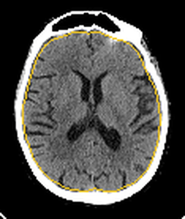
Aynı bölgenin, kaynak veri setinde patoloji içermediği belirtilen bir BT incelemesini kesit kesit kaydırın; organ sınırlarını açarak anatomiyi hasta görüntüsüyle karşılaştırın. Küçük rastlantısal bulgular tümüyle dışlanmamıştır.
Kaynak: TotalSegmentator v2.01 veri seti (Wasserthal ve ark., Radiology: Artificial Intelligence 2023), CC BY 4.0, vaka s0103; organ sınırları veri setinin otomatik segmentasyonundan, birleştirilerek sadeleştirildi. Görüntüler 2,5 mm kesit aralığına yeniden örneklendi.
Ayırıcı tanı
- Akut pontin enfarkt
- gerçek hipoattenüasyon pons içinde anatomik yerleşim gösterir ve uygun klinikte komşu kesitlerde devam eder; erken BT normal olabilir.
- Beyin sapı tümörü
- parankim genişlemesi veya kontur bozulması yapar; salt ince çizgisel bant paterni beklenmez.
- Serebellar kanama
- fokal hiperdens kan ürünü ve çevre dokuda kitle etkisi oluşturur, petroz kemikler arasındaki çizgiyle sınırlı kalmaz.
- Hareket artefaktı
- kafa konturlarında çiftlenme ve keskin kenarların tekrarı görülebilir; ışın sertleşmesi gibi kemik çiftleri arasında sabit doğrultulu kararma tipik değildir.
Tuzaklar
- Pons ortasındaki koyu bandı enfarkt sayma; görünümün kemiklerin arasından geçtiğini ve farklı seviyelerde kaybolduğunu doğrula.
- Arka çukurda BT bulguları artefakt nedeniyle belirsizse veya negatifse, klinik şüppe devam sürecekte infra-tentoryal enfarkt dışlanamaz ve BT'nin bu bölgede sınırlı olduğu kabul edilmelidir.
- Tek bir gürültülü kesitteki çizgiyi kitle sınırı kabul etme; yumuşak doku konturu, sistern açıklığı ve ardışık görüntülerde süreklilik ara.
Kendini dene
Petroz kemikler arasında ponsu çaprazlayan koyu bant üst-alt kesitlerde biçim değiştiriyor ve damar dağılımına uymuyor. En olası açıklama nedir?
Cevabı göster
Kemik kaynaklı ışın sertleşmesi/kısmi hacim artefaktı. Petroz kemikler arası bantlar ışın sertleşmesiyle birlikte kısmi hacim etkilerinden de kaynaklanabilir; farklı kesitlerde biçim değiştirmesi artefaktı destekler. Enfarkt damar alanına uyan parankimal değişiklik yapar; gliom genişleme, kanama ise fokal hiperdensite ve kitle etkisi oluşturabilir.
Arka çukurda BT/CTP değerlendirmesi yapılmış ve artefakt nedeniyle bulgu belirsiz kalmıştır; klinik şüppe devam etmektedir. Bu durumda BT bulgusunun yorumu nasıl olmalıdır?
Cevabı göster
Negatif/Belirsiz bulgu, klinik şüphe devam sürecekte infra-tentoryal enfarkti dışlamaz. S6, arka çukurda artefaktların algılanabilirliği azalttığını ve negatif/belirsiz BT/CTP bulgularının klinik şüphe devam sürecekte infra-tentoryal enfarkti dışlamayacağını belirtir.
İlgili konular
Kaynaklar
Bu sayfadaki 32 cümle ve 2 quiz sorusu aşağıdaki kaynaklardan alıntılarla tek tek karşılaştırıldı; 2 cümle bu karşılaştırmada düzeltildi (2026-10-08).
- Submandibular fossa depth in relation to the mandibular canal and alveolar crest: a retrospective cross-sectional CBCT study analyzing side-specific and sex-based variationspmc.ncbi.nlm.nih.gov
- Three-dimensional finite element analysis of temporomandibular joint stresses following botulinum toxin injection and occlusal splint therapy in bruxismpmc.ncbi.nlm.nih.gov
- Brazilian Society of Otology Task Force – bone-anchored hearing system – recommendations based on strength of evidencepmc.ncbi.nlm.nih.gov
- Magnetic resonance imaging in oral and maxillofacial surgery: a structured guide for traineespmc.ncbi.nlm.nih.gov
- Cranial anatomy, palaeoneurology, palaeobiology and stratigraphic age of the large-bodied ornithopod, Muttaburrasaurus langdoni Bartholomai and Molnar, 1981, from the mid-Cretaceous of Australiapmc.ncbi.nlm.nih.gov
- Where Does the Signal Go? Technical Challenges of CT Perfusion in Lacunar and Infratentorial Stroke.pubmed.ncbi.nlm.nih.gov
- Neuroimaging with photon-counting computed tomography: A review of clinical applications.pubmed.ncbi.nlm.nih.gov
- Tradeoffs between Radiation Exposure to the Lens of the Eyes and Diagnostic Image Quality in Pediatric Brain Computed Tomography.pubmed.ncbi.nlm.nih.gov
- CT Angiography of the Upper Extremities: Review of Acute Arterial Entities.pubmed.ncbi.nlm.nih.gov
- State of the art: noninvasive imaging and management of neurovascular trauma.pubmed.ncbi.nlm.nih.gov
- Methodological standards for body composition assessment-an expert-endorsed guide for research and clinical applications: bioimpedance, dual-energy X-ray absorptiometry, computerized tomography, and ultrasound methods.pubmed.ncbi.nlm.nih.gov
- AAPM Task Group Report 238: 3D C-arms with volumetric imaging capability.pubmed.ncbi.nlm.nih.gov
- A review on Deep Learning approaches for low-dose Computed Tomography restoration.pubmed.ncbi.nlm.nih.gov
- Mega Cisterna Magna: Current Perspectives and Future Directions-A Literature Review.pubmed.ncbi.nlm.nih.gov
- An anatomical study of the subarachnoid space surrounding the trigeminal ganglion in horses-in preparation for a controlled glycerol rhizotomy in equids.pubmed.ncbi.nlm.nih.gov
- Magnetic Resonance Imaging of Unusual Neoplasms Related to Foramen of Luschka: A Review for Differential Diagnosis.pubmed.ncbi.nlm.nih.gov
- Neuroimaging in traumatic brain imaging.pubmed.ncbi.nlm.nih.gov
- Imaging Ischemic and Hemorrhagic Disease of the Brain in Dogs.pubmed.ncbi.nlm.nih.gov
- International Harmonization of Nomenclature and Diagnostic Criteria (INHAND): Non-proliferative and Proliferative Lesions of the Non-human Primate ( M. fascicularis ).pubmed.ncbi.nlm.nih.gov
- Commentary: an eye on PET quantification.pubmed.ncbi.nlm.nih.gov
- A case of an immunoglobulin G4 related inflammatory pseudotumor involving the clivus and the nasopharynx: A challenging case of a perplexing entity with an exhaustive overview.pubmed.ncbi.nlm.nih.gov
- Laser interstitial thermal therapy in pediatric neurosurgery: a prospective nationwide study on indications, safety, and early outcomes.pubmed.ncbi.nlm.nih.gov
- Understanding Photon-Counting CT: Physics, Detector Technology, and Image Reconstructions.pubmed.ncbi.nlm.nih.gov
- A survey of transformer-based architectures in medical image analysis: models, applications, and challenges.pubmed.ncbi.nlm.nih.gov
- Imaging of Sacroiliac Pain: The Current State-of-the-Art.pubmed.ncbi.nlm.nih.gov
- High-affinity alphavbeta3 integrin targeted optical probe as a new imaging biomarker for early atherosclerosis: initial studies in Watanabe rabbits.pubmed.ncbi.nlm.nih.gov
- Anatomical and quantitative analysis of safe entry zones to the lower brainstem through the far-lateral approach.pubmed.ncbi.nlm.nih.gov
- Analysis of Pontine cavernous malformation resection based on 3D microanatomical study.pubmed.ncbi.nlm.nih.gov
- Operative Corridors in Endoscopic Skull Base Tumor Surgery.pubmed.ncbi.nlm.nih.gov
- Effect of blending Jersey and Holstein-Friesian milk on Cheddar cheese processing, composition, and quality.pubmed.ncbi.nlm.nih.gov
- Proceedings of the 15th International Congress on Circumpolar Health.pubmed.ncbi.nlm.nih.gov
- 2.5-mm articulated endoluminal forceps using a compliant mechanism.pubmed.ncbi.nlm.nih.gov
- Deep Learning-Based Versus Iterative Image Reconstruction for Unenhanced Brain CT: A Quantitative Comparison of Image Quality.pubmed.ncbi.nlm.nih.gov
- Need for Routine Brain Magnetic Resonance Imaging for Unilateral Facial Palsy in Emergency Department.pubmed.ncbi.nlm.nih.gov
- Safety and efficacy of extracranial vertebral artery stenting in elderly patients with symptomatic vertebral artery stenosis.pubmed.ncbi.nlm.nih.gov
- Continuum topological derivative - A novel application tool for segmentation of CT and MRI images.pubmed.ncbi.nlm.nih.gov
- Role of Low-Field MRI in Acute Stroke.pubmed.ncbi.nlm.nih.gov
- Multimodal imaging in neuro-ophthalmology.pubmed.ncbi.nlm.nih.gov
- Submandibular fossa depth in relation to the mandibular canal and alveolar crest: a retrospective cross-sectional CBCT study analyzing side-specific and sex-based variations.pubmed.ncbi.nlm.nih.gov
- Three-dimensional finite element analysis of temporomandibular joint stresses following botulinum toxin injection and occlusal splint therapy in bruxism.pubmed.ncbi.nlm.nih.gov
- Brazilian Society of Otology Task Force - bone-anchored hearing system - recommendations based on strength of evidence.pubmed.ncbi.nlm.nih.gov
- Cerebellum and neurodegenerative diseases: Beyond conventional magnetic resonance imaging.pubmed.ncbi.nlm.nih.gov
- Imaging Evaluation of Acute Traumatic Brain Injury.pubmed.ncbi.nlm.nih.gov
- Imaging of Neurotrauma in Acute and Chronic Settings.pubmed.ncbi.nlm.nih.gov
- Luteolin ameliorates steroid-induced osteonecrosis of the femoral head via a gut microbiota-L-Carnitine-IFIH1 axis.pubmed.ncbi.nlm.nih.gov
- Incidental elastofibroma dorsi on chest computed tomography scan: A cross-sectional study of prevalence and diagnostic oversight among radiologists.pubmed.ncbi.nlm.nih.gov
- Obesity and Cardiovascular Disease: An Obesity Medicine Association (OMA) Clinical Practice Statement (CPS) 2026.pubmed.ncbi.nlm.nih.gov
- Acute Ischemic Stroke in 2026: From Time to Penumbra-An Updated Narrative Review of Reperfusion Strategies with a Latin American Implementation Perspective.pubmed.ncbi.nlm.nih.gov
- Obesity and Skull Base Manifestations: Clinical Implications and Management.pubmed.ncbi.nlm.nih.gov
- Intentional Self-Inflicted Transcranial Screwdriver Injury: A Case Report.pubmed.ncbi.nlm.nih.gov
- Detection of phase-binning and interpolation artifacts in 4-dimensional computed tomography imaging using deep learning and rule-based approaches.pubmed.ncbi.nlm.nih.gov
- Virtual photon-counting micro-CT platform for simulation of head and neck cancer imaging in mice.pubmed.ncbi.nlm.nih.gov
- Enhancing CBCT-based CT synthesis using planning MRI in adaptive proton therapy for head cancer: A deep learning approach.pubmed.ncbi.nlm.nih.gov
- Venous-significant intracranial hemorrhage in neonatal HIE treated with therapeutic hypothermia: imaging phenotypes, diagnostic pitfalls, and prognostic implications.pubmed.ncbi.nlm.nih.gov
- Macrovascular blood flow and microvascular cerebrovascular reactivity are regionally coupled in adolescence.pubmed.ncbi.nlm.nih.gov
- From intravascular imaging to adaptive vascular care: intelligent photonics and digital twins in panvascular disease.pubmed.ncbi.nlm.nih.gov
- A high-resolution pediatric female whole-body numerical model with comparison to a male model.pubmed.ncbi.nlm.nih.gov
- In vivo mapping of the human locus coeruleus.pubmed.ncbi.nlm.nih.gov
- Twenty classic signs in neuroradiology: A pictorial essay.pubmed.ncbi.nlm.nih.gov
- The interrelationship of TMJ morphology, craniofacial structure, and airway dimensions in patients with disc displacement.pubmed.ncbi.nlm.nih.gov
- Reference Ranges of Maximum Diameter and Volume of the Fetal Large Intestine During the Second-to Third-Trimester: An MRI Study.pubmed.ncbi.nlm.nih.gov
- EACR 2025 Congress: Innovative Cancer Science, 16-19 June 2025.pubmed.ncbi.nlm.nih.gov
- Patterns of injury and violence in Yaoundé Cameroon: an analysis of hospital data.pubmed.ncbi.nlm.nih.gov
- Grid-based Methods in Relativistic Hydrodynamics and Magnetohydrodynamics.pubmed.ncbi.nlm.nih.gov
- Distinct manifestations of excitatory-inhibitory imbalance associated with amyloid-β and tau in patients with Alzheimer's disease.pubmed.ncbi.nlm.nih.gov
- Potentiation of cortico-spinal output via targeted electrical stimulation of the motor thalamus.pubmed.ncbi.nlm.nih.gov
- Monitoring the autophagy-endolysosomal system using monomeric Keima-fused MAP1LC3B.pubmed.ncbi.nlm.nih.gov
- Brazilian Society of Otology task force - Vestibular Schwannoma ‒ evaluation and treatment.pubmed.ncbi.nlm.nih.gov
- Ophthalmologic assessment and intracranial pressure in children: diagnostic methods, clinical correlations, and future directions.pubmed.ncbi.nlm.nih.gov
- Developmental venous anomalies and cerebral cavernous malformations: Partners in crime.pubmed.ncbi.nlm.nih.gov
- Calcified Prolactinoma of the Pituitary Gland: Illustrative Case Reports Highlighting Medical versus Surgical Intervention.pubmed.ncbi.nlm.nih.gov
- International Society of Paediatric Surgical Oncology (IPSO) Surgical Practice Guidelines.pubmed.ncbi.nlm.nih.gov
- Long-term safety and effectiveness of the "OptEase" vena cava filter.pubmed.ncbi.nlm.nih.gov
- Use of perfusion imaging and other imaging techniques to assess risks/benefits of acute stroke interventions.pubmed.ncbi.nlm.nih.gov
- Acute Traumatic Aortic Injury: What the Radiologist Needs to Know.pubmed.ncbi.nlm.nih.gov
- Intracranial venous thrombosis imaging by anatomical location: an educational review with mimics and diagnostic pitfalls.pubmed.ncbi.nlm.nih.gov
- Dual‑Energy CT in Inflammatory Arthritis: A Comprehensive Review.pubmed.ncbi.nlm.nih.gov
- Effect of Phantom Containing Implants Angulation on the Amount of Metal Artifacts Around the Implants in CBCT Images.pubmed.ncbi.nlm.nih.gov
- Assessment of metal artifacts from titanium wrist prostheses: photon-counting versus energy-integrating detector CT.pubmed.ncbi.nlm.nih.gov
- Cardiac CT blooming artifacts: clinical significance, root causes and potential solutions.pubmed.ncbi.nlm.nih.gov
- Effect of Additional Aluminum Filtration on the Image Quality in Cone Beam Computed Tomographic Studies of Equine Distal Limbs Using Visual Grading Characteristics Analysis: A Pilot Study.pubmed.ncbi.nlm.nih.gov
- Metal artifact reduction and contrast agent performance as a function of source spectral shape.pubmed.ncbi.nlm.nih.gov
- AI-based modality-agnostic classification system for vascular calcifications.pubmed.ncbi.nlm.nih.gov
- Multi-modality parathyroid imaging: A shifting paradigm.pubmed.ncbi.nlm.nih.gov
- Cranial anatomy, palaeoneurology, palaeobiology and stratigraphic age of the large-bodied ornithopod, <i>Muttaburrasaurus langdoni</i> Bartholomai and Molnar, 1981, from the mid-Cretaceous of Australia.pubmed.ncbi.nlm.nih.gov
- Vascular computed tomography angiography technique and indications.pubmed.ncbi.nlm.nih.gov
- Current Management and Image Review of Skull Base Chordoma: What the Radiologist Needs to Know.pubmed.ncbi.nlm.nih.gov
- Generation of an Age-Dependent and Harmonized <sup>18</sup>F-FDG Brain PET Atlas Using a High-Sensitivity Short-Axial FOV PET/CT System.pubmed.ncbi.nlm.nih.gov
- Case Report: severe <i>Mycoplasma pneumoniae</i>-associated acute disseminated encephalomyelitis in an adult: challenges in diagnosis and management.pubmed.ncbi.nlm.nih.gov
- Leptomeninges: Anatomy, Mechanisms of Disease and Neuroimaging.pubmed.ncbi.nlm.nih.gov
- Image Quality Standardization in Radiomics: A Systematic Review of Artifacts, Variability, and Feature Stability.pubmed.ncbi.nlm.nih.gov
- Pediatric Posterior Reversible Encephalopathy Syndrome: A Review With Emphasis on Neuroimaging Characteristics.pubmed.ncbi.nlm.nih.gov
- Diagnosis and treatment of Watershed strokes: a narrative review.pubmed.ncbi.nlm.nih.gov
- Neurological manifestations and neuroimaging findings in patients with SARS-CoV2-a systematic review.pubmed.ncbi.nlm.nih.gov
- Dental metal artifacts in magnetic resonance-based synthetic computed tomography for brain radiotherapy: Impact on dose, patient setup, and geometric distortion.pubmed.ncbi.nlm.nih.gov
- Multi-fidelity Monte Carlo fusion for BNCT dose calculation using a dual-branch network.pubmed.ncbi.nlm.nih.gov
- Factors requiring adaptive re-planning in carbon ion radiotherapy for head and neck cancers.pubmed.ncbi.nlm.nih.gov
- Differentiating pulmonary embolism from contrast accumulation-induced beam hardening artifacts using dual-energy CT quantitative parameters.pubmed.ncbi.nlm.nih.gov
- CT, MRI, and FDG PET/CT in the Assessment of Lymph Node Involvement in Pediatric Hodgkin Lymphoma: An Expert Consensus Definition by an International Collaboration on Staging Evaluation and Response Criteria Harmonization for Children, Adolescent, and Young Adult Hodgkin Lymphoma (SEARCH for CAYAHL).pubmed.ncbi.nlm.nih.gov
- Development of a magnetic resonance imaging protocol to visualize encapsulated contrast agent markers in prostate brachytherapy recipients: initial patient experience.pubmed.ncbi.nlm.nih.gov
- Quantitative assessment of carotid plaque morphology (geometry and tissue composition) using computed tomography angiography.pubmed.ncbi.nlm.nih.gov
- Comprehensive review on rhino-neurosurgery.pubmed.ncbi.nlm.nih.gov
- CT and MR imaging findings of systemic complications occurring during pregnancy and puerperal period, adversely affected by natural changes.pubmed.ncbi.nlm.nih.gov
- Slice-Level Deep Learning Classification of Acute Cholecystitis on Contrast-Enhanced CT: A Single-Center Benchmark of Six CNN Architectures.pubmed.ncbi.nlm.nih.gov
- Phantom-based assessment of a modified 4D-MRI protocol for abdominal tumor motion tracking: Effects of sequence parameters and modality comparison.pubmed.ncbi.nlm.nih.gov
Eğitim amaçlıdır; hasta başında tanı ve tedavi kararının yerine geçmez. İçerik yapay zekâ destekli bir süreçle güncel literatüre göre cümle cümle kaynaklarla karşılaştırıldı, ancak uzman radyolog incelemesinden geçmedi. Klinik kararlarda güncel kılavuzları, kurum protokollerini ve radyoloji raporunu esas alın.