Özet
Çocukta litik kemik odağı gelişimsel lezyon, enfeksiyon veya iyi ya da kötü huylu tümör olabilir; yaş ve kemik içindeki yerleşim ayırıcı tanıyı değiştirir. Şüpheli fokal kemik lezyonunda ilk inceleme direkt grafidir. BT korteks bütünlüğünü, periostal kemiği ve mineralize matriksi ayrıntılandırır; kemik iliği ve yumuşak doku yayılımında MR tamamlayıcıdır. Geçiş zonu, anatomik merkez ve korteks dışı bileşen birlikte tanımlanmalı, görüntü paterni histolojik tanı yerine kullanılmamalıdır. Agresif enfeksiyonu tümör ya da maligniteyi enfeksiyon sanmak tanı ve evrelemeyi geciktirebilir.
Faz ve pencere
- Kemik algoritması tanısal
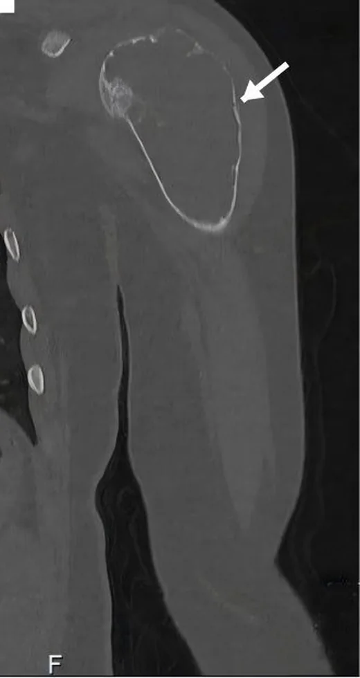
- Kontrastsız ince kesit kemik rekonstrüksiyonunda litik odağın medüller, kortikal ya da yüzey merkezini; geçiş zonunu, kortikal incelme veya kesintiyi, periostal yeni kemiği ve osteoid ya da kondroid matriks mineralizasyonunu gösterir.
Önerilen pencereler: Kemik (G 1800 / M 400), Yumuşak doku (G 400 / M 40).
BT bulguları
- Dar ve keskin geçiş zonu, özellikle çevresinde sklerotik kenar varsa daha yavaş büyüyen coğrafi litik paterni tarif eder; benignlik kanıtı değildir.
- Geniş, belirsiz geçiş zonuna eşlik eden güve yeniği ya da permeatif yıkım, agresif kemik davranışını gösterir; enfeksiyon ve malignite örtüşebilir.
- Metafiz, diafiz, epifiz, medüller kanal, korteks ve kemik yüzeyinden hangisinin odağın merkezi olduğu ayırıcı tanıyı daraltır.
- Kortikal incelme ve dışa bombeleşme ekspansiyonu; kortikal kesinti ise lezyonun kemik kabuğunu aşmış olabileceğini gösterir.
- Periostal yanıtın türü ayırıcı tanıyı daraltmaya yardımcı olabilir.
- Matriks mineralizasyonunun biçimi ayırıcı tanıyı daraltmaya yardımcı olabilir.
- Korteksi aşan solid yumuşak doku, özellikle agresif kemik yıkımıyla komşuysa lokal tümör yayılımı açısından kayda değerdir.
- Uzun kemiklerde litik odağın medüller, kortikal veya yüzey merkezini belirlemek; geçiş zonunu, kortikal bütünlüğü ve periostal yanıtı değerlendirmek ayırıcı tanıyı daraltır.
Ölçütler ve sınıflamalar
- Lodwick-Madewell paterni ve önerilen Caracciolo revizyonu
- Lodwick-Madewell sınıflaması: I coğrafi yıkım; IA sklerotik kenarlı, IB keskin sınırlı fakat sklerotik kenarsız, IC belirsiz kenarlı ve geniş geçiş zonlu; II güve yeniği; III permeatif paterndir. Caracciolo'nun revizyonunda IC ile II birleştirilir; tip III A seri görüntülerde sınır değişikliği veya ilerleyen endosteal kazıma, B güve yeniği ve permeatif desenler, C radyografik olarak gizli olarak sınıflandırılır. Bu radyografik paternler agresifliği tarif eder; histolojik tanı yerine geçmez.
Normalde
Kemik lezyonu değerlendirilirken yerleşim, sınır ve yoğunluk özellikleri tanımlanmalıdır. Kemik lezyonunun yerleşimi ve sınırları, ayırıcı tanıyı daraltmaya yardımcı olur.
Normal BT ile kıyasla
Aynı bölgenin, kaynak veri setinde patoloji içermediği belirtilen bir BT incelemesini kesit kesit kaydırın; organ sınırlarını açarak anatomiyi hasta görüntüsüyle karşılaştırın. Küçük rastlantısal bulgular tümüyle dışlanmamıştır.
Kaynak: TotalSegmentator v2.01 veri seti (Wasserthal ve ark., Radiology: Artificial Intelligence 2023), CC BY 4.0, vaka s0685; organ sınırları veri setinin otomatik segmentasyonundan, birleştirilerek sadeleştirildi. Görüntüler 2,5 mm kesit aralığına yeniden örneklendi.
Ayırıcı tanı
- Osteomiyelit litik kemik lezyonlarının ayırıcı tanısında yer alır.
- Ewing sarkomu
- diafizer permeatif yıkım, lameller periostal yanıt ve solid ekstraosseöz kitle birlikte görülebilir; bu bulgular enfeksiyondan kesin ayrım sağlamaz.
- Osteosarkom
- çoğunlukla metafizer yerleşim, agresif kortikal değişiklik ve bulutsu osteoid mineralizasyonu görülebilir.
- Langerhans hücreli histiyositoz
- çocuklarda çoklu litik kemik lezyonlarına yol açabilir; eşlik eden bulgular ve diğer kemik odaklarının varlığı ayırıcı tanıda yardımcı olur.
- Basit kemik kisti
- uzun kemiğin merkezî medüllasında, çoğunlukla iyi sınırlı ekspansil litik lezyon oluşturur; agresif periostal kılıf veya solid dış kitle tipik değildir.
- Anevrizmal kemik kisti
- ekspansil, septalı litik lezyon görünümü verebilir; sıvı-sıvı seviyelerini BT güvenilir biçimde karakterize edemez, MR daha uygundur.
Tuzaklar
- Geniş geçiş zonu enfeksiyonda da görülebilir; ateş veya inflamasyon bulgusu eşlik etse bile agresif tümörü tek başına dışlamaz.
- Açık fizis, apofiz ve gelişen ossifikasyon merkezleri düzensiz görünebilir; gerçek litik odağın çevresinde medüller trabekül kaybı ve karşı tarafla fark aranır.
- Matriks mineralizasyonunun görülmemesi tümörü dışlamaz; küçük veya az mineralize lezyonlarda BT’de matriks belirgin olmayabilir.
- Periostal yanıtın katmanlı ya da ışınsal olması tümör türünü tek başına adlandırmaz; kırık, enfeksiyon ve tümör benzer yanıt oluşturabilir.
Kendini dene
Adölesanda distal femur metafizinde agresif litik kitle ve osteoid matriks mineralizasyonu var. En olası tanı hangisidir?
Cevabı göster
Osteosarkom. S1'e göre osteosarkom metafizer yerleşimli ve osteoid matriks üreten klasik bir tümördür. Ewing sarkomu ise sıklıkla diafizleri tutar. Anevrizmal kemik kisti ve osteomiyelit osteoid matriks üretmez.
Diafizer litik lezyonda permeatif yıkım ve katmanlı periostal yanıt var. Hangi tümör öncelikle düşünülür?
Cevabı göster
Ewing sarkomu. S1'e göre Ewing sarkomu sıklıkla diafizleri tutar ve permeatif/güve yenmiş yıkım ile periostal reaksiyon yapar. Bu bulgular enfeksiyonla örtüşebilir ancak malignite açısından Ewing sarkomu akla gelir.
İlgili konular
Kaynaklar
Bu sayfadaki 45 cümle ve 2 quiz sorusu aşağıdaki kaynaklardan alıntılarla tek tek karşılaştırıldı; 12 cümle bu karşılaştırmada düzeltildi (2026-10-08).
- Conventional radiography for the assessment of focal bone lesions of the appendicular skeleton: fundamental concepts in the modern imaging erapmc.ncbi.nlm.nih.gov
- Proximal Tibial Giant Cell Tumor in a Skeletally Immature Child: A Diagnostic Dilemma and Review of the Literaturepmc.ncbi.nlm.nih.gov
- Recurrent Mandibular Ewing sarcoma in an 11-year-old boy: a rare pediatric case and review of the literaturepmc.ncbi.nlm.nih.gov
- Skeletal coccidioidomycosis: an atypical pediatric presentationpmc.ncbi.nlm.nih.gov
- Transphyseal Proximal Humeral Aneurysmal Bone Cyst with Pathologic Fracture in a Child: A Case Reportpmc.ncbi.nlm.nih.gov
- Metastatic pulmonary dissemination as differential diagnosis of COVID-19 disease.pubmed.ncbi.nlm.nih.gov
- Multifocal Osseous Tuberculosis Mimicking Langerhans' Cell Histiocytosis: A Case Series.pubmed.ncbi.nlm.nih.gov
- Desmoplastic fibroma of the pediatric cranium: case report and review of the literature.pubmed.ncbi.nlm.nih.gov
- Imaging of pediatric skull lytic lesions: A review.pubmed.ncbi.nlm.nih.gov
- Transphyseal Proximal Humeral Aneurysmal Bone Cyst with Pathologic Fracture in a Child: A Case Report.pubmed.ncbi.nlm.nih.gov
- Solitary Diaphyseal Langerhans Cell Histiocytosis of Femur in an Infant - A Case Report.pubmed.ncbi.nlm.nih.gov
- Proximal Tibial Giant Cell Tumor in a Skeletally Immature Child: A Diagnostic Dilemma and Review of the Literature.pubmed.ncbi.nlm.nih.gov
- The solitary skull mass: An atypical presentation of anaplastic large cell lymphoma.pubmed.ncbi.nlm.nih.gov
- Intravascular papillary endothelial hyperplasia presenting as a lytic frontal bone lesion in a pediatric male.pubmed.ncbi.nlm.nih.gov
- Distal tibial tuberculous osteitis in a child: a case report.pubmed.ncbi.nlm.nih.gov
- Bilateral Knee Osteonecrosis in an Adolescent With Leukemia on Prolonged Corticosteroids: Discordant Femoral-Tibial Findings on Bone Scan SPECT-CT.pubmed.ncbi.nlm.nih.gov
- Atypical Case of Presumed <i>Bartonella henselae</i> Osteomyelitis in a Child with Cat-Scratch Disease Mimicking a Malignant Bone Tumor.pubmed.ncbi.nlm.nih.gov
- hNET-targeted [<sup>18</sup>F]MFBG PET as baseline functional imaging in newly diagnosed neuroblastoma.pubmed.ncbi.nlm.nih.gov
- Whole-Body MRI Versus <sup>18</sup>F-FDG-PET/CT for the Detection of Nodal and Distant Metastases in Pediatric Patients with Solid Tumors.pubmed.ncbi.nlm.nih.gov
- Screw Versus Suture Bridge Fixation for Medial Femoral Condyle Osteochondritis Dissecans Demonstrate Comparable Healing Rates.pubmed.ncbi.nlm.nih.gov
- Ultra-High-Frequency Ultrasound of Oral Cavity Lesions: A Retrospective Pictorial Study with Histopathologic and Clinical Correlation.pubmed.ncbi.nlm.nih.gov
- Two-Stage Conservative Management of Mandibular Unicystic Ameloblastoma with an Intraluminal Pattern: A Case Report.pubmed.ncbi.nlm.nih.gov
- A Huge Intraosseous Hemangioma in the Maxillary Sinus Misdiagnosed as Osteosarcoma: A Case Report.pubmed.ncbi.nlm.nih.gov
- Skeletal coccidioidomycosis: an atypical pediatric presentation.pubmed.ncbi.nlm.nih.gov
- Oral Carcinoma Cuniculatum: Diagnostic Challenges and Emerging Perspectives-Case Report.pubmed.ncbi.nlm.nih.gov
- The quiet flame: whole-body MRI and the underestimated burden of pediatric chronic non-bacterial osteomyelitis (CNO).pubmed.ncbi.nlm.nih.gov
- Where Does the Signal Go? Technical Challenges of CT Perfusion in Lacunar and Infratentorial Stroke.pubmed.ncbi.nlm.nih.gov
- Case Report: Synchronous lingual osseous choristoma and suprahyoid thyroglossal duct cyst: insights into the embryological thyroglossal-hyoid axis.pubmed.ncbi.nlm.nih.gov
- A Rare Case of Giant Cell tumor of Bone in Distal Tibia.pubmed.ncbi.nlm.nih.gov
- Clinicoradiological Outcomes after Percutaneous Radiofrequency Ablation of Benign Bone Tumors: A Prospective Observational Study.pubmed.ncbi.nlm.nih.gov
- High-Grade Undifferentiated Pleomorphic Sarcoma of the Distal Tibia Presenting as a Radiographically Benign-Appearing Lesion: A Case Report and Review of the Literature.pubmed.ncbi.nlm.nih.gov
- Giant neglected cutaneous squamous cell carcinoma of the scalp with full-thickness calvarial erosion and early fatal outcome: CT findings and treatment-planning implications.pubmed.ncbi.nlm.nih.gov
- Primary osseous kaposiform hemangioendothelioma confined to long bones: a retrospective imaging analysis of 12 pediatric cases.pubmed.ncbi.nlm.nih.gov
- Musculoskeletal Manifestations of T-Cell Lymphomas: Clinical Presentation, Imaging Features, Pathobiology, and Orthopedic Considerations-A Narrative Review.pubmed.ncbi.nlm.nih.gov
- Unicameral Bone Cyst: Diagnosis, Treatment, and Follow-Up Methods, a Multicenter Study.pubmed.ncbi.nlm.nih.gov
- Chondrosarcoma arising from long-standing Dysplasia Epiphysealis Hemimelica of the proximal humerus: A case report.pubmed.ncbi.nlm.nih.gov
- Giant expansile humeral lesion with soap-bubble appearance mimicking a primary bone tumor: Multiple myeloma as the final diagnosis.pubmed.ncbi.nlm.nih.gov
- Radiographic evolution of a dentigerous cyst associated with a deeply impacted mandibular first molar in a pediatric patient: a CBCT-based longitudinal case report with minimal-visit orthodontic management.pubmed.ncbi.nlm.nih.gov
- Low-grade osteogenic tumor with secondary aneurysmal bone cyst: Distinguishing osteoblastoma from low-grade osteosarcoma in the setting of noncanonical molecular findings.pubmed.ncbi.nlm.nih.gov
- [Primary bone tumors of the spine].pubmed.ncbi.nlm.nih.gov
- Computed Tomographic Findings of Primary Hepatic Extraskeletal Osteosarcoma With Peritoneal Metastasis via Periportal Space in a Dog.pubmed.ncbi.nlm.nih.gov
- Beyond Chordoma: A Comprehensive Review of Sacral Lesions.pubmed.ncbi.nlm.nih.gov
- Secondary distal femoral osteosarcoma in a non-irradiated survivor of bilateral retinoblastoma.pubmed.ncbi.nlm.nih.gov
- Bilateral nasolabial cysts: MRI findings in a rare case report.pubmed.ncbi.nlm.nih.gov
- Purely osteolytic bone lesions in the early osteoarticular phase of synovitis, acne, pustulosis, hyperostosis and osteitis syndrome.pubmed.ncbi.nlm.nih.gov
- Chronic Recurrent Multifocal Osteomyelitis in a Pediatric Patient: An Uncommon Case With Diagnostic Challenge.pubmed.ncbi.nlm.nih.gov
- Anatomy-driven automated subsegmentation of pelvic and proximal femoral CT into clinically relevant subregions and landmarks.pubmed.ncbi.nlm.nih.gov
- Rare Cause of Congenital Unilateral Facial Palsy: High-Resolution Computed Tomography (HRCT) Findings of Isolated Facial Canal Hypoplasia and Aplasia.pubmed.ncbi.nlm.nih.gov
- Kinematic Alignment in TKA Using the Contralateral Healthy Knee as Reference: A Combined Finite Element and Clinical Analysis.pubmed.ncbi.nlm.nih.gov
- Pediatric Distal Femoral Surface Tumor With Extraosseous Lesion Mimicking Parosteal Osteosarcoma: A Diagnostic Challenge.pubmed.ncbi.nlm.nih.gov
- Arthroscopic Management of Patellar Instability in Skeletally Immature Patients: Current Concepts and Future Directions.pubmed.ncbi.nlm.nih.gov
- Indications and Timing of Guided Growth Techniques for Pediatric Upper Extremity Deformities: A Literature Review.pubmed.ncbi.nlm.nih.gov
- Conventional radiography for the assessment of focal bone lesions of the appendicular skeleton: fundamental concepts in the modern imaging era.pubmed.ncbi.nlm.nih.gov
- Salmonella Typhi Osteomyelitis of the Ulnar Diaphysis in an Immunocompetent Adult: A Case Report.pubmed.ncbi.nlm.nih.gov
- Candida Femoral Osteomyelitis in a Young Adult Without Known Risk Factors for Immunosuppression: A Case Report.pubmed.ncbi.nlm.nih.gov
- A Case Report of Acute on Chronic Osteomyelitis of Distal Femur Managed with Sequestrectomy, Saucerization, and Stimulan Placement.pubmed.ncbi.nlm.nih.gov
- Case Report: CBFA2T3::GLIS2-positive myeloid sarcoma with focal bone marrow involvement mimicking Ewing sarcoma in an infant.pubmed.ncbi.nlm.nih.gov
- Primary Mediastinal Osteosarcoma: A Rare and Aggressive Tumour in an Unusual Location.pubmed.ncbi.nlm.nih.gov
- Low-grade Central Osteosarcoma with High-grade Transformation in the Distal Tibia: A Rare Case Report.pubmed.ncbi.nlm.nih.gov
- Hybrid Prosthetic and Biological Reconstruction Using Extracorporeal Radiotherapy for Long-Segment Humeral Osteosarcoma: A Case Report.pubmed.ncbi.nlm.nih.gov
- Contrast-enhanced sonography in the diagnosis of Langerhans cell hyperplasia in children with isolated liver involvement: A case report and review of the literature.pubmed.ncbi.nlm.nih.gov
- An Unusual Presentation of Multisystem Langerhans Cell Histiocytosis in a Child: A Case Report.pubmed.ncbi.nlm.nih.gov
- Enigmatic Case of Adult-Onset Langerhans Cell Histiocytosis with Aggressive Bony Involvement.pubmed.ncbi.nlm.nih.gov
- How the anatomic location affects the surgical outcomes of simple bone cysts in children: a single-center retrospective cohort study.pubmed.ncbi.nlm.nih.gov
- Unveiling a Silent Bone Lesion: Pathological Fracture in a Child With Non-Ossifying Fibroma.pubmed.ncbi.nlm.nih.gov
- Imaging features and differential diagnosis of benign osseous cystic lesions: Aneurysmal bone cyst versus femoral neck herniation pit.pubmed.ncbi.nlm.nih.gov
- Use of Fibrin Sealant Combined With Demineralized Freeze-Dried Bone Allograft in the Management of a Recurrent Mandibular Aneurysmal Bone Cyst: A Case Report.pubmed.ncbi.nlm.nih.gov
- Primary denosumab therapy for a three-column pediatric cervical aneurysmal bone cyst: illustrative case.pubmed.ncbi.nlm.nih.gov
- Osteitis Pubis in Athletes in the Era of Pubic-Related Groin Pain: Contemporary Concepts in Diagnosis, Rehabilitation, and Surgical Decision-Making.pubmed.ncbi.nlm.nih.gov
- The Role of Sex Hormone Receptors in the Squamous Cell Carcinoma of the Uterine Ectocervix: A Review and Future Directions.pubmed.ncbi.nlm.nih.gov
- Engineering high-fidelity bone organoids: Operational classification, multilineage crosstalk, biofabrication evidence, and translational validation.pubmed.ncbi.nlm.nih.gov
- Multimodal AI fusion: integrating MRI with PET/CT, histopathology, and liquid biopsy for bone tumor diagnosis.pubmed.ncbi.nlm.nih.gov
- Phosphaturic mesenchymal tumor in the temporalis muscle causing tumor-induced osteomalacia: a case report and literature review.pubmed.ncbi.nlm.nih.gov
- Moth-Eaten Osteolysis of Multiple Foot Bones After a Conservatively Treated "Stable" Ankle Fracture: A Case Report and Literature Review.pubmed.ncbi.nlm.nih.gov
- Case Report: Arthroscopic management of a giant Baker cyst containing numerous rice body-like loose bodies in a patient with rheumatoid arthritis.pubmed.ncbi.nlm.nih.gov
- Management of Tibial Adamantinoma: Balancing Oncologic Cure and Reconstructive Longevity.pubmed.ncbi.nlm.nih.gov
- Conservative Treatment of Pediatric Fractures-Narrative Review of Acceptable Deformity and Remodeling Potential.pubmed.ncbi.nlm.nih.gov
- Computed Tomographic Lesion Descriptors in Foals With Hematogenous Osteomyelitis: Interobserver Agreement and Association With Outcome.pubmed.ncbi.nlm.nih.gov
- The Vital Illusion: Cone-Beam Computed Tomography (CBCT)-Tracked Evolution of Periapical Cemento-Osseous Dysplasia.pubmed.ncbi.nlm.nih.gov
- Understanding Diagnostic Challenges in Ewing Sarcoma - A Report of Two Cases.pubmed.ncbi.nlm.nih.gov
- Cortical Window Osteotomy for Diagnostic Biopsy in Iliac Osteomyelitis: A Case Report and Technical Note.pubmed.ncbi.nlm.nih.gov
- Proximal Phalanx Enchondroma in a Pediatric Patient: Curettage, Liquid Nitrogen Cryotherapy, Iliac Crest Autograft, and Microfragment Plate Fixation.pubmed.ncbi.nlm.nih.gov
- Posterior Femoral Neck Exostosis in a Young Adult: Open Resection and Prophylactic Dynamic Hip Screw Fixation - A Case Report.pubmed.ncbi.nlm.nih.gov
- When plasmacytoma defies expectations: An unusual maxillary presentation with mixed imaging features in a young patient.pubmed.ncbi.nlm.nih.gov
- Periostitis of the Pubic Symphysis in Inguinal and Femoral Hernia: An Uninterpreted Source of Chronic Groin Pain.pubmed.ncbi.nlm.nih.gov
- Focal periphyseal edema with atypical MRI features: Biopsy findings and imaging follow-up.pubmed.ncbi.nlm.nih.gov
- A Novel 3D-Printed Strut-Type Prosthesis Reconstructs the Hemicortical Defect After Distal Femur Parosteal Osteosarcoma Resection: The Mid-Term Follow-Up Outcome.pubmed.ncbi.nlm.nih.gov
- Cryptococcal Osteomyelitis in a Virally Suppressed Adolescent Living With HIV: A Case Report.pubmed.ncbi.nlm.nih.gov
- Treatment trends of benign bone lesions in a suburban New York healthcare system.pubmed.ncbi.nlm.nih.gov
- Diffusion weighted imaging of extremity bone tumors-inter-reader analysis and incremental value over conventional MR imaging.pubmed.ncbi.nlm.nih.gov
- Systematic approach to musculoskeletal benign tumors.pubmed.ncbi.nlm.nih.gov
- Management and Outcomes of Osteochondritis Dissecans of the Knee in the Pediatric and Adolescent Population.pubmed.ncbi.nlm.nih.gov
- Bibliometric Analysis of Whole-Body MRI from 2015 to 2025 Across Clinical Applications, Quantitative Imaging, and Artificial Intelligence.pubmed.ncbi.nlm.nih.gov
- Recurrent Mandibular Ewing sarcoma in an 11-year-old boy: a rare pediatric case and review of the literature.pubmed.ncbi.nlm.nih.gov
- Multi-detector computed tomography in evaluating locally aggressive and malignant bone tumours.pubmed.ncbi.nlm.nih.gov
- A Rare Occurrence of Chronic Recurrent Multifocal Osteomyelitis in Clavicle: Case Report.pubmed.ncbi.nlm.nih.gov
- Unveiling Primary Bone Tumors of the Spine: A Review of Essential Imaging Clues.pubmed.ncbi.nlm.nih.gov
- Sclerotherapy for Aneurysmal Bone Cyst in Canine.pubmed.ncbi.nlm.nih.gov
- Multidisciplinary approach to the accurate diagnosis of parosteal osteosarcoma.pubmed.ncbi.nlm.nih.gov
- Multimodal Imaging of Osteosarcoma: From First Diagnosis to Radiomics.pubmed.ncbi.nlm.nih.gov
- From Static Scaffolds to Responsive Implants: 3D-Printed Field-Active Bioceramics for Adaptive Bone Regeneration.pubmed.ncbi.nlm.nih.gov
- Photo-Responsive In Situ Forming Hydrogels for Drug Delivery: A Critical Review of Polymer Matrices, Photoinitiators, and the Gap Towards Clinical Translation.pubmed.ncbi.nlm.nih.gov
- Synchronizing degradation with regeneration: a model-driven framework for designing biodegradable biomaterials in bone tissue engineering.pubmed.ncbi.nlm.nih.gov
- Bone and joint quality remains impaired in long-term controlled acromegaly: an HR-pQCT study.pubmed.ncbi.nlm.nih.gov
- Assessment of Radiographic Image Texture in the Maxilla and Mandible Around Titanium Inserts Used for Osteosynthesis of Dentofacial Deformities.pubmed.ncbi.nlm.nih.gov
- Axial Spondyloarthritis: Mimics and Pitfalls of Imaging Assessment.pubmed.ncbi.nlm.nih.gov
- Chitosan and Chitin-Derived Biomaterials in Orthopedics: A Structured Narrative Review of Polymer Design, Quantitative Performance, and Clinical Translation.pubmed.ncbi.nlm.nih.gov
- Phylogeography of Bone Metastasis: Clonal Evolution, Skeletal Niche Adaptation, and Clinical Implications.pubmed.ncbi.nlm.nih.gov
- Beyond Cartilage Regeneration: Targeting the Subchondral Bone with Extracorporeal Shock Wave Therapy and Regenerative Medicine in Knee Osteoarthritis.pubmed.ncbi.nlm.nih.gov
- Diagnostic Pitfalls of Dental Follicles and Cyst-like Lesions in Juvenile Patients: An Early Odontogenic Myxoma Mimicking a Follicular Cyst.pubmed.ncbi.nlm.nih.gov
- Tumors of the Posterior Elements of the Spine in Children-an Overview.pubmed.ncbi.nlm.nih.gov
- RADS classification systems for bone tumors: current status and where do we go from here?pubmed.ncbi.nlm.nih.gov
- Squamous Cell Carcinoma Arising from Chronic Osteomyelitis: A Single-Center Case Series of Six Patients.pubmed.ncbi.nlm.nih.gov
- Osteomalacia Following Iron Infusion Therapy in a Patient With Rendu-Osler-Weber Syndrome: F-18-FDG PET/CT Discrimination of a Stress Fracture in the Setting of a Musculoskeletal Tumor Mimic.pubmed.ncbi.nlm.nih.gov
- Calcified Chondroid Mesenchymal Neoplasm of the Temporomandibular Joint: A Rare and Recently Described Benign Tumor.pubmed.ncbi.nlm.nih.gov
Eğitim amaçlıdır; hasta başında tanı ve tedavi kararının yerine geçmez. İçerik yapay zekâ destekli bir süreçle güncel literatüre göre cümle cümle kaynaklarla karşılaştırıldı, ancak uzman radyolog incelemesinden geçmedi. Klinik kararlarda güncel kılavuzları, kurum protokollerini ve radyoloji raporunu esas alın.