Özet
Çocuklarda LCH kemikte tek veya çok odaklı tutulum yapabilir; iskelet lezyonları kafatası, pelvis, omurga ve uzun kemiklerde görülebilir. BT kemik yıkımını gösterir; MRI kemik iliği tutulumunu, yumuşak doku uzanımını ve nöral basıyı değerlendirmede tamamlayıcıdır. Beveled-edge görünümü LCH'yi düşündürebilir ancak tek başına tanı koydurmaz; kemik LCH'si osteomiyelit ve Ewing sarkomu gibi durumları taklit edebilir. Kemik LCH'sinde ağırlık taşıyan kemikte patolojik kırık riski veya lezyonun neden olduğu nörolojik bası bulunabilir; kapsamlı evreleme tek odaklı hastalığı multisistem hastalıktan ayırmaya yardımcı olur.
Faz ve pencere
- Kontrastsız tanısal
- Kontrastsız kemik pencereli BT'de litik kemik yıkımı, kafatasında beveled-edge görünümü ve vertebra korpus çökmesi değerlendirilebilir.
- Kemik algoritması tanısal
- BT kemik yıkımını ve vertebra korpus çökmesini değerlendirmede; MRI ise lezyonun yumuşak doku bileşeni ve lokal uzanımını göstermede tamamlayıcıdır.
Önerilen pencereler: Kemik (G 1800 / M 400), Yumuşak doku (G 400 / M 40).
BT bulguları
- Litik kemik odağı — tek veya çoklu, yuvarlak/oval, iyi ya da silik sınırlı osteolitik destrüksiyon.
- Beveled-edge görünümü — kafatasında iç ve dış tabula farklı derecede yıkıldığı için lezyon kenarı eğimli görünür.
- Kafatasında çoklu litik odaklar sık görülür; bu odaklar genellikle kötü sınırlıdır ve yaygın kalvaryal tutulum oluşturabilir.
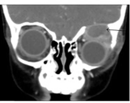
- Kortikal destrüksiyon ve yumuşak doku — özellikle uzun kemik veya temporal kemikte kortikal kesintiyle komşu kitle eşlik edebilir.
- Periostal reaksiyon — tek katmanlı veya buttress biçimli yeni kemik oluşumu görülebilir.
- Vertebra plana — vertebra korpusu ileri derecede çökerken komşu disk aralıkları korunabilir.
- Çoklu odak — mevcut BT görüş alanındaki diğer kemik odaklarını inceleyin; bölgesel BT başka alanlardaki hastalığı dışlamaz. İskelet yaygınlığı uygun evreleme yöntemiyle ayrıca belirlenir.
Normalde
Normal kalvaryumda iç ve dış tabula kesintisiz, diploik aralık düzenlidir; vertebra korpusu yüksekliği ve korteksi korunur, uzun kemik medüller trabekülleri yıkılmış odak içermez. Lezyon düzeyini karşı tabula, komşu kemik ve aynı incelemedeki diğer iskelet odaklarıyla kıyaslayarak yıkımın yayılımını, kortikal kırılmayı ve eşlikçi yumuşak dokuyu belirleyin.
Normal BT ile kıyasla
Aynı bölgenin, kaynak veri setinde patoloji içermediği belirtilen bir BT incelemesini kesit kesit kaydırın; organ sınırlarını açarak anatomiyi hasta görüntüsüyle karşılaştırın. Küçük rastlantısal bulgular tümüyle dışlanmamıştır.
Kaynak: TotalSegmentator v2.01 veri seti (Wasserthal ve ark., Radiology: Artificial Intelligence 2023), CC BY 4.0, vaka s0103; organ sınırları veri setinin otomatik segmentasyonundan, birleştirilerek sadeleştirildi. Görüntüler 2,5 mm kesit aralığına yeniden örneklendi.
Ayırıcı tanı
- Osteomiyelit
- kortikal destrüksiyon ve yumuşak doku eşlik edebilir; sekestrum özgül değildir, klinik enfeksiyon bulguları ve MRI'da apse ayırıcıdır.
- Ewing sarkomu
- uzun kemikte agresif permeatif yıkım ve lameller/ışınsal periostal reaksiyon, yumuşak doku kitlesiyle birlikte olabilir.
- Dermoid veya epidermoid kist
- özellikle orta hat yerleşiminde kemik remodeling/defekti ve içeriğe bağlı yağ ya da sıvı dansitesi görülür.
- Anevrizmal kemik kisti
- ekspansil litik lezyon ve kortikal incelme yapabilir; BT tek başına LCH'yi kesinleştirmez, MRI'daki sıvı-sıvı seviyeleri destekleyebilir.
Tuzaklar
- Beveled-edge ve düğme sekestrumu patognomonik değildir; osteomiyelit de sekestrum yapabilir ve tanı klinik, diğer görüntüleme ve gerektiğinde doku örneklemesiyle bütünlenir.
- Kafatası lezyonunun yumuşak doku bileşeni ve çevre yumuşak dokuyla ilişkisi MRI ile değerlendirilebilir.
- Vertebra plana LCH düşündürse de enfeksiyon, lösemi ve diğer tümörler benzer çökme yapabilir; posterior eleman ve epidural uzanım için MRI gerekir.
Kendini dene
Çocukta parietal kemikte iç ve dış tabulayı farklı derecede yıkan litik odak var. Hangi görünüm LCH'yi destekler?
Cevabı göster
Beveled-edge. Tabulaların asimetrik yıkımı LCH'de beveled-edge görünümü oluşturabilir. Fibröz displazi ve LCH, kraniofasiyal lezyonların ayırıcısında karşılaştırılan önemli fibro-osseus patolojilerdir.
Litik kalvaryal lezyon içinde küçük korunmuş kemik parçası görülüyor. Bu bulgu için hangi ifade doğrudur?
Cevabı göster
Düğme sekestrumu tek başına LCH tanısını doğrulamaz. Kaynak, düğme sekestrumunun özellikle endemik bölgelerde multifokal kemik tüberkülozuna işaret edebileceğini bildirir; bu nedenle bulgu tek başına LCH tanısını doğrulamaz.
Kaynaklar
Bu sayfadaki 35 cümle ve 2 quiz sorusu aşağıdaki kaynaklardan alıntılarla otomatik olarak karşılaştırıldı; 14 cümle bu karşılaştırmada düzeltildi (2026-10-09).
- What to do to beat Langerhans cell histiocytosis of bone? A narrative review and case series of radiofrequency ablationpmc.ncbi.nlm.nih.gov
- Dabrafenib or trametinib in pediatric Langerhans cell histiocytosis with bone lesions: a retrospective efficacy and safety studypmc.ncbi.nlm.nih.gov
- Intravenous zoledronate for pediatric langerhans cell histiocytosis with bone involvementpmc.ncbi.nlm.nih.gov
- Adult-Onset Multisystem Langerhans Cell Histiocytosis With Bone and Hepatic Involvement: A Rare Case With Radiologic-Pathologic Correlationpmc.ncbi.nlm.nih.gov
- Scurvy, abnormal MRI, and gelatinous bone marrow in an adolescent with avoidant restrictive food intake disorderpmc.ncbi.nlm.nih.gov
- Langerhans Cell Histiocytosis of Bone:Report of Eight Cases and Review of the Literature.pubmed.ncbi.nlm.nih.gov
- Erdheim-Chester disease.pubmed.ncbi.nlm.nih.gov
- [Skeletal sarcoidosis].pubmed.ncbi.nlm.nih.gov
- Rosai-Dorfman-Destombes (RDD) disease presenting as palindromic rheumatism.pubmed.ncbi.nlm.nih.gov
- Resection of benign vertebral tumors by minimally invasive techniques.pubmed.ncbi.nlm.nih.gov
- Hematopoietic tumors and metastases involving bone.pubmed.ncbi.nlm.nih.gov
- Computed tomography-based navigation system-assisted surgery for primary spine tumor.pubmed.ncbi.nlm.nih.gov
- Clinical application of "black bone" imaging in paediatric craniofacial disorders.pubmed.ncbi.nlm.nih.gov
- 18F-FDG PET/CT in Xanthoma Disseminatum.pubmed.ncbi.nlm.nih.gov
- FDG-PET/CT in pediatric solid tumors.pubmed.ncbi.nlm.nih.gov
- The histiocytosis Erdheim-Chester disease is an inflammatory myeloid neoplasm.pubmed.ncbi.nlm.nih.gov
- Pulmonary Langerhans' cell histiocytosis in adults.pubmed.ncbi.nlm.nih.gov
- Imaging in Erdheim-Chester disease: classic features and new insights.pubmed.ncbi.nlm.nih.gov
- Petrous apex lesions in the pediatric population.pubmed.ncbi.nlm.nih.gov
- Case Report: Pediatric Erdheim-Chester disease with cerebral ventricular system infiltration and epiphyseal involvement.pubmed.ncbi.nlm.nih.gov
- The solitary skull mass: An atypical presentation of anaplastic large cell lymphoma.pubmed.ncbi.nlm.nih.gov
- Paediatric Langerhans Cell Histiocytosis: A 20-Year Single-Centre Retrospective Study of 35 Cases.pubmed.ncbi.nlm.nih.gov
- An Unusual Presentation of Multisystem Langerhans Cell Histiocytosis in a Child: A Case Report.pubmed.ncbi.nlm.nih.gov
- Langerhans cell sarcoma of the right calf: a case report.pubmed.ncbi.nlm.nih.gov
- ERDHEIM-Chester Disease: Two Fatal Cases With Unusual Presentations.pubmed.ncbi.nlm.nih.gov
- International Society of Paediatric Surgical Oncology (IPSO) Surgical Practice Guidelines.pubmed.ncbi.nlm.nih.gov
- Imaging of back pain in children.pubmed.ncbi.nlm.nih.gov
- Rare giant cell tumour of the left parieto-occipital skull in an 8-year-old female patient: a case report.pubmed.ncbi.nlm.nih.gov
- Solitary Diaphyseal Langerhans Cell Histiocytosis of Femur in an Infant - A Case Report.pubmed.ncbi.nlm.nih.gov
- A rare case of Langerhans cell histiocytosis with unexplained endocrine dysfunction and extensive skeletal involvement in a Pediatric patient.pubmed.ncbi.nlm.nih.gov
- Review of imaging modalities and radiological findings of calvarial lesions.pubmed.ncbi.nlm.nih.gov
- Multidisciplinary Exploration of Computed-Tomographic and Ancient-DNA Data of an Iron Age Skull From Latvia With Multiple Lytic Bone Lesions: Differential Diagnosis Between Metastatic Carcinoma, Multiple Myeloma and Skeletal Tuberculosis.pubmed.ncbi.nlm.nih.gov
- Spontaneous resolution of unifocal Langerhans cell histiocytosis of the skull: potential role of ultrasound in detection and imaging follow-up.pubmed.ncbi.nlm.nih.gov
- The Challenge of Differentiating Mixed Histiocytosis: A Case Report.pubmed.ncbi.nlm.nih.gov
- Xanthogranulomatous Osteomyelitis as a Bone Tumor Mimic: A Systematic Review of Pre-biopsy Diagnostic Clues and Management Decision Points.pubmed.ncbi.nlm.nih.gov
- Langerhans Cell Histiocytosis in Adults: A Canadian Multicenter Case Series.pubmed.ncbi.nlm.nih.gov
- Low Bone Mass in Probable CD1a-negative Langerhans Cell Histiocytosis: A Diagnostic Challenge.pubmed.ncbi.nlm.nih.gov
- Multifocal Skeletal Tuberculosis Mimicking Langerhans Cell Histiocytosis in a Child: a Case Report With a Long-Term Follow-Up.pubmed.ncbi.nlm.nih.gov
- Bone up on spinal osseous lesions: a case review series.pubmed.ncbi.nlm.nih.gov
- Isolated Lesser Trochanter Fractures in Adults: A Systematic Review of Malignant, Infectious, and Benign Etiologies.pubmed.ncbi.nlm.nih.gov
- Langerhans cell histiocytosis in children: the value of ultrasound in diagnosis and follow-up.pubmed.ncbi.nlm.nih.gov
- Whole-body magnetic resonance imaging in pediatric oncology - recommendations by the Oncology Task Force of the ESPR.pubmed.ncbi.nlm.nih.gov
- Age-Stratified Imaging Selection in Langerhans Cell Histiocytosis: Towards a Clinical Decision Framework.pubmed.ncbi.nlm.nih.gov
- Watchful waiting in children with unifocal osseous Langerhans cell histiocytosis.pubmed.ncbi.nlm.nih.gov
- Whole-Body MRI Versus <sup>18</sup>F-FDG-PET/CT for the Detection of Nodal and Distant Metastases in Pediatric Patients with Solid Tumors.pubmed.ncbi.nlm.nih.gov
- Skull bone marrow channels as immune gateways to the central nervous system.pubmed.ncbi.nlm.nih.gov
- Bioactive Scaffolds as a Promising Alternative for Enhancing Critical-Size Bone Defect Regeneration in the Craniomaxillofacial Region.pubmed.ncbi.nlm.nih.gov
- <i>Ex vivo</i>, <i>in situ</i> perfusion protocol for human brain fixation compatible with microscopy, MRI techniques, and anatomical studies.pubmed.ncbi.nlm.nih.gov
- Anatomical Step-by-Step Dissection of Complex Skull Base Approaches for Trainees: Surgical Anatomy of the Posterior Petrosal Approach.pubmed.ncbi.nlm.nih.gov
- Skeletal coccidioidomycosis: an atypical pediatric presentation.pubmed.ncbi.nlm.nih.gov
- The quiet flame: whole-body MRI and the underestimated burden of pediatric chronic non-bacterial osteomyelitis (CNO).pubmed.ncbi.nlm.nih.gov
- Primary osseous kaposiform hemangioendothelioma confined to long bones: a retrospective imaging analysis of 12 pediatric cases.pubmed.ncbi.nlm.nih.gov
- Imaging of ewing sarcoma: an updated analysis including presenting features, prognostic imaging biomarkers, and treatment response assessment.pubmed.ncbi.nlm.nih.gov
- The most common bone tumors of the upper extremity in childhood and adolescence and their treatment: a review of the current literature.pubmed.ncbi.nlm.nih.gov
- A Rare Hybrid Skin Cyst of the Scalp With Pilar and Apocrine Features.pubmed.ncbi.nlm.nih.gov
- Sellar, suprasellar, and parasellar masses: Imaging features and neurosurgical approaches.pubmed.ncbi.nlm.nih.gov
- Imaging of skull vault tumors in adults.pubmed.ncbi.nlm.nih.gov
- Diagnostic and Treatment Strategies for Adult Non-Ossifying Fibromas: A Case Report and Literature Review.pubmed.ncbi.nlm.nih.gov
- Management of Tibial Adamantinoma: Balancing Oncologic Cure and Reconstructive Longevity.pubmed.ncbi.nlm.nih.gov
- Prolyl 3-hydroxylase 1 (P3H1) deficient osteogenesis imperfecta with vascular malformations: a rare disorder with atypical features.pubmed.ncbi.nlm.nih.gov
- Multimodal imaging in neuro-ophthalmology.pubmed.ncbi.nlm.nih.gov
- Primary central nervous system lymphoma: Imaging features and differential diagnosis.pubmed.ncbi.nlm.nih.gov
- Tumors and infections of the growing spine.pubmed.ncbi.nlm.nih.gov
- Giant Cell Tumor of the Cranial Vault: A Rare Frontotemporal Case With Complete Neurological Recovery and Narrative Review.pubmed.ncbi.nlm.nih.gov
- A systematic approach in the diagnosis of paediatric skull lesions: what radiologists need to know.pubmed.ncbi.nlm.nih.gov
- Pediatric Langerhans cell histiocytosis of the temporal bone: clinical and imaging studies of 27 cases.pubmed.ncbi.nlm.nih.gov
- Imaging of pediatric calvarial and skull base tumors: A COG Diagnostic Imaging Committee/SPR Oncology Committee/ASPNR White Paper.pubmed.ncbi.nlm.nih.gov
- Non-odontogenic tumors of the facial bones in children and adolescents: role of multiparametric imaging.pubmed.ncbi.nlm.nih.gov
- Fluorodeoxyglucose Positron Emission Tomography-Computed Tomography (FDG PET/CT) Findings in an Unusual Case of Multiple Myeloma Presenting with a Large Extra-Axial Intracranial Mass.pubmed.ncbi.nlm.nih.gov
- Calvarial Langerhans cell histiocytosis in an Adult: Typical imaging findings in an atypical age group.pubmed.ncbi.nlm.nih.gov
- A case report of delayed-diagnosed adult multisystem Langerhans cell histiocytosis with isolated MAP2K1 somatic mutation and systematic literature review.pubmed.ncbi.nlm.nih.gov
- Contrast-enhanced sonography in the diagnosis of Langerhans cell hyperplasia in children with isolated liver involvement: A case report and review of the literature.pubmed.ncbi.nlm.nih.gov
- Imaging of Benign Tumors of the Osseous Spine.pubmed.ncbi.nlm.nih.gov
- Growing intradiploic epidermoid cyst presenting as a diagnostic challenge in the setting of Langerhans cell histiocytosis: A case report.pubmed.ncbi.nlm.nih.gov
- Beyond Chordoma: A Comprehensive Review of Sacral Lesions.pubmed.ncbi.nlm.nih.gov
- Radiologic findings that aid in the reduction of misdiagnoses of Langerhans cell histiocytosis of the bone: a retrospective study.pubmed.ncbi.nlm.nih.gov
- Pediatric Cranial Vault Lesions: A Tailored Approach According to Bony Involvement.pubmed.ncbi.nlm.nih.gov
- Imaging of Skull Base Tumors.pubmed.ncbi.nlm.nih.gov
- Clinical features, diagnostic difficulties and therapeutic enlightenment of adult-onset multisystem Langerhans cell histiocytosis complicated with panhypopituitarism and central diabetes insipidus: a case report and systematic literature review.pubmed.ncbi.nlm.nih.gov
- Temporal bone mass as the initial presentation of peripheral T-cell lymphoma in an infant: a case report.pubmed.ncbi.nlm.nih.gov
- Disease response criteria in Langerhans cell histiocytosis: a global view.pubmed.ncbi.nlm.nih.gov
- Literature review of pediatric Erdheim-Chester disease (ECD) and mixed histiocytosis (Langerhans cell histiocytosis/ECD).pubmed.ncbi.nlm.nih.gov
- Multifocal osseous and cutaneous Rosai-Dorfman-Destombes disease presenting as an aggressive intramedullary femoral lesion.pubmed.ncbi.nlm.nih.gov
- Isolated gastric Langerhans cell histiocytosis: a case report and literature review.pubmed.ncbi.nlm.nih.gov
- Patterns and Outcomes of Recurrence in Pediatric Langerhans Cell Histiocytosis.pubmed.ncbi.nlm.nih.gov
- Operative diagnosis of a meningioma-like intraosseous skull lesion revealing multiple myeloma: Case report and literature review.pubmed.ncbi.nlm.nih.gov
- Atypical fibrous histiocytoma of the skull: a case report of temporal bone involvement and comprehensive literature review.pubmed.ncbi.nlm.nih.gov
- Pulmonary langerhans cell histiocytosis following resection of lung adenocarcinoma: a case report and diagnostic challenges.pubmed.ncbi.nlm.nih.gov
- Unveiling Primary Bone Tumors of the Spine: A Review of Essential Imaging Clues.pubmed.ncbi.nlm.nih.gov
- Imaging of Central Nervous System Involvement in Pediatric Hematologic Disorders.pubmed.ncbi.nlm.nih.gov
- Retinoblastoma and beyond: pediatric orbital mass lesions.pubmed.ncbi.nlm.nih.gov
- Multifocal Osseous Tuberculosis Mimicking Langerhans' Cell Histiocytosis: A Case Series.pubmed.ncbi.nlm.nih.gov
Eğitim amaçlıdır; hasta başında tanı ve tedavi kararının yerine geçmez, tıbbi tavsiye değildir. Metinler yapay zekâ desteğiyle yazıldı ve otomatik bir adımda kaynak alıntılarıyla karşılaştırıldı; bu bir tıbbi doğrulama değildir ve içerik uzman radyolog incelemesinden geçmedi. Klinik kararlarda güncel kılavuzları, kurum protokollerini ve radyoloji raporunu esas alın.