Özet
Orbita ile paranazal sinüs arasında travma veya mevcut kemik açıklığıyla hava geçişi oluşabilir; basit orbital amfizem sıklıkla duvar yaralanmasının işaretidir. Kontrastsız BT, hava kabarcıklarını ve olası sinüs-orbita ileti yolunu kemik defektiyle aynı incelemede gösterir. Basınç altında hapsolan hava globu ve optik siniri gererek görmeyi tehdit eden kompartman etkisine dönüşebilir. Havanın dağılımı, glob konumu ve optik sinir seyri incelenmezse az sayıdaki ciddi tansiyon amfizemi olgusu fark edilmeyebilir.
Okuma sırası
- medial ekstrakonal orbita. Aksiyel kesitte siyah hava hücrelerinden laterale ilerleyip etmoid ile orbital yağ arasındaki sınırda beklenmeyen hava olup olmadığını bulun.
- lamina papyracea. Kemik penceresine geçerek en yakın lamina papyracea parçasının sürekliliğini ve hava odağının komşu sinüsle hizasını gösterin.
- glob. Havanın globu hangi yöne ittiğini, karşı globla aynı anatomik düzeyde kıyaslayarak tarif edin.
- optik sinir ve intrakonal alan. Tepeye doğru ilerlerken optik sinir gerilmesi, küre çadırlaşması ve retrobulber hematom bulguları değerlendirilir.
Faz ve pencere
- Kontrastsız tanısal
- Orbital yağ içinde çok düşük yoğunluklu, siyah odaklar/şeritler kemik ve yumuşak-doku pencerelerinde izlenir; ekstrakonal veya intrakonal yerleşim, komşu sinüs duvarındaki kırık/dehisans ve glob-yağ dokusu yer değiştirmesi değerlendirilir. İyotlu kontrast hava varlığını göstermek için gerekmez.
Önerilen pencereler: Kemik (G 1800 / M 400), Yumuşak doku (G 400 / M 40).
BT bulguları
- Ekstrakonal yağda, çoğunlukla medial duvar veya tabana komşu, keskin sınırlı çok düşük yoğunluklu hava odakları.
- Yaygın orbital hava intraorbital basıyı artırarak globun öne çıkmasını, posterior glob çadırlaşmasını ve optik sinir gerilmesini tetikleyebilir.
- Lamina papyracea veya orbital tabandaki kemik bütünlüğünün bozulması (kırık hattı veya dehisans), etmoid veya maksiller sinüsle direkt iletişim kurarak havanın orbitaya geçiş yolunu oluşturur.
- Orbital yağın sinüs içine sarkması veya kırık olmaksızın düzgün kenarlı kemik dehisansı.
- Yaygın orbital hava ile aynı tarafta proptoz, globun öne/alta itilmesi veya optik sinir gerginliği.
- Eşlik eden sinüs kanaması, orbital hematom veya göz kapağı/preseptal yumuşak dokuda hava.
Normalde
Orbital BT’de hava düşük atenüasyonlu (siyah) görünür ve sinüs ile orbita arasında iletişimi düşündürür. Normal görünümde globun arka kutbunda çadırlaşma ve optik sinirde gerilme izlenmez. Orbital hava görüldüğünde, iletişime yol açabilecek orbital duvar kırığı veya dehisansını ve havanın sinüsle ilişkisini değerlendirin.
Normal BT ile kıyasla
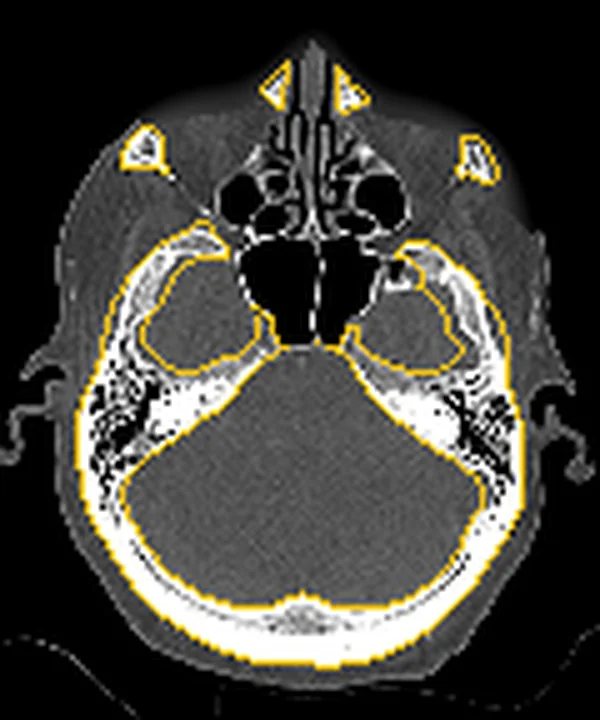
Aynı bölgenin, kaynak veri setinde patoloji içermediği belirtilen bir BT incelemesini kesit kesit kaydırın; organ sınırlarını açarak anatomiyi hasta görüntüsüyle karşılaştırın. Küçük rastlantısal bulgular tümüyle dışlanmamıştır.
Kaynak: TotalSegmentator v2.01 veri seti (Wasserthal ve ark., Radiology: Artificial Intelligence 2023), CC BY 4.0, vaka s0849; organ sınırları veri setinin otomatik segmentasyonundan, birleştirilerek sadeleştirildi. Görüntüler 2,5 mm kesit aralığına yeniden örneklendi.
Ayırıcı tanı
- Kuru tahta yabancı cisimler BT'de genellikle görünmez veya belirsiz olup tümör ve enfeksiyon odaklarıyla karışabilir.
- Lamina papyracea gelişimsel dehisansı
- kemik açıklığı düzgün olabilir; akut travmatik kenar, yumuşak doku prolapsusu veya sinüs kanaması eşlik etmeyebilir.
Tuzaklar
- Tahta yabancı cisimler BT'de görünmeyebilir veya tümör/yangı ile karışabilir; klinik şüphe devam ettiğinde MRG eklenmeli ve BT'nin sınırlı duyarlılığı belirtilmelidir.
- İnce lamina papyracea açıklığı kırık yerine anatomik dehisans olabilir; düzgün kortike kenar ve sinüse yağ prolapsusu olmaması varyantı destekler.
- Orbital hava tek başına kompartman sendromu anlamına gelmez; glob öne çıkıntısı, posterior glob deformasyonu ve optik sinir gerilmesini ayrıca arayın.
Kendini dene
Etmoid hücre ile orbita arasında hava görülüyor; lamina papyracea boyunca düzgün, kortike kenarlı bir açıklık var ve akut kırık hattı ya da deplasman yok. En olası açıklama nedir?
Cevabı göster
Duvar dehisansı. Düzgün kortike kenarlı kemik açıklığı, akut deplase kırık yerine önceden var olan dehisansı destekler; travma öyküsü ve komşu yumuşak doku bulguları birlikte değerlendirilir. BT'de lamina papyracea duvarında aralık ve orbital doku prolapsusu olmaması, kırığa kıyasla dehisansı destekler.
Orbital hava ile birlikte posterior globun optik sinir girişinde çadırlaşması ve optik sinirin gerilmesi izleniyor. Bu görüntüleme örüntüsü hangi durumu destekler?
Cevabı göster
Tansiyon amfizemi. Posterior glob çadırlaşması ve optik sinir gerilmesi orbital kompartman basısını ve tansiyon amfizemini destekleyen BT bulgularıdır; sendrom oküler muayene bulgularıyla korele edilmelidir. Posterior glob çadırlaşması BT'de orbital kompartman sendromunu destekler; orbital amfizemde yüksek intraorbital basınç optik siniri etkileyip görmeyi tehdit edebilir.
İlgili konular
Kaynaklar
Bu sayfadaki 33 cümle ve 2 quiz sorusu aşağıdaki kaynaklardan alıntılarla tek tek karşılaştırıldı; 8 cümle bu karşılaştırmada düzeltildi (2026-10-08).
- Traumatic Orbital Emphysema Following Blunt Trauma and Nose Blowingpmc.ncbi.nlm.nih.gov
- Orbital compartment syndrome: Pearls and pitfalls for the emergency physicianpmc.ncbi.nlm.nih.gov
- Radiological findings of orbital blowout fractures: a review.pubmed.ncbi.nlm.nih.gov
- Sudden orbital emphysema occurred during rhinoplasty operation.pubmed.ncbi.nlm.nih.gov
- Orbital Emphysema: A Case Report and Comprehensive Review of the Literature.pubmed.ncbi.nlm.nih.gov
- Pneumocephalus and orbital emphysema associated with the use of an autoinflation device: a unique case report and review of the literature.pubmed.ncbi.nlm.nih.gov
- A case of orbital emphysema associated with frontal sinus pneumocele.pubmed.ncbi.nlm.nih.gov
- Orbital Emphysema Secondary to Frontal Osteoma.pubmed.ncbi.nlm.nih.gov
- Unilateral Orbital Emphysema Secondary to Vitreoretinal Surgery.pubmed.ncbi.nlm.nih.gov
- Case Report: Atraumatic Orbital Emphysema after Bronchoscopy with Endobronchial Biopsy.pubmed.ncbi.nlm.nih.gov
- Orbital emphysema after transcanalicular laser-assisted dacryocystorhinostomy treated with needle decompression.pubmed.ncbi.nlm.nih.gov
- [Orbital emphysema: radiologic and ophthalmologic findings].pubmed.ncbi.nlm.nih.gov
- Post periorbital carboxytherapy orbital emphysema: a case report.pubmed.ncbi.nlm.nih.gov
- Orbital Emphysema as a Consequence of Forceful Nose-Blowing: Report of a Case.pubmed.ncbi.nlm.nih.gov
- Sub-Tenon Emphysema From Compressed Air Injury in a Patient With Marfan Syndrome.pubmed.ncbi.nlm.nih.gov
- Reconstruction of an orbital fracture in a mare using a 3D-printed patient-specific implant.pubmed.ncbi.nlm.nih.gov
- Long-term clinical outcome of maxillofacial fractures in horses: A retrospective study of 30 cases (2020-2024).pubmed.ncbi.nlm.nih.gov
- Orbital Compartment Syndrome: An Update With Review Of The Literature.pubmed.ncbi.nlm.nih.gov
- Orbital apex disorders: Imaging findings and management.pubmed.ncbi.nlm.nih.gov
- Pearls of orbital trauma management.pubmed.ncbi.nlm.nih.gov
- A review of the revisions and complications management procedure in sinus surgery.pubmed.ncbi.nlm.nih.gov
- Danger points, complications and medico-legal aspects in endoscopic sinus surgery.pubmed.ncbi.nlm.nih.gov
- Coexistence of Autoantibodies against the Golgi Complex and Ro52 Antigen in a Patient with Nonspecific Interstitial Pneumonia.pubmed.ncbi.nlm.nih.gov
- Orbital and periorbital migration of silicone oil associated with emphysema development after retinal detachment repair - Case report and literature review.pubmed.ncbi.nlm.nih.gov
- Revisiting rhino-orbito-cerebral acute invasive fungal sinusitis in the era of COVID-19: pictorial review.pubmed.ncbi.nlm.nih.gov
- Orbital and Intracranial Emphysema Causing Orbital Compartment Syndrome: A Rare Case Report and Literature Review.pubmed.ncbi.nlm.nih.gov
- Clinical and imaging manifestations of metastases from rare sites.pubmed.ncbi.nlm.nih.gov
- Role of pulmonary perfusion magnetic resonance imaging for the diagnosis of pulmonary hypertension: A review.pubmed.ncbi.nlm.nih.gov
- Human biomarker navigator.pubmed.ncbi.nlm.nih.gov
- Spontaneous Orbital Emphysema: A Case Report.pubmed.ncbi.nlm.nih.gov
- Orbital Defect and Emphysema After Nose Blowing: A Case Report and Literature Review.pubmed.ncbi.nlm.nih.gov
- Computed tomography in traumatic orbital emergencies: a pictorial essay-imaging findings, tips, and report flowchart.pubmed.ncbi.nlm.nih.gov
- Imaging in orbital trauma.pubmed.ncbi.nlm.nih.gov
- Traumatic Orbital Emphysema Following Blunt Trauma and Nose Blowing.pubmed.ncbi.nlm.nih.gov
- Analysis of the Lamina Papyracea Dehiscence Based on Computed Tomography Findings.pubmed.ncbi.nlm.nih.gov
- Chinese Guidelines for Diagnosis and Treatment of Chronic Rhinosinusitis (2024).pubmed.ncbi.nlm.nih.gov
- Intracranial Hemorrhage and Facial Fractures After Nose Blowing and Sternutation: A Case Report.pubmed.ncbi.nlm.nih.gov
- Surgical Approaches to Orbital Fractures: A Practical and Systematic Review.pubmed.ncbi.nlm.nih.gov
- Radiological Insights of a Complex Orbital Blow-Out Fracture.pubmed.ncbi.nlm.nih.gov
- Treatment and Survival Outcomes in Rhino-Orbital Mucormycosis with and without Orbital Exenteration: A Retrospective Case Series and Literature Review.pubmed.ncbi.nlm.nih.gov
- Guidelines for standard operation of imaging modalities in orbital diseases (2024).pubmed.ncbi.nlm.nih.gov
- Penetrating orbital floor injury by an undetected foreign body: a case report.pubmed.ncbi.nlm.nih.gov
- Intraorbital Foreign Body- Endoscope Assisted Removal.pubmed.ncbi.nlm.nih.gov
- Penetrating Orbital Injury: A Narrative Review for Emergency Clinicians.pubmed.ncbi.nlm.nih.gov
- Superonasal orbital traumatic neuroma mimicking a dermoid cyst in a young patient: A case report.pubmed.ncbi.nlm.nih.gov
- Imaging features of lacrimal gland disease.pubmed.ncbi.nlm.nih.gov
- Explosive multi-site metastasis temporally associated with acute exacerbation of idiopathic pulmonary fibrosis in a patient with surgically resected lung squamous cell carcinoma: a case report.pubmed.ncbi.nlm.nih.gov
- Critical anatomical variants in preoperative computed tomography of paranasal sinuses in a tertiary care center: a cross-sectional study.pubmed.ncbi.nlm.nih.gov
- Anatomical Variations of the Nasal Cavities and Paranasal Sinuses: A Systematic Review.pubmed.ncbi.nlm.nih.gov
- Imaging of Skull Base Tumors.pubmed.ncbi.nlm.nih.gov
- Modern management of maxillofacial trauma in the emergency department.pubmed.ncbi.nlm.nih.gov
- A Comprehensive Look at Maxillofacial Traumas: On the Basis of Orbital Involvement.pubmed.ncbi.nlm.nih.gov
- Management of Mechanical Strabismus After Complicated Functional Endoscopic Sinus Surgery (FESS).pubmed.ncbi.nlm.nih.gov
- Sino-orbital foreign body caused by a slingshot injury in a young boy.pubmed.ncbi.nlm.nih.gov
- Rhinosinusitis: Evidence and experience - 2024.pubmed.ncbi.nlm.nih.gov
- Dental Procedure-Related Emphysema: A Scoping Review of Clinical Presentation, Anatomical Distribution, Management, and Outcomes.pubmed.ncbi.nlm.nih.gov
- Beyond benign swelling: fascial plane spread of dental therapy-related subcutaneous emphysema to critical cranio-cervicothoracic compartments - Clinical predictors and a management algorithm.pubmed.ncbi.nlm.nih.gov
- Clinical diagnosis and treatment of intraorbital wooden foreign bodies.pubmed.ncbi.nlm.nih.gov
- Commentary: an eye on PET quantification.pubmed.ncbi.nlm.nih.gov
- Imaging for Pediatric Head and Neck Emergencies: Critical Insights.pubmed.ncbi.nlm.nih.gov
- Nasoethmoid Ancient Schwannoma with Bilateral Intracranial Extension: A Case Report with Review of the Literature.pubmed.ncbi.nlm.nih.gov
- Review of imaging modalities and radiological findings of calvarial lesions.pubmed.ncbi.nlm.nih.gov
- Pediatric intraorbital wooden foreign body mimicking an orbital mass: Delayed diagnosis and imaging considerations on MRI.pubmed.ncbi.nlm.nih.gov
- Clinical Analysis of Perforating Globe Injuries with Intraorbital Foreign Bodies: An 18-Year Retrospective Study.pubmed.ncbi.nlm.nih.gov
- Multimodal imaging in neuro-ophthalmology.pubmed.ncbi.nlm.nih.gov
- Sphenoid sinus hyperpneumatization: anatomical variants, molecular blueprints, and AI-augmented roadmaps for skull base surgery.pubmed.ncbi.nlm.nih.gov
- Early adipose tissue wasting in a preclinical model of human lung cancer cachexia.pubmed.ncbi.nlm.nih.gov
- Orbital computed tomography biomarkers for the assessment of activity of Thyroid Eye Disease.pubmed.ncbi.nlm.nih.gov
- Restricted Diffusion on Diffusion-Weighted MRI in a Cat With Orbital Lymphoma and Presumptive Pulmonary Lymphoma.pubmed.ncbi.nlm.nih.gov
- Understanding Photon-Counting CT: Physics, Detector Technology, and Image Reconstructions.pubmed.ncbi.nlm.nih.gov
- Computed Tomography Findings in Pathology-Confirmed Rhino-Orbital Mucormycosis During the COVID-19 Era: A Retrospective Cross-Sectional Study.pubmed.ncbi.nlm.nih.gov
- Occult recurrence of sinonasal inverted papilloma masked by a mucocele: a case report.pubmed.ncbi.nlm.nih.gov
- Kadish C/Hyams II Esthesioneuroblastoma With Atypical Immunophenotype Mimicking NK/T-Cell Lymphoma: Endoscope-Assisted Transcranial Resection and Adjuvant Intensity-Modulated Radiotherapy.pubmed.ncbi.nlm.nih.gov
- Ocular ultrasound: a clinical review for the medicine-trained intensivist.pubmed.ncbi.nlm.nih.gov
- Ocular Misalignment and Motility Limitation in Orbital Tumors.pubmed.ncbi.nlm.nih.gov
- Morphological and Imaging Characterization of the Eye and Adnexa of the Lesser Anteater (Tamandua tetradactyla).pubmed.ncbi.nlm.nih.gov
- Unusual Manifestations of Sinonasal Osteomas: A Narrative Review.pubmed.ncbi.nlm.nih.gov
- Assessment of the Relationship Between the Risk for Orbital Blowout Fracture After Trauma and Ethmoidal Sinus Morphometry Using the 3D Slicer Application.pubmed.ncbi.nlm.nih.gov
- Lost in the Woods: A Case Report of a Radiolucent Wooden Foreign Body in a Child After Negative CT, Detected on Repeat Ultrasound.pubmed.ncbi.nlm.nih.gov
- Successful lung-preserving tractotomy to treat intrathoracic wood impalement after high-fall trauma: a case report.pubmed.ncbi.nlm.nih.gov
- Transorbital Penetrating Intracranial Injury by Wooden Foreign Body: A Rare Case Report.pubmed.ncbi.nlm.nih.gov
- Comparative Diagnostic Value of Computed Tomography Lung and Bone Window Settings for the Detection of Nasal Foreign Bodies in 47 Dogs Presented to Two UK Referral Hospitals (2015-2023).pubmed.ncbi.nlm.nih.gov
- Anatomical variations of lamina papyracea, internal carotid artery, and optic canal dehiscence and protrusion: a computed tomography retrospective cohort study.pubmed.ncbi.nlm.nih.gov
- Atypical Presentation of Undiagnosed Crouzon's Syndrome With Pansinusitis, Orbital Cellulitis, and Altered Mentation Successfully Managed With Functional Endoscopic Sinus Surgery: A Case Report.pubmed.ncbi.nlm.nih.gov
- Small ethmoid mucocele with intracranial extension associated with fibrous dysplasia: A rare case report.pubmed.ncbi.nlm.nih.gov
- Isolated sphenoid sinus foreign body following slingshot injury: a case report.pubmed.ncbi.nlm.nih.gov
- Hybrid adjuvant therapy with 3D-printed orbital brachytherapy and stereotactic body radiotherapy for recurrent adenoid cystic carcinoma: A technical note.pubmed.ncbi.nlm.nih.gov
- Compression Syndromes of the Stylohyoid Complex: Neural, Arterial, and Venous Phenotypes in a Dynamic Neurovascular Corridor.pubmed.ncbi.nlm.nih.gov
- Recognizing Descending Necrotizing Mediastinitis in Adults: A Narrative Review of Diagnostic Dilemmas and Barriers.pubmed.ncbi.nlm.nih.gov
- Ophthalmic Complications Following Sinus Lifting Surgery: Case Report and Review of Literature.pubmed.ncbi.nlm.nih.gov
- Enophthalmospubmed.ncbi.nlm.nih.gov
- Transorbital neuroendoscopic approach for recurrent sinonasal inverted papilloma.pubmed.ncbi.nlm.nih.gov
- Contiguous orbital inflammation from paranasal sinus abnormalities in etiology of thyroid-associated ophthalmopathy.pubmed.ncbi.nlm.nih.gov
- Atypical imaging characteristics in patients with epithelial tumors of lacrimal gland and impact on initial surgical planning.pubmed.ncbi.nlm.nih.gov
- A-eye: Automated 3D MRI segmentation and morphometric feature extraction for eye and orbit atlas construction.pubmed.ncbi.nlm.nih.gov
- Personalized 3D-Printed Hybrid PDMS and PEEK Implants for Revisional Orbitomaxillary Reconstruction: A Translational Case-Based Technical Note.pubmed.ncbi.nlm.nih.gov
- Anatomical Study and CT Scan of the Scleral Ring in the Little Owl (Athene noctua).pubmed.ncbi.nlm.nih.gov
- Rare Bilateral Massive Pneumothorax, Pneumomediastinum, Pneumoperitoneum, and Diffuse Subcutaneous Emphysema During Colonoscopy: Multidisciplinary Collaboration: Case Report.pubmed.ncbi.nlm.nih.gov
- The Comprehensive AOCMF Classification System: Radiological Issues and Systematic Approach.pubmed.ncbi.nlm.nih.gov
- Photon-counting detector of the ophthalmic artery: impact of virtual monoenergetic levels, reconstruction kernel, and quantum iterative reconstruction (QIR) on image quality.pubmed.ncbi.nlm.nih.gov
- Minimally invasive resection of a spheno-orbital tumor: Review of surgical techniques and a case report.pubmed.ncbi.nlm.nih.gov
- Isolated trigeminal neuropathy as an initial manifestation of chronic inflammatory demyelinating polyradiculoneuropathy: A case report.pubmed.ncbi.nlm.nih.gov
- Indirect traumatic optic neuropathy in a high-risk diabetic adolescent: A rare clinical presentation.pubmed.ncbi.nlm.nih.gov
- Long-term safety and effectiveness of the "OptEase" vena cava filter.pubmed.ncbi.nlm.nih.gov
Eğitim amaçlıdır; hasta başında tanı ve tedavi kararının yerine geçmez. İçerik yapay zekâ destekli bir süreçle güncel literatüre göre cümle cümle kaynaklarla karşılaştırıldı, ancak uzman radyolog incelemesinden geçmedi. Klinik kararlarda güncel kılavuzları, kurum protokollerini ve radyoloji raporunu esas alın.