Özet
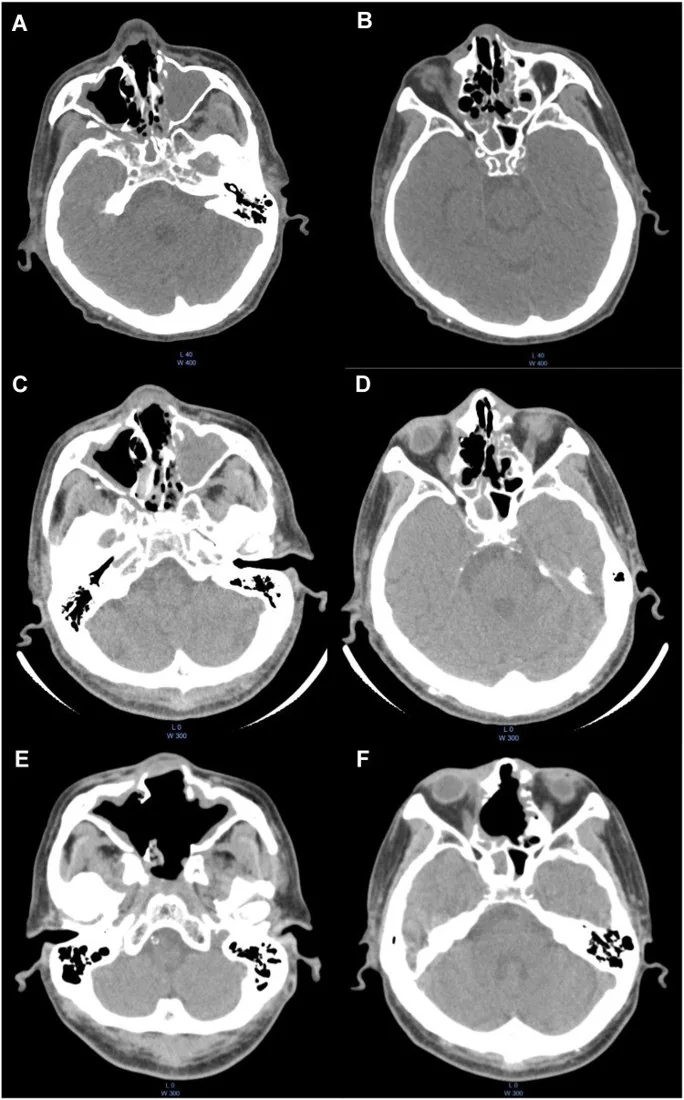
Kronik invaziv fungal rinosinüzit, haftalar-aylar içinde ilerleyebilen ve bazen bağışıklığı normal kişilerde de görülen, sinüs mukozasını aşarak komşu dokulara yayılan fungal enfeksiyondur. BT; tek taraflı hastalık yükünü, kemik erozyonunu ve periantral, orbital ya da kafa tabanı uzanımını haritalar; erken yumuşak doku yayılımı kemik sağlamken de bulunabilir. Kontrastlı inceleme sinüs dışı inflamatuvar dokuyu ve olası koleksiyonları göstermeye yardım eder, ancak etkeni veya doku invazyonunu tek başına kanıtlamaz. Yayılımın gözden kaçması görme kaybı, kraniyal nörovasküler tutulum veya intrakraniyal komplikasyonların geç fark edilmesine yol açabilir.
Faz ve pencere
- Kontrastsız tanısal
- İnce kesitlerde asimetrik mukozal kalınlaşma/opasifikasyon, kronik invaziv hastalıkta hiperdens fungal içerik, sinüs duvarında erozyon ya da skleroz ve periantral yağ planı silinmesi aranır.
- Kontrastlı tanısal
- IV kontrast sonrası yumuşak doku serisinde sinüs dışı infiltratif dokunun sınırı, periantral alan, pterigopalatin fossa, orbita ve kafa tabanı komşuluğundaki kontrastlanma değerlendirilir; bu, standart postkontrast sinonazal seridir.
Önerilen pencereler: Kemik (G 1800 / M 400), Yumuşak doku (G 400 / M 40).
BT bulguları
- Tek taraflı, düzensiz mukozal kalınlaşma veya sinüsleri dolduran infiltratif yumuşak doku.
- Hiperdens, heterojen sinüs içeriği kronik fungal materyali destekler; tek başına invazyon kanıtı değildir.
- Retroantral yağ planında çizgilenme veya silinme, kemik korteksinin bütünlüğü bozulmadan erken sinüs dışı yayılımı düşündürür.
- Maksiller duvar, lamina papyracea veya kafa tabanında fokal erozyon/destrüksiyon görülebilir.
- Retroantral yağ veya pterigopalatin fossa çevresine infiltrasyon yayılımı gösterir.
- Orbital yağ, ekstraoküler kas veya intrakraniyal komşulukta infiltratif doku ileri yayılımı gösterir.
- Kontrastlanan solid inflamatuvar doku ve nekrotik/az kontrastlanan alanlar birlikte bulunabilir.
Normalde
Maksiller sinüs çevresindeki yumuşak doku planları ve kemik bütünlüğü karşılaştırılarak değerlendirilir. Aynı düzeyde karşı tarafla mukoza kalınlığını, yağ planlarının görünürlüğünü ve orbita tabanı/lamina papyracea korteksini karşılaştır; tek taraflı plan kaybını yalnız sinüs opasifikasyonuna bağlama.
Normal BT ile kıyasla
Aynı bölgenin, kaynak veri setinde patoloji içermediği belirtilen bir BT incelemesini kesit kesit kaydırın; organ sınırlarını açarak anatomiyi hasta görüntüsüyle karşılaştırın. Küçük rastlantısal bulgular tümüyle dışlanmamıştır.
Kaynak: TotalSegmentator v2.01 veri seti (Wasserthal ve ark., Radiology: Artificial Intelligence 2023), CC BY 4.0, vaka s0849; organ sınırları veri setinin otomatik segmentasyonundan, birleştirilerek sadeleştirildi. Görüntüler 2,5 mm kesit aralığına yeniden örneklendi.
Ayırıcı tanı
- Akut invaziv fungal rinosinüzit
- benzer ekstrasinüs yağ infiltrasyonu; klinik seyir günler içinde daha hızlıdır.
- Sinonazal skuamöz hücreli karsinom
- solid kitle ve düzensiz kemik yıkımı; histoloji olmadan ayrılamaz.
- Granülomatöz invaziv fungal rinosinüzit
- belirgin solid granulomatöz kitle ve ekspansil orbital uzanım görülebilir.
- Bakteriyel komplike sinüzit
- koleksiyon ve çevresel inflamasyon olabilir; doku invazyonu klinik ve MRI ile ayrıştırılır.
- Noninvaziv fungal top
- sinüs içinde hiperdens materyal olabilir, çevre yağ planları ve kemik genellikle korunur.
Tuzaklar
- Hiperdens sinüs içeriğini tek başına invaziv fungal enfeksiyon sayma; fungal top da hiperdens olabilir.
- Erken invazyonda kemik erozyonu görülmeyebilir; periantral yağ planlarını kemik penceresiyle sınırlı kalmadan incele.
Kendini dene
Tek taraflı maksiller opasifikasyonun yanında retroantral yağ çizgili ve silinmiş; kemik duvar henüz sağlam. En kaygı verici yorum hangisi?
Cevabı göster
İnvaziv fungal rinosinüzit. Periantral yağ infiltrasyonu erken ekstrasinüs yayılımını gösterebilir ve kemik erozyonundan önce ortaya çıkabilir; yalnız sinüs içi fungal top çevre yağını infiltre etmez.
Kronik invaziv fungal rinosinüzit şüphesinde kemik erozyonunu ve periantral yumuşak dokuyu birlikte hangi inceleme en iyi haritalar?
Cevabı göster
Sinüs BT kemik ve kontrastlı yumuşak doku serileri. İnce kesit sinüs BT kemik korteksini gösterir; postkontrast yumuşak doku serisi sinüs dışı infiltrasyonu sınırlar. Diğer seçenekler sinonazal uzanımı uygun biçimde kapsamaz.
İlgili konular
Kaynaklar
Bu sayfadaki 33 cümle ve 2 quiz sorusu aşağıdaki kaynaklardan alıntılarla tek tek karşılaştırıldı; 4 cümle bu karşılaştırmada düzeltildi (2026-10-08).
- Chronic Invasive Fungal Rhinosinusitis Managed With Surgical Debridement and Topical Amphotericin B Without Systemic Antifungal Therapy: A Report of Two Casespmc.ncbi.nlm.nih.gov
- Chronic invasive fungal rhinosinusitis in a patient with chronic migrainepmc.ncbi.nlm.nih.gov
- Chronic invasive Aspergillus Fumigatus rhinosinusitis with intracranial extensionpmc.ncbi.nlm.nih.gov
- From delay to diagnosis: Chronic invasive fungal rhinosinusitis presenting with facial and orbital complicationspmc.ncbi.nlm.nih.gov
- An Atypical Case of Chronic Fungal Rhinosinusitis: Temporary Symptom Relief Resulted in a Delay of Diagnosis and Brain Abscesspmc.ncbi.nlm.nih.gov
- Imaging Features of Invasive Fungal Rhinosinusitis: A Systematic Review.pubmed.ncbi.nlm.nih.gov
- Fungal rhinosinusitis.pubmed.ncbi.nlm.nih.gov
- Orbital Involvement In Sinonasal Diseases.pubmed.ncbi.nlm.nih.gov
- Definition and management of invasive fungal rhinosinusitis: a single-centre retrospective study.pubmed.ncbi.nlm.nih.gov
- Chronic invasive rhinosinusitis by Conidiobolus coronatus, an emerging microorganism.pubmed.ncbi.nlm.nih.gov
- Scedosporium apiospermum invasive sinusitis presenting as extradural abscess.pubmed.ncbi.nlm.nih.gov
- Chronic invasive fungal rhinosinusitis vs sinonasal squamous cell carcinoma: the differentiating value of MRI.pubmed.ncbi.nlm.nih.gov
- A Case of Chronic Invasive Fungal Sinusitis With Orbital Abscess.pubmed.ncbi.nlm.nih.gov
- [Endoscopic sinus surgery for the treatment of invasive fungal sinusitis].pubmed.ncbi.nlm.nih.gov
- [Clinical analysis of chronic invasive fungal rhinosinusitis].pubmed.ncbi.nlm.nih.gov
- Fungal Rhinosinusitis: An integrated diagnostic approach.pubmed.ncbi.nlm.nih.gov
- Chronic Invasive Fungal Rhinosinusitis with Atypical Clinical Presentation in an Immunocompromised Patient.pubmed.ncbi.nlm.nih.gov
- [ALLERGIC FUNGAL RHINOSINUSITIS].pubmed.ncbi.nlm.nih.gov
- Allergic fungal sinusitis in children.pubmed.ncbi.nlm.nih.gov
- Paresthesia and Facial Bone Destruction due to Chronic Granulomatous Invasive Aspergillosis in the Left Maxillary Sinus.pubmed.ncbi.nlm.nih.gov
- <i>Saksenaea oblongispora</i> Rhinosinusitis in Advanced HIV: A Rare and Lethal Mucormycosis.pubmed.ncbi.nlm.nih.gov
- Unprecedented Finding of Isolated Sphenoid Fungal Ball in a Child: A Case Report.pubmed.ncbi.nlm.nih.gov
- Clinical and Radiological Predictors of Invasive Fungal Sinusitis.pubmed.ncbi.nlm.nih.gov
- Rhinosinusitis: Evidence and experience - 2024.pubmed.ncbi.nlm.nih.gov
- Sinonasal Inflammation or Neoplasm: Raise the Red Flagsǃ-A Pictorial Review.pubmed.ncbi.nlm.nih.gov
- Behind the Unilateral Rhinorrhea: Delayed Pediatric Intranasal Foreign Bodies Presenting as Chronic Sinonasal Disease and Severe Complications.pubmed.ncbi.nlm.nih.gov
- Detection to Disruption: A Comprehensive Review of Bacterial Biofilms and Therapeutic Advances.pubmed.ncbi.nlm.nih.gov
- An Unveiling of the Misdiagnosis of Granulomatosis with Polyangiitis as Acute Sinusitis: A Case Report.pubmed.ncbi.nlm.nih.gov
- Critical infections in the head and neck: A pictorial review of acute presentations and complications.pubmed.ncbi.nlm.nih.gov
- Extrasinonasal infiltrative process associated with a sinonasal fungus ball: does it mean invasive fungal sinusitis?pubmed.ncbi.nlm.nih.gov
- Sphenoid fungal rhinosinusitis across the disease spectrum: a case series of non-invasive fungal ball and fatal acute invasive fungal rhinosinusitis.pubmed.ncbi.nlm.nih.gov
- Radiologic Characterization of Invasive Fungal Infections of the Paranasal Sinuses and Skull Base: A Prospective Analysis.pubmed.ncbi.nlm.nih.gov
- Using the Hounsfield Units Derived From Computed Tomography (CT) Scans to Differentiate Between the Subtypes of Allergic Fungal Rhinosinusitis: A Retrospective Study.pubmed.ncbi.nlm.nih.gov
- The Radiological Spectrum of Rhino-Oculo-Cerebral Mucormycosis.pubmed.ncbi.nlm.nih.gov
- Invasive Cryptococcal Meningitis Presenting as a Skull Base Mass in An Immunocompetent Host: A Case Report.pubmed.ncbi.nlm.nih.gov
- Fungal rhinosinusitis and imaging modalities.pubmed.ncbi.nlm.nih.gov
- Orbital complications in allergic fungal rhinosinusitis: a case series from a tertiary center hospital experience.pubmed.ncbi.nlm.nih.gov
- ESR Essentials: acute infections of the head and neck-practice recommendations by the European Society of Head and Neck Radiology.pubmed.ncbi.nlm.nih.gov
- Rhino-Orbital-Cerebral Mucormycosis Complicated by Vision Loss in a Patient with Uncontrolled Diabetes: A Case Report.pubmed.ncbi.nlm.nih.gov
- Application and prognostic analysis of endoscopic sinus surgery combined with multidisciplinary team management in rhino-orbito-cerebral mucormycosis.pubmed.ncbi.nlm.nih.gov
- Chronic Invasive Fungal Rhinosinusitis Managed With Surgical Debridement and Topical Amphotericin B Without Systemic Antifungal Therapy: A Report of Two Cases.pubmed.ncbi.nlm.nih.gov
- Imaging for Pediatric Head and Neck Emergencies: Critical Insights.pubmed.ncbi.nlm.nih.gov
- Post-COVID-19 Jaw Osteonecrosis: A Narrative Review.pubmed.ncbi.nlm.nih.gov
- The Importance of MRI in the Early Diagnosis of Acute Invasive Fungal Rhinosinusitis.pubmed.ncbi.nlm.nih.gov
- What's that smell? A pictorial review of the olfactory pathways and imaging assessment of the myriad pathologies that can affect them.pubmed.ncbi.nlm.nih.gov
- Comprehensive review on endonasal endoscopic sinus surgery.pubmed.ncbi.nlm.nih.gov
- ESR Essentials: imaging in nasal obstruction and epistaxis-practice recommendations by the European Society of Head and Neck Radiology.pubmed.ncbi.nlm.nih.gov
- Uncommon Nasal Mass Presentation: A Radiological Case Series.pubmed.ncbi.nlm.nih.gov
- Sphenoid sinus anatomical variants and pathologies: pictorial essay.pubmed.ncbi.nlm.nih.gov
- Review of Fusariosis in Patients with Hematologic Malignancies.pubmed.ncbi.nlm.nih.gov
- Varicella-zoster virus infection in pregnancy: a case of aseptic meningitis before 20 weeks gestation and review of the literature.pubmed.ncbi.nlm.nih.gov
- Rathke's cleft cyst: From history to molecular genetics.pubmed.ncbi.nlm.nih.gov
- A Retrospective Study of Imaging of Invasive Rhino-Orbital-Cerebral Mucormycosis in the COVID-19 Pandemic in a Tertiary Care Center.pubmed.ncbi.nlm.nih.gov
- Revisiting rhino-orbito-cerebral acute invasive fungal sinusitis in the era of COVID-19: pictorial review.pubmed.ncbi.nlm.nih.gov
- Post Covid-19 Acute Invasive Fungal Rhinosinusitis (AIFR): A Study of Histopathological Findings After FESS in Radiologically Diagnosed AIFR.pubmed.ncbi.nlm.nih.gov
- A case series of 99mTc-MDP bone scintigraphy (planar and SPECT CT) in mucormycosis in the era of COVID 19.pubmed.ncbi.nlm.nih.gov
- Cranial anatomy, palaeoneurology, palaeobiology and stratigraphic age of the large-bodied ornithopod, <i>Muttaburrasaurus langdoni</i> Bartholomai and Molnar, 1981, from the mid-Cretaceous of Australia.pubmed.ncbi.nlm.nih.gov
- Revisiting the "Puffed Cheek" Technique: Advantages, Fallacies, and Potential Solutions.pubmed.ncbi.nlm.nih.gov
- Chronic Rhinosinusitis at the Interface of Type 2 Inflammation, Epithelial Barrier Dysfunction, and Microbiome Dysbiosis.pubmed.ncbi.nlm.nih.gov
- A Systematic Classification of Surgical Approaches for the Sphenoid Sinus: Establishing a Standardized Nomenclature for Endoscopic Sphenoid Sinus Surgery.pubmed.ncbi.nlm.nih.gov
- Rare Diseases of the Orbit.pubmed.ncbi.nlm.nih.gov
- Acute Invasive Fungal Rhino-Orbital Sinusitis: Current Concepts and Controversies.pubmed.ncbi.nlm.nih.gov
- Morphological and Epidemiological Analysis of the Sphenoid Sinus Based on Computed Tomography and Magnetic Resonance Imaging: A Narrative Review.pubmed.ncbi.nlm.nih.gov
- Probable invasive <i>Alternaria</i> rhinosinusitis in a previously healthy patient diagnosed with heterozygous CARD9 deficiency and combined immunodeficiency.pubmed.ncbi.nlm.nih.gov
- Fungal Sinusitispubmed.ncbi.nlm.nih.gov
- Spectrum of magnetic resonance imaging findings in post-COVID-19 patients presenting with rhino-orbito-cerebral mucormycosis in a teaching hospital in Malwa region of Punjab.pubmed.ncbi.nlm.nih.gov
- CT Imaging Features in Acute Invasive Fungal Rhinosinusitis- Recalling the Oblivion in the COVID Era.pubmed.ncbi.nlm.nih.gov
- The Relevance and Potential Role of Orbital Fat in Inflammatory Orbital Diseases: Implications for Diagnosis and Treatment.pubmed.ncbi.nlm.nih.gov
- Detection of maxillary sinusitis of endodontic origin in cone-beam CT images using deep learning algorithms.pubmed.ncbi.nlm.nih.gov
- Adaptation and validation of the Wylie cephalometric analysis for three-dimensional assessment of canine occlusion in Labrador retrievers.pubmed.ncbi.nlm.nih.gov
- Left Atrial Appendage: Mere Spectator or Protagonist? A Narrative Review of Its Multimodal Imaging Assessment and Its Closure Techniques.pubmed.ncbi.nlm.nih.gov
- Pictorial review: a step-by-step guide to coronary CT angiography with photon-counting detector CT.pubmed.ncbi.nlm.nih.gov
- Sequential Helical-Axial-Helical Triple-Rule-Out CT Angiography: Technical Feasibility and Territory-Specific Image Quality in the Emergency Department.pubmed.ncbi.nlm.nih.gov
Eğitim amaçlıdır; hasta başında tanı ve tedavi kararının yerine geçmez. İçerik yapay zekâ destekli bir süreçle güncel literatüre göre cümle cümle kaynaklarla karşılaştırıldı, ancak uzman radyolog incelemesinden geçmedi. Klinik kararlarda güncel kılavuzları, kurum protokollerini ve radyoloji raporunu esas alın.