
Özet
Sinonazal metastaz nadirdir; renal hücreli karsinom başta olmak üzere meme, tiroid ve akciğer tümörleri sinüs veya burun boşluğunda kitle oluşturabilir. Tek taraflı tıkanıklık ya da epistaksis, bilinen primer tümörün yıllar önce tedavi edildiği hastada bile metastazın ilk ipucu olabilir. BT, kemik destrüksiyonunun yerini, sinüs duvarları boyunca yayılımı ve kontrastlanan kitlenin damar zenginliğini değerlendirmeye yardım eder; görüntü görünümü metastazı primer sinonazal kanserden tek başına ayıramaz. Lezyonu primer tümör sanmak sistemik hastalık araştırmasını geciktirebilir; hipervasküler odağı tanımamak biyopsi öncesi kanama riskinin gözden kaçmasına neden olabilir.
Faz ve pencere
- Kontrastsız tanısal
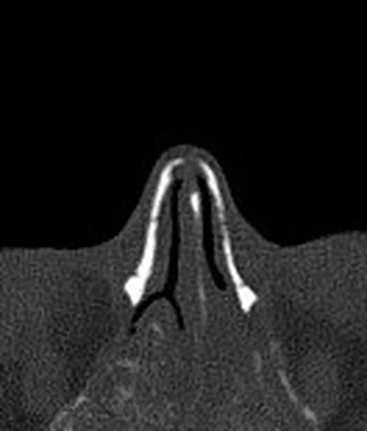
- İnce kesit kemik algoritmasında maksilla, sinüs duvarları ve kafa tabanındaki litik veya düzensiz destrüktif odak; sinüs lümenine uzanan yumuşak doku ile birlikte gösterilir. Tek odaklılık ya da birden fazla kemik odağının dağılımı bu seride haritalanır.
- Arteriyel tanısal
- İntravenöz kontrast sonrası yumuşak doku penceresinde kitlenin damar zenginliği ve ekstrasinüs uzanımı incelenir. Renal hücreli karsinom gibi hipervasküler metastazlarda belirgin, bazen heterojen kontrastlanma görülebilir; bu bulgu primeri tek başına belirlemez.
Önerilen pencereler: Kemik (G 1800 / M 400), Yumuşak doku (G 400 / M 40).
BT bulguları
- Maksiller sinüs, etmoid hücreler veya nazal kavitede tek taraflı, düzensiz yumuşak doku kitlesi.
- Kitlenin komşuluğundaki maksilla ya da sinüs duvarında fokal kortikal kayıp ve destrüksiyon.
- Renal hücreli karsinom kaynaklı odaklarda yoğun kontrastlanma görülebilir; damar zenginliği biyopsi öncesi görüntülemede önem taşır.
- Maksiller sinüs tabanı veya infraorbital kanal çevresindeki kemik kaybı, tümörün kemik boyunca komşu boşluklara ilerlediğini gösterebilir.
- Kitle orbitaya, pterigopalatin fossaya, retroantral yağ planına veya kafa tabanına uzanabilir.
- Sinonazal metastaz, tek bir bölgede sınırlı olabileceği gibi başka organ metastazlarıyla birlikte de görülebilir.
- Kontrastlanma ve kemik yıkımı metastazı düşündürebilir; BT morfolojisi metastatik kökeni histolojik olarak kanıtlamaz.
Normalde
Aynı aksiyel ve koronal düzeyde karşı tarafı referans alarak kemik korteksindeki fokal kaybı, sinüs içindeki yumuşak dokuyu ve kontrastlanmanın dağılımını karşılaştır.
Normal BT ile kıyasla
Aynı bölgenin, kaynak veri setinde patoloji içermediği belirtilen bir BT incelemesini kesit kesit kaydırın; organ sınırlarını açarak anatomiyi hasta görüntüsüyle karşılaştırın. Küçük rastlantısal bulgular tümüyle dışlanmamıştır.
Kaynak: TotalSegmentator v2.01 veri seti (Wasserthal ve ark., Radiology: Artificial Intelligence 2023), CC BY 4.0, vaka s0849; organ sınırları veri setinin otomatik segmentasyonundan, birleştirilerek sadeleştirildi. Görüntüler 2,5 mm kesit aralığına yeniden örneklendi.
Ayırıcı tanı
- Sinonazal skuamöz hücreli karsinom
- benzer infiltratif kitle ve kemik yıkımı yapabilir; görüntü tek başına primer ile metastatik tümörü ayırmaz.
- Sinonazal lenfoma
- kemik destrüksiyonu olmadan veya sınırlı kemik değişikliğiyle geniş yumuşak doku oluşturabilir; doku tanısı gerekir.
- Sinonazal osteosarkom
- tümör matriksinde bulutumsu osteoid mineralizasyonu ve kemik merkezli kitle metastazdan ayrılmasına yardım eder.
- İnvaziv fungal rinosinüzit
- retroantral yağ ve orbita çevresinde infiltrasyon yapabilir; sinüs içeriği ve klinik enfeksiyon bağlamı farklılaştırır.
- Juvenil nazofaringeal anjiofibrom
- yoğun kontrastlanabilir; posterior nazal kavite ve pterigopalatin fossa kaynaklı büyüme paterni tipiktir.
Tuzaklar
- Öyküde aktif kanser bulunmamasını metastazı dışlayan bulgu sayma; sinonazal odak primer tümörden önce ortaya çıkabilir veya gecikmiş nüks olabilir.
- Yoğun kontrastlanmayı renal hücreli karsinom için özgül kabul etme; başka hipervasküler metastazlar ve primer sinonazal tümörler de kontrastlanabilir.
- Yalnız sinüs lümenindeki opasiteye odaklanıp maksiller duvarı ve infraorbital kanalı incelememek kemik merkezli başlangıcı kaçırabilir.
- Hipervasküler kitleyi kontrastsız BT ile sınırlı değerlendirip damar zenginliğini fark etmemek, sonraki örnekleme planında kanama riskini olduğundan az düşündürebilir.
Kendini dene
Nefrektomi öyküsü bulunan hastada tek taraflı, kanlanan maksiller sinüs kitlesi görülüyor. En olası köken hangisidir?
Cevabı göster
Berrak hücreli böbrek tümörü metastazı. Böbrek tümörleri, özellikle berrak hücreli tip, sinonazal bölgeye metastaz yapabilir ve belirgin damar zenginliği gösterebilir; eski nefrektomi öyküsü de anlamlıdır. Papillom, mukozal melanom ve olfaktör nöroblastom primer sinonazal tümörlerdir ve bu klinik öyküyle metastaz kadar uyumlu değildir.
Önceden tedavi edilmiş tiroid kanseri olan hastada kemik yıkımıyla birlikte kontrastlanan burun boşluğu kitlesi saptandı. Hangi tanı hesaba katılmalıdır?
Cevabı göster
Tiroid karsinomu metastazı. Tiroid maligniteleri sinonazal bölgeye metastaz yapabilir; kanser öyküsü ve kontrastlanan destrüktif kitle bu olasılığı gündeme getirir. Antrokoanal polip ve osteom genellikle bu destrüktif tümör görünümünü oluşturmaz; invaziv mantar hastalığı ise enfeksiyon bağlamı ve sinüs çevresi doku bulgularıyla değerlendirilir.
İlgili konular
Kaynaklar
Bu sayfadaki 38 cümle ve 2 quiz sorusu aşağıdaki kaynaklardan alıntılarla tek tek karşılaştırıldı (2026-10-08).
- Sinonasal Metastasis of a Pancreatic Mucinous Adenocarcinoma: An Unusual Case Reportpmc.ncbi.nlm.nih.gov
- Primary orbital solitary fibrous tumor with an 18-year course of five recurrences and extensive sinonasal metastasis following eyeball enucleation: a case reportpmc.ncbi.nlm.nih.gov
- Sinonasal metastasis as the initial presentation of renal clear cell carcinoma: a dual-center retrospective study and proposed diagnostic algorithmpmc.ncbi.nlm.nih.gov
- Stereotactic Body Radiotherapy of a Solitary Metachronous Sphenoid Metastasis from Renal Cell Cancer: A Case Reportpmc.ncbi.nlm.nih.gov
- Occult renal cell carcinoma manifesting with epistaxis in a woman: a case reportpmc.ncbi.nlm.nih.gov
- Renal cell carcinoma metastatic to the sinonasal cavity: A review and report of 8 cases.pubmed.ncbi.nlm.nih.gov
- Orbit and sinonasal metastasis as presenting sign of renal cell carcinoma.pubmed.ncbi.nlm.nih.gov
- Renal cell carcinoma metastasis to the paranasal sinuses and orbit.pubmed.ncbi.nlm.nih.gov
- Nasal congestion and nasal obstruction: An unusual presentation of renal cell carcinoma.pubmed.ncbi.nlm.nih.gov
- Metastatic hepatocellular carcinoma presenting as a sphenoid sinus mass and meningeal carcinomatosis.pubmed.ncbi.nlm.nih.gov
- Occult renal cell carcinoma manifesting with epistaxis in a woman: a case report.pubmed.ncbi.nlm.nih.gov
- Stereotactic Body Radiotherapy of a Solitary Metachronous Sphenoid Metastasis from Renal Cell Cancer: A Case Report.pubmed.ncbi.nlm.nih.gov
- Sinonasal Metastasis of a Pancreatic Mucinous Adenocarcinoma: An Unusual Case Report.pubmed.ncbi.nlm.nih.gov
- Sinonasal metastasis as the initial presentation of renal clear cell carcinoma: a dual-center retrospective study and proposed diagnostic algorithm.pubmed.ncbi.nlm.nih.gov
- Primary orbital solitary fibrous tumor with an 18-year course of five recurrences and extensive sinonasal metastasis following eyeball enucleation: a case report.pubmed.ncbi.nlm.nih.gov
- 18 FDG-PET/CT computer-assisted biopsies for suspected persistent or recurrent malignant skull base disease.pubmed.ncbi.nlm.nih.gov
- Outcome of sinonasal melanoma: clinical experience and review of the literature.pubmed.ncbi.nlm.nih.gov
- [Clinicopathologic and molecular genetic characterizations of biphenotypic sinonasal sarcoma].pubmed.ncbi.nlm.nih.gov
- Case Series on Four Treated Cases of Embryonal Rhabdomyosarcoma in Children.pubmed.ncbi.nlm.nih.gov
- Cancer Metastasis to the Sinonasal Cavity: Clinical Characteristics and Survival Analysis in 35 Patients.pubmed.ncbi.nlm.nih.gov
- Head and Neck Metastases From Infraclavicular Primary Tumors: Analysis of 136 Cases.pubmed.ncbi.nlm.nih.gov
- Head-to-head comparison of <sup>18</sup>F-PSMA and <sup>18</sup>F-FDG PET/CT in locoregionally advanced head and neck squamous cell carcinoma: a pilot study.pubmed.ncbi.nlm.nih.gov
- Primary Unilateral Ethmoidal Ewing Sarcoma With Orbital Extension: A Report of a Rare Case.pubmed.ncbi.nlm.nih.gov
- Sinonasal osteosarcoma of the ethmoid bone with decade-later recurrence presenting as early orbital apex syndrome: an 18-year case report.pubmed.ncbi.nlm.nih.gov
- Extramedullary Plasmacytomas of the Nasal Cavity: Case-Based Perspectives into Optimizing the Diagnostic Differentiation from Inflammatory Polyps.pubmed.ncbi.nlm.nih.gov
- A silent vascular mimicker of Sinonasal disease: Nasal cavity angioma presenting with progressive epistaxis and obstruction.pubmed.ncbi.nlm.nih.gov
- Solitary Fibrous Tumor of the Nasal Vestibule: A Case Report.pubmed.ncbi.nlm.nih.gov
- Leiomyosarcoma of the maxillary sinus presenting with epiphora: a case report.pubmed.ncbi.nlm.nih.gov
- Mycotic Sphenopalatine Sinusitis With Concurrent Compression of the Optic Nerves and Chiasm and Severe Visual Impairment in A Horse.pubmed.ncbi.nlm.nih.gov
- Comprehensive review of post-treatment imaging in head and neck cancers: from expected to unexpected and beyond.pubmed.ncbi.nlm.nih.gov
- A practical overview of CT and MRI features of developmental, inflammatory, and neoplastic lesions of the sphenoid body and clivus.pubmed.ncbi.nlm.nih.gov
- Parapharyngeal Schwannoma: Limitations Regarding Diagnosis and Surgical Intervention-A Case Report and Focused Literature Review.pubmed.ncbi.nlm.nih.gov
- PET-CT in Clinical Adult Oncology-VI. Primary Cutaneous Cancer, Sarcomas and Neuroendocrine Tumors.pubmed.ncbi.nlm.nih.gov
- Updated WHO nomenclature of head and neck lesions and associated imaging findings.pubmed.ncbi.nlm.nih.gov
- Chronic Invasive Fungal Sinusitis Concerning for Sinonasal Malignancy With Unique Intracranial Extension.pubmed.ncbi.nlm.nih.gov
- Sphenoid fungal rhinosinusitis across the disease spectrum: a case series of non-invasive fungal ball and fatal acute invasive fungal rhinosinusitis.pubmed.ncbi.nlm.nih.gov
- Sinonasal Inflammation or Neoplasm: Raise the Red Flagsǃ-A Pictorial Review.pubmed.ncbi.nlm.nih.gov
- Solitary fibrous tumors from A to Z: a pictorial review with radiologic-pathologic correlation.pubmed.ncbi.nlm.nih.gov
- Chinese expert consensus on imaging examination and diagnosis of nasal cavity and paranasal sinus tumors.pubmed.ncbi.nlm.nih.gov
- Case Report: Sinonasal plasmablastic lymphoma in a human immunodeficiency virus-negative elderly woman diagnosed by immunohistochemistry.pubmed.ncbi.nlm.nih.gov
- Cavernous venous malformation of the frontal sinus: CT and MRI imaging findings.pubmed.ncbi.nlm.nih.gov
- Pyogenic Granuloma in the Sinonasal Cavity: A Potential 38-Year Latency Following Foreign Body Entrapment.pubmed.ncbi.nlm.nih.gov
- Small ethmoid mucocele with intracranial extension associated with fibrous dysplasia: A rare case report.pubmed.ncbi.nlm.nih.gov
- Nasopharyngeal carcinoma masquerading as a primary sinonasal mass: a case report.pubmed.ncbi.nlm.nih.gov
- Orbital Exenteration in Maxillary Osteosarcoma: A Case Report.pubmed.ncbi.nlm.nih.gov
- Craniofacial surgery for removing tumors in advanced stages that invade the anterior skull base.pubmed.ncbi.nlm.nih.gov
- Surgical Management of a Large Dentigerous Cyst Associated With an Impacted Supernumerary Tooth: A Rare Case Report.pubmed.ncbi.nlm.nih.gov
- Operative Corridors in Endoscopic Skull Base Tumor Surgery.pubmed.ncbi.nlm.nih.gov
- Imaging of Skull Base Tumors.pubmed.ncbi.nlm.nih.gov
- Perineural spread in head and neck malignancies: imaging findings - an updated literature review.pubmed.ncbi.nlm.nih.gov
- Persistent Long-Term Risk After Primary Surgery for Head and Neck Adenoid Cystic Carcinoma: Competing-Risk and Conditional Estimates.pubmed.ncbi.nlm.nih.gov
- Post-COVID-19 Jaw Osteonecrosis: A Narrative Review.pubmed.ncbi.nlm.nih.gov
- Prognostic significance of PET/CT false-positive cervical lymph nodes in head and neck cancer.pubmed.ncbi.nlm.nih.gov
- Skull base inverted papilloma: a comprehensive review.pubmed.ncbi.nlm.nih.gov
- Explainable hybrid CNN-transformer with self-supervised learning for structural analysis of paranasal sinus CT.pubmed.ncbi.nlm.nih.gov
- The cavernous sinus: An anatomic study with clinical implication.pubmed.ncbi.nlm.nih.gov
- Magnetic resonance imaging of the orbit, Part 1: Basic principles and radiological approach.pubmed.ncbi.nlm.nih.gov
- How Clinical and Radiological Findings in Chronic Mandibular Osteomyelitis Do Not Always Correlate: Diagnostic Dilemmas in Dental-Related Bone Inflammations.pubmed.ncbi.nlm.nih.gov
- Extended applications of chest wall perforator flaps in breast surgery: case series and systematic literature review.pubmed.ncbi.nlm.nih.gov
- Surgical Measures Improving Functional Limitations of the Masticatory System, Aesthetic Deficits, and Skeletal Malformations in Neurofibromatosis Type 1-associated Hemifacial Diffuse Plexiform Neurofibroma Complicated by Rapidly Growing Midfacial Peripheral Nerve Sheath Tumor.pubmed.ncbi.nlm.nih.gov
- Radiological and Histological Findings of Primary Pleomorphic Rhabdomyosarcoma Arising from the Mandibular Gingiva: A Case Report and Literature Review.pubmed.ncbi.nlm.nih.gov
- Poorly Differentiated Non-keratinizing Sinonasal Squamous Cell Carcinoma Masquerading as Chronic Sinusitis: A Case Report.pubmed.ncbi.nlm.nih.gov
- Olfactory neuroblastoma in a goat.pubmed.ncbi.nlm.nih.gov
- Sinonasal Phosphaturic Mesenchymal Tumor: A Case Report.pubmed.ncbi.nlm.nih.gov
- Malignant Bone-Forming Neoplasm With NIPBL::BEND2 Fusion.pubmed.ncbi.nlm.nih.gov
- The Importance of MRI in the Early Diagnosis of Acute Invasive Fungal Rhinosinusitis.pubmed.ncbi.nlm.nih.gov
- Magnetic resonance imaging in COVID-19-associated acute invasive fungal rhinosinusitis - Diagnosis and beyond.pubmed.ncbi.nlm.nih.gov
- The Relevance and Potential Role of Orbital Fat in Inflammatory Orbital Diseases: Implications for Diagnosis and Treatment.pubmed.ncbi.nlm.nih.gov
- Juvenile Nasopharyngeal Angiofibroma in an Adult Patient: A Rare Presentation with Fahr Syndrome and Multiple Comorbidities-A Case Report and Literature Review.pubmed.ncbi.nlm.nih.gov
- Unilateral proptosis as the primary presentation of Stage IVb Juvenile Nasopharyngeal Angiofibroma with direct external carotid artery supply: Case report.pubmed.ncbi.nlm.nih.gov
- The management of extensive juvenile nasopharyngeal angiofibroma involving the carotid artery: An illustrative case series and literature review original.pubmed.ncbi.nlm.nih.gov
- Orbital rhabdomyosarcoma in an adult: a misleading presentation.pubmed.ncbi.nlm.nih.gov
- Bartholin Gland Carcinoma: A State-of-the-Art Review of Epidemiology, Histopathology, Molecular Testing, and Clinical Management.pubmed.ncbi.nlm.nih.gov
- Isocitrate Dehydrogenase Mutations in Cancer: From Bench to Bedside Applications.pubmed.ncbi.nlm.nih.gov
- Clinical and imaging manifestations of metastases from rare sites.pubmed.ncbi.nlm.nih.gov
- Giant Pleomorphic Adenoma of the Lacrimal Sac Initially Suspected to Represent a Sinonasal Malignancy 16 Years After Dacryocystorhinostomy.pubmed.ncbi.nlm.nih.gov
- Alveolar Recess of the Maxillary Sinus Mimicking a Radicular Cyst: A Diagnostic Challenge and Systematic Literature Search.pubmed.ncbi.nlm.nih.gov
- Integrating CT radiomics and morphologic features for preoperative risk stratification of gastrointestinal stromal tumours.pubmed.ncbi.nlm.nih.gov
- Nasoethmoid Ancient Schwannoma with Bilateral Intracranial Extension: A Case Report with Review of the Literature.pubmed.ncbi.nlm.nih.gov
- Computed tomography in traumatic orbital emergencies: a pictorial essay-imaging findings, tips, and report flowchart.pubmed.ncbi.nlm.nih.gov
- V Brazilian Consensus on Rhinitis - 2024.pubmed.ncbi.nlm.nih.gov
- Sinonasal giant cell tumour. Report of four cases and review of the literature.pubmed.ncbi.nlm.nih.gov
- Disseminated Metastatic Renal Cell Carcinoma Manifesting With Recurrent Epistaxis: A Case Report and Comprehensive Literature Review.pubmed.ncbi.nlm.nih.gov
- Late-Onset Metastasis of Renal Cell Carcinoma: A Rare Case of Sinonasal Malignancy Post-Nephrectomy.pubmed.ncbi.nlm.nih.gov
- AI-Powered TAT-Net Hyperspectral Imaging for Rapid and Accurate Differentiation of Sinonasal Inverted Papilloma and Malignancy.pubmed.ncbi.nlm.nih.gov
- Comprehensive analysis of recurrence patterns and timing in sinonasal malignancies.pubmed.ncbi.nlm.nih.gov
- Imaging features of lacrimal gland disease.pubmed.ncbi.nlm.nih.gov
- A Rare Case of Biopsy-Proven Sacral Mass Metastatic From Follicular Thyroid Cancer With a Literature Review.pubmed.ncbi.nlm.nih.gov
- ITAC and Non-ITAC Sinonasal Adenocarcinoma: Classification, Etiopathogenesis, Diagnosis and Therapy Focusing on Interdisciplinarity.pubmed.ncbi.nlm.nih.gov
- Prostate Cancer Recurrence Mimicking a Primary Paranasal Sinus Tumor on <sup>18</sup>F-FDG PET/CT During Treatment for Metastases After 10 Years of Remission.pubmed.ncbi.nlm.nih.gov
- SMARCB1-deficient sinonasal carcinoma in a pediatric patient: Imaging findings and differential diagnosis of a rare entity.pubmed.ncbi.nlm.nih.gov
- Delayed Diagnosis of Primary Sinonasal Mucosal Melanoma.pubmed.ncbi.nlm.nih.gov
- Systematic anatomical validation of the endoscopic mononostril transethmoid-paraseptal approach to the central skull base.pubmed.ncbi.nlm.nih.gov
- Chondromyxoid Fibroma: An Updated Review.pubmed.ncbi.nlm.nih.gov
- Sinonasal Mucosal Epithelioid Melanoma with Rapid Skull-Base and Orbital Progression.pubmed.ncbi.nlm.nih.gov
- Clival and paraclival pathologies: imaging features and differential diagnosis.pubmed.ncbi.nlm.nih.gov
Eğitim amaçlıdır; hasta başında tanı ve tedavi kararının yerine geçmez. İçerik yapay zekâ destekli bir süreçle güncel literatüre göre cümle cümle kaynaklarla karşılaştırıldı, ancak uzman radyolog incelemesinden geçmedi. Klinik kararlarda güncel kılavuzları, kurum protokollerini ve radyoloji raporunu esas alın.