Özet
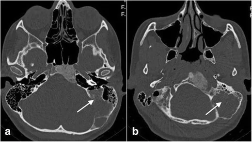
Temporal kemikte yavaş büyüyen kabarıklık, iletim tipi işitme kaybı veya tesadüfi kemik genişlemesi olan genç hastada fibröz displazi olasılığı değerlendirilir. Kontrastsız yüksek çözünürlüklü BT, intradiploik ekspansil lezyonu ve buzlu cam matriksini gösterir; temporal kemikteki yayılımı ve komşu yapıların etkilenmesini değerlendirmeye yardımcı olur. Dış kulak yolu stenozuna sekonder kolesteatom gelişebilir; temporal kemik fibröz displazisinde fasiyal kanal tutulumu da BT’de görülebilir. Paget hastalığı, osteoma veya sınırları keskin başka bir fibro-osseöz lezyonla karıştırmak ise lezyonun yayılım örüntüsünü yanlış yorumlatır.
Faz ve pencere
- Kontrastsız tanısal
- BT’de temporal kemikte intradiploik ekspansil lezyon ve buzlu cam matriksi değerlendirilir.
- Kemik algoritması tanısal
- Aksiyel ve koronal ince kesit rekonstrüksiyonlarda lezyonun dış kulak yolu, timpanik kavite, mastoid, koklea çevresi ve kafa tabanı foraminalarıyla ilişkisi gösterilir; otik kapsül korteksinin korunması ayrıca belirtilir.
Önerilen pencereler G genişlik, M merkez; değerler Hounsfield birimi (HU)
- KemikG 1800 / M +400
- Yumuşak dokuG 400 / M +40
BT bulguları
- Ekspansil lezyon — temporal kemikte kemik genişlemesine yol açabilir ve timpanik yarığı daraltabilir.
- Buzlu cam matriksi — intradiploik ekspansil lezyonun tümünde veya bir bölümünde görülebilir.
- Pagetoid örüntü — fibröz displazide Pagetoid/buzlu cam radyografik örüntüsü görülebilir.
- BT görünümü karma litik-sklerotik, homojen sklerotik veya ağırlıklı kistik/litik olabilir; homojen sklerotik buzlu cam örüntüsü genellikle görülür.
- Dış kulak yolu stenozu — temporal kemik fibröz displazisine eşlik edebilir; sekonder kolesteatom gelişebilir.
- Orta kulak ve kemikçik komşuluğu — lezyon orta kulağa uzanabilir; kemikçik zincirinde erozyon veya fiksasyon görülebilir.
- Otik kapsül — displastik kemik labirenti çevreleyebilir; labirenti saran yoğun kemik dokusunun korunup korunmadığı değerlendirilir.
- Kraniofasiyal fibröz displazide BT görünümü homojen sklerotik, buzlu cam paternden karma litik-sklerotik ya da ağırlıklı kistik/litik paterne değişebilir; lezyon kemiği genişletip korteksi inceltebilir.
Normalde
Fibröz displazi lezyonu labirenti çevrelese bile labirenti saran yoğun kemik doku korunabilir. Kıyaslamada kanalın daralıp daralmadığını, kemik genişlemesinin iç-dış konturunu, matriksin karşı taraf normal kemiğinden buzlu cam görünümüyle ayrılıp ayrılmadığını ve otik kapsül korteksinin korunmasını değerlendirin.
Normal BT ile kıyasla
Aynı bölgenin, kaynak veri setinde patoloji içermediği belirtilen bir BT incelemesini kesit kesit kaydırın; organ sınırlarını açarak anatomiyi hasta görüntüsüyle karşılaştırın. Küçük rastlantısal bulgular tümüyle dışlanmamıştır.
Kaynak: TotalSegmentator v2.01 veri seti (Wasserthal ve ark., Radiology: Artificial Intelligence 2023), CC BY 4.0, vaka s0103; organ sınırları veri setinin otomatik segmentasyonundan, birleştirilerek sadeleştirildi. Görüntüler 2,5 mm kesit aralığına yeniden örneklendi.
Ayırıcı tanı
- Paget hastalığında kafatasında kortikal ve trabeküler kalınlaşma, kemik genişlemesi ve pamuk yünü görünümü görülebilir.
- Osteoma
- BT’de genellikle iyi sınırlı, homojen sklerotik ve kortekse komşu bir lezyondur.
- Ossifiye fibrom
- BT’de ekspansil litik lezyon veya kistik değişiklik alanları içeren solid lezyon şeklinde görülebilir.
- Langerhans hücreli histiyositoz
- BT’de kenarları eğimli litik lezyon görülebilir; yumuşak doku bileşeni eşlik edebilir.
- Sekonder kolesteatom
- dış kulak yolu stenozuna bağlı gelişebilir ve orta kulağa uzanarak kemikçik zincirinde erozyon veya fiksasyona eşlik edebilir; ekspansil buzlu cam matriks fibröz displiziyi destekler.
Tuzaklar
- Dış kulak yolu daralmasını lezyonun tek bulgusu gibi raporlamayın; kanalın gerisinde keratin birikimi, kolesteatom ve orta kulak havalanmasını ayrıca kontrol edin.
- Buzlu cam matriksi her olguda homojen değildir; karma veya pagetoid patern fibröz displaziyi dışlamaz, lezyonun genişlemesi ve kemiğe geçiş sınırıyla birlikte yorumlanır.
- Lezyonun koklea çevresini sarması otik kapsülün yıkıldığı anlamına gelmez; yoğun kortikal labirent halkasının devamlılığını ayrı takip edin.
Kendini dene
Temporal kemik BT’sinde buzlu cam görünümü ve dış kulak yolu stenozu birlikte en çok hangi tanıyı destekler?
Cevabı göster
Fibröz displazi. Temporal kemik fibröz displazisinde BT’de buzlu cam görünümü tanıyı destekler; dış kulak yolu stenozu sık görülen bulgulardandır.
Fibröz displazi dış kulak yolunu belirgin daraltmış. Görüntülemede hangi eşlikçi sorun özellikle aranmalıdır?
Cevabı göster
Sekonder kolesteatom. Kanal stenozu keratin debrisinin birikmesine ve sekonder kolesteatoma zemin hazırlayabilir; stenozun medialindeki yumuşak doku ile erozyon kontrol edilmelidir. Diğer seçenekler kanal daralmasının doğrudan sekeli değildir.
İlgili konular
Kaynaklar
Bu sayfadaki 35 cümle ve 2 quiz sorusu aşağıdaki kaynaklardan alıntılarla otomatik olarak karşılaştırıldı; 11 cümle bu karşılaştırmada düzeltildi (2026-10-10).
- Radiological review of skull lesionspmc.ncbi.nlm.nih.gov
- Fibrous dysplasia: rare manifestation in the temporal bonepmc.ncbi.nlm.nih.gov
- Different clinical presentation and management of temporal bone fibrous dysplasia in childrenpmc.ncbi.nlm.nih.gov
- Minimally invasive resection of a spheno-orbital tumor: Review of surgical techniques and a case reportpmc.ncbi.nlm.nih.gov
- Small ethmoid mucocele with intracranial extension associated with fibrous dysplasia: A rare case reportpmc.ncbi.nlm.nih.gov
- Fibrous Dysplasia of Temporal Bone.pubmed.ncbi.nlm.nih.gov
- Characteristics and management of superior semicircular canal dehiscence.pubmed.ncbi.nlm.nih.gov
- Otosclerosis and Dysplasias of the Temporal Bone.pubmed.ncbi.nlm.nih.gov
- Craniofacial Fibrous Dysplasia Involvements of Mccune-Albright Syndrome: A Review with an Additional Case.pubmed.ncbi.nlm.nih.gov
- Labyrinthine Sequestrum: A Case Report and Review of the Literature.pubmed.ncbi.nlm.nih.gov
- Orbitocranial Fibrous Dysplasia: Immediate Reconstruction with Titanium 3-dimensional Printing.pubmed.ncbi.nlm.nih.gov
- Otalgia revealing McCune-Albright syndrome: A case report.pubmed.ncbi.nlm.nih.gov
- Protuberant fibro-osseous lesions of the temporal bone: two additional case reports.pubmed.ncbi.nlm.nih.gov
- Fibrous dysplasia of the temporal bone: A case from the Glen Williams Ossuary, Ontario, Canada.pubmed.ncbi.nlm.nih.gov
- Fibrous Dysplasia Mimicking Malignancy on 68Ga-DOTATATE PET/CT.pubmed.ncbi.nlm.nih.gov
- Utility of intraoperative computed tomography for cochlear implantation in patients with difficult anatomy.pubmed.ncbi.nlm.nih.gov
- Solitary fibrous tumor with aneurysmal bone cyst-like change in the middle cranial fossa: illustrative case.pubmed.ncbi.nlm.nih.gov
- Craniofacial Fibrous Dysplasia: Clinical and Therapeutic Implications.pubmed.ncbi.nlm.nih.gov
- Fibrous dysplasia of the head and neck in Southern Finland: a retrospective study on clinical characteristics, diagnostics, and treatment.pubmed.ncbi.nlm.nih.gov
- Imaging of Skull Base Tumors.pubmed.ncbi.nlm.nih.gov
- Clinical Outcomes of the U-Linear Split Thickness Skin Graft Technique for Reconstruction for Ear Canal Stenosis.pubmed.ncbi.nlm.nih.gov
- Review of imaging modalities and radiological findings of calvarial lesions.pubmed.ncbi.nlm.nih.gov
- Coexistence of meningioma and craniofacial fibrous dysplasia: a case series of clinicopathological study and literature review.pubmed.ncbi.nlm.nih.gov
- Non-invasive imaging modalities for diagnosing pulsatile tinnitus: a comprehensive review and recommended imaging algorithm.pubmed.ncbi.nlm.nih.gov
- Jansen metaphyseal chondrodysplasia: analysis of craniofacial manifestations.pubmed.ncbi.nlm.nih.gov
- A rare calvarial aneurysmal bone cyst in an adult: Case report and literature review.pubmed.ncbi.nlm.nih.gov
- Intracranial aneurysmal bone cysts in association with fibrous dysplasia: Case report and scoping review of case reports.pubmed.ncbi.nlm.nih.gov
- Severe Manifestation of Hyperparathyroidism With Sagliker Syndrome: A Case Report.pubmed.ncbi.nlm.nih.gov
- Radiological and Surgical Findings of Patients with Intradiploic Mass Lesion: Case Series and Systematic Review.pubmed.ncbi.nlm.nih.gov
- Brazilian Society of Otology task force - Otosclerosis: evaluation and treatment.pubmed.ncbi.nlm.nih.gov
- Rare Diseases of the Middle Ear and Lateral Skull Base.pubmed.ncbi.nlm.nih.gov
- Current Trends, Controversies, and Future Directions in the Evaluation and Management of Superior Canal Dehiscence Syndrome.pubmed.ncbi.nlm.nih.gov
- Key pre-operative CT parameters for cochlear implantation: a focused radiologic review with surgical relevance.pubmed.ncbi.nlm.nih.gov
- Correlation analysis between CT values of cochlear bone labyrinth and auditory function and disease progression in patients with chronic suppurative otitis media.pubmed.ncbi.nlm.nih.gov
- Cochlear Implantation After Temporal Bone Fracture: A Systematic Review of Preoperative Predictors and Timing.pubmed.ncbi.nlm.nih.gov
- Disrupted bone remodeling leads to cochlear overgrowth and hearing loss in a mouse model of fibrous dysplasia.pubmed.ncbi.nlm.nih.gov
- High-affinity alphavbeta3 integrin targeted optical probe as a new imaging biomarker for early atherosclerosis: initial studies in Watanabe rabbits.pubmed.ncbi.nlm.nih.gov
- Invited review: Recommendations for reporting intervention studies on reproductive performance in dairy cattle: Improving design, analysis, and interpretation of research on reproduction.pubmed.ncbi.nlm.nih.gov
- Calcium Phosphates: A Key to Next-Generation In Vitro Bone Modeling.pubmed.ncbi.nlm.nih.gov
- Update from the 5th Edition of the World Health Organization Classification of Head and Neck Tumours: Tumours of the Ear.pubmed.ncbi.nlm.nih.gov
- Mastoid osteoma: A rare incidental finding in an orthodontic patient.pubmed.ncbi.nlm.nih.gov
- Cemento-ossifying fibroma: A South American multicenter study.pubmed.ncbi.nlm.nih.gov
- Cemento-Ossifying Fibroma of the Craniofacial Region: A Diagnostically Challenging Case Report.pubmed.ncbi.nlm.nih.gov
- Ossifying fibroma of the maxilla: A case report with literature review.pubmed.ncbi.nlm.nih.gov
- Operative diagnosis of a meningioma-like intraosseous skull lesion revealing multiple myeloma: Case report and literature review.pubmed.ncbi.nlm.nih.gov
- Langerhans Cell Histiocytosis in Adults: A Canadian Multicenter Case Series.pubmed.ncbi.nlm.nih.gov
- Xanthogranulomatous Osteomyelitis as a Bone Tumor Mimic: A Systematic Review of Pre-biopsy Diagnostic Clues and Management Decision Points.pubmed.ncbi.nlm.nih.gov
- Middle ear malformations combined with cerebrospinal fluid otorrhea: A case report.pubmed.ncbi.nlm.nih.gov
- Postoperative recurrence of traumatic acquired external auditory canal atresia: a case report.pubmed.ncbi.nlm.nih.gov
- A Case of McCune-Albright Syndrome with External Auditory Canal Stenosis Treated with Image-Guided Surgery System-Assisted Temporal Bone Surgery.pubmed.ncbi.nlm.nih.gov
- Clival fibrous dysplasia in which short interval disease progression posed a diagnostic challenge in a skeletally mature patient: a case report.pubmed.ncbi.nlm.nih.gov
- A Case Series and Literature Review of Craniofacial Fibrous Dysplasia.pubmed.ncbi.nlm.nih.gov
- Rare Cause of Congenital Unilateral Facial Palsy: High-Resolution Computed Tomography (HRCT) Findings of Isolated Facial Canal Hypoplasia and Aplasia.pubmed.ncbi.nlm.nih.gov
- Assessment of the Oval Window-Facial Canal Corridor and Mastoid Pneumatization: A Preliminary HRCT-Based Radiological-Anatomical Framework.pubmed.ncbi.nlm.nih.gov
- Different clinical presentation and management of temporal bone fibrous dysplasia in children.pubmed.ncbi.nlm.nih.gov
- External and middle ear diseases: radiological diagnosis based on clinical signs and symptoms.pubmed.ncbi.nlm.nih.gov
- Secondary aneurysmal bone cyst arising from polyostotic craniofacial fibrous dysplasia.pubmed.ncbi.nlm.nih.gov
- Imaging of skull vault tumors in adults.pubmed.ncbi.nlm.nih.gov
- Navigation-Assisted Surgery for Temporal Bone Fibrous Dysplasia With Simultaneous Improvement of Facial Asymmetry and External Auditory Canal Stenosis.pubmed.ncbi.nlm.nih.gov
- Cholesterol granuloma of the sphenoid wing with long-term growth dynamics: illustrative case.pubmed.ncbi.nlm.nih.gov
- Impact of endoscopy and related technologies on indications for transsphenoidal pituitary surgery for growth hormone-producing pituitary neuroendocrine tumors in McCune-Albright syndrome: illustrative case.pubmed.ncbi.nlm.nih.gov
- Small ethmoid mucocele with intracranial extension associated with fibrous dysplasia: A rare case report.pubmed.ncbi.nlm.nih.gov
- Fibrous dysplasia: Rare sphenoidal and parietal involvement.pubmed.ncbi.nlm.nih.gov
- The Convergence of Fibrosis and Granulomatous Inflammation: A Case Series on the Diagnostic and Therapeutic Challenges of Systemic Sclerosis and Sarcoidosis Overlap Syndrome.pubmed.ncbi.nlm.nih.gov
- Diffuse cystic lung diseases-a primer for radiologists.pubmed.ncbi.nlm.nih.gov
- Revisiting Myositis Ossificans: A Comprehensive Stage-by-Stage Imaging Review.pubmed.ncbi.nlm.nih.gov
- Pediatric triad of craniofacial fibrous dysplasia, Chiari malformation type I and syringomyelia: a case report.pubmed.ncbi.nlm.nih.gov
- Severe hypocalcemia in a patient with extensive cranial Paget disease: a diagnostic challenge.pubmed.ncbi.nlm.nih.gov
- Trichoepithelioma of the External Auditory Canal Mimicking Cholesteatoma.pubmed.ncbi.nlm.nih.gov
- Fibroepithelial polyp as an unexpected pathology in the external auditory canal: a case report.pubmed.ncbi.nlm.nih.gov
- Update on diagnostic procedures in third window syndromes.pubmed.ncbi.nlm.nih.gov
- Subtotal petrosectomy in cochlear implantation: Gruppo Otologico experience after 348 cases.pubmed.ncbi.nlm.nih.gov
- Cochlear Implantation by Veria Technique in Abnormal Cochlea: Our Experience.pubmed.ncbi.nlm.nih.gov
- Vestibular Atelectasis: A Narrative Review and Our Experience.pubmed.ncbi.nlm.nih.gov
- Craniofacial disorders and dysplasias: Molecular, clinical, and management perspectives.pubmed.ncbi.nlm.nih.gov
- A Rare Case of Mastoid Process Osteoma Presenting During Puberty: A Case Report.pubmed.ncbi.nlm.nih.gov
- Exploring Atypical Origins of Trismus: Surgical Solutions for Rare Pathologies-Insights from Rare Clinical Cases.pubmed.ncbi.nlm.nih.gov
- Musculoskeletal tumors and tumor-like lesions with "dark" signal intensity on T2-weighted MR images: A pictorial review.pubmed.ncbi.nlm.nih.gov
- Apparent diffusion coefficient can assist in differentiating between benign and malignant primary bone tumors in pediatric patients.pubmed.ncbi.nlm.nih.gov
- Diagnostic Performance of a Locally Deployed Vision Language Model for Bone Tumor Diagnosis Using Smartphone-Captured Images: Exploratory Retrospective Study.pubmed.ncbi.nlm.nih.gov
- Decoding calvarial lesions: Contemporary neurosurgical management integrating radiological and histopathological features.pubmed.ncbi.nlm.nih.gov
- Facial Bone Defects Associated with Lateral Facial Clefts Tessier Type 6, 7 and 8 in Syndromic Neurocristopathies: A Detailed Micro-CT Analysis on Historical Museum Specimens.pubmed.ncbi.nlm.nih.gov
- Imaging of Temporal Bone Mass Lesions: A Pictorial Review.pubmed.ncbi.nlm.nih.gov
- Exploring Prevalence Trends of Jaw Bone Pathologies: A Three-Year Institutional Study.pubmed.ncbi.nlm.nih.gov
- Fibrous Dysplasia of Parietal Bone: A Case Report and Review of Literature.pubmed.ncbi.nlm.nih.gov
- High-resolution computed tomography predicts optimal cochlear implantation strategy in patients with chronic otitis media.pubmed.ncbi.nlm.nih.gov
- Degeneration rather than hypoplasia may be the pathologic feature of the endolymphatic sac in patients with ipsilateral delayed endolymphatic hydrops.pubmed.ncbi.nlm.nih.gov
- Fibrous Dysplasia Developed at the Site of a Previous Central Giant Cell Granuloma in a Middle-Aged Patient: A Case Report.pubmed.ncbi.nlm.nih.gov
- Benign and malignant lesions of the mandible and maxilla: imaging findings and systematic approach.pubmed.ncbi.nlm.nih.gov
- Paget's bone disease in a nonendemic region: Exploring novel therapeutic strategies.pubmed.ncbi.nlm.nih.gov
- Enlivening the radiographic features of Paget disease.pubmed.ncbi.nlm.nih.gov
- Osteosarcoma associated with cemento-osseous dysplasia: co-incidence or two related entities?pubmed.ncbi.nlm.nih.gov
- Eight months of misdiagnosis: Intra-articular osteoid osteoma in a 48-year-old woman.pubmed.ncbi.nlm.nih.gov
- An Unusual Skull With Bilateral Multiple Bony Lesions on the Parietal and Frontal Bones of the Superior Surface of the Calvaria: A Case Report.pubmed.ncbi.nlm.nih.gov
- Intraosseous osteoid osteoma of the temporomandibular joint: a case report and review of the literature.pubmed.ncbi.nlm.nih.gov
- Cystic bone Lesions: Diagnostic pitfalls and therapeutic considerations.pubmed.ncbi.nlm.nih.gov
- Growing intradiploic epidermoid cyst presenting as a diagnostic challenge in the setting of Langerhans cell histiocytosis: A case report.pubmed.ncbi.nlm.nih.gov
- Unveiling Langerhans cell histiocytosis: A rare cause of scalp lesions in pediatrics.pubmed.ncbi.nlm.nih.gov
- Characteristics of the Facial Canal-Lateral Canal Dehiscence: The Under-recognized Third Window.pubmed.ncbi.nlm.nih.gov
Eğitim amaçlıdır; hasta başında tanı ve tedavi kararının yerine geçmez, tıbbi tavsiye değildir. Metinler yapay zekâ desteğiyle yazıldı ve otomatik bir adımda kaynak alıntılarıyla karşılaştırıldı; bu bir tıbbi doğrulama değildir ve içerik uzman radyolog incelemesinden geçmedi. Klinik kararlarda güncel kılavuzları, kurum protokollerini ve radyoloji raporunu esas alın.