Özet
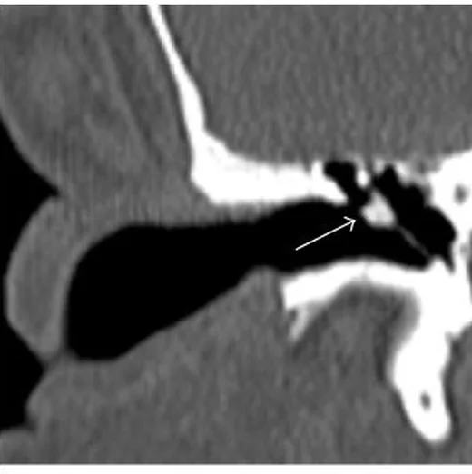
Timpanoskleroz, kronik inflamasyon sonrasında timpanik membran veya orta kulak mukozasında gelişen hiyalinize ve kalsifiye plaklarla karakterizedir. Temporal kemik BT, plakların zar içinde mi orta kulak boşluğunda mı olduğunu ve ossiküler zincir ya da oval pencereyle ilişkisini gösterir. Kemik algoritmasında hiperdens plaklar çevre havalı orta kulakla kontrast oluşturabilir; iletim tipi işitme kaybının kemikçik fiksasyonuyla ilişkisini anatomik olarak açıklar. Kalsifik plaklar kitle veya kolesteatom ile karıştırılabilir ve yanlış tanıya yol açabilir.
Faz ve pencere
- Kontrastsız tanısal
- İnce kesit kemik algoritmalı BT'de timpanik membran veya orta kulakta plak/web tarzı hiperdens kalsifikasyon, ossiküler bağ ve eklemler çevresindeki tutulum ile oval pencere nişi değerlendirilir; kontrast fazı tanısal değildir.
Önerilen pencereler: Kemik (G 1800 / M 400), Yumuşak doku (G 400 / M 40).
BT bulguları
- Timpanik membran boyunca çizgisel veya plak tarzı hiperdens kalınlaşma; zar içinde sınırlı olabilir.
- Orta kulak boşluğunda yuvarlak kitle oluşturmayan, düzensiz veya ağsı kalsifiye plaklar.
- Malleus-incus eklemi, ossiküler bağlar veya stapes çevresinde plak; zincirde fiksasyonla ilişkili anatomik dağılım.
- Oval pencere nişinde kalsifikasyon ve stapes tabanı çevresinde hiperdensite.
- Plak çevresindeki havalı orta kulak boşluğu, kalsifikasyonu sıvı veya yaygın yumuşak dokudan ayırmaya yardım eder; orta kulakta havalı alan yoksa kalsifikasyonun tespiti zorlaşabilir.
- Uzun süreli orta kulak hastalığına bağlı mastoid hipopnömatizasyon eşlik edebilir; tek başına tanısal değildir.
Normalde
Normal timpanik membran ince olup belirgin hiperdens plak içermez; orta kulak boşluğu havalı, ossiküler zincir serbest konturlu ve oval pencere nişi açıktır. Ossiküler zincirde olası timpanoskleroz tutulumunu, ardışık aksiyel kesitlerde kaudokraniyal yönde izle.
Ayırıcı tanı
- Kolesteatom
- genellikle yumuşak doku opasifikasyonu ve basınç/erozyon paterni gösterir; timpanosklerozda plaklar belirgin hiperdens ve kalsifiyedir.
- Otoskleroz
- oval pencerenin önündeki fissula ante fenestram çevresinde demineralizasyon odağı görülür; orta kulak mukozal plakları beklenmez.
- Orta kulak efüzyonu
- hava boşluğunda yumuşak doku/sıvı dansitesi oluşturur, kalsifiye hiperdens plak içermaz.
- Kemikçik anomalisi
- kemikçik şekil veya süreklilik kusuru vardır; çevre mukozada kalsifiye plak eşlik etmez.
Tuzaklar
- Kalsifikasyon yalnız timpanik membran içinde sınırlıysa (myringoskleroz) orta kulak ossiküler tutulum varmış gibi genelleme yapma; plakı anatomik kompartımanıyla tarif et.
- Stapes çevresindeki hiperdens lezyonların oval pencere ilişkisi ve ossiküler tutulum derecesi, ardışık aksiyel kesitler üzerinde izlenerek doğrulanmalıdır.
- Mastoid hipopnömatizasyon eşlik edebilir ama timpanoskleroz tanısını tek başına kanıtlamaz; belirleyici bulgu kalsifiye mukozal/timpanik plak dağılımıdır.
Kendini dene
Havalı orta kulak içinde stapes çevresinde düzensiz hiperdens plaklar var; yumuşak doku kitlesi ve erozyon yok. Tanı nedir?
Cevabı göster
Timpanoskleroz. Plak tarzı hiperdens kalsifikasyon ve korunmuş çevre kemik konturları timpanosklerozu destekler. Kolesteatom yumuşak doku ve olası erozyonla, efüzyon sıvı dansitesiyle, paragangliom ise kitle paterniyle ayrılır.
Oval pencerenin önünde küçük demineralizasyon odağı, orta kulakta kalsifiye plak olmaması hangi tanıyı destekler?
Cevabı göster
Otoskleroz. Fissula ante fenestram çevresindeki demineralizasyon otoskleroz için tipiktir. Timpanoskleroz orta kulak veya membranda hiperdens plaklarla, attik kolesteatom ise Prussak aralığı kitlesi ve scutum değişikliğiyle seyreder.
İlgili konular
- Kronik otitis media
- Edinsel kolesteatom, attik tipi
- Edinsel kolesteatom, pars tensa/sinus timpani tipi
Kaynaklar
Bu sayfadaki 32 cümle ve 2 quiz sorusu aşağıdaki kaynaklardan alıntılarla tek tek karşılaştırıldı; 18 cümle bu karşılaştırmada düzeltildi (2026-10-08).
- Reliability of high‐resolution CT scan in diagnosis of ossicular tympanosclerosispmc.ncbi.nlm.nih.gov
- Clinical Significance of Körner's Septum in Relation to Occurrence of Squamous Chronic Otitis Media.pubmed.ncbi.nlm.nih.gov
- Tophaceous Gout of the Middle Ear: Case Reports and Review of the Literature.pubmed.ncbi.nlm.nih.gov
- Middle Ear Tophi: A Case Series of Two Unusual Lesions and a Report of Facial Weakness and Review of the Literature.pubmed.ncbi.nlm.nih.gov
- Reliability of high-resolution CT scan in diagnosis of ossicular tympanosclerosis.pubmed.ncbi.nlm.nih.gov
- The bone conduction implant--first implantation, surgical and audiologic aspects.pubmed.ncbi.nlm.nih.gov
- Do the angle and length of the eustachian tube influence the development of chronic otitis media?pubmed.ncbi.nlm.nih.gov
- Tympanosclerosis Presenting as Mass: Workup and Differential.pubmed.ncbi.nlm.nih.gov
- Outcomes Following Revision Stapes Surgery in Children: A Single-Surgeon, Single-Center 15-Year Experience.pubmed.ncbi.nlm.nih.gov
- Körner's septum (petrosquamosal lamina): the anatomical variant or clinical problem?pubmed.ncbi.nlm.nih.gov
- [A multi-center retrospective study of ossiculoplasty surgery under oto-endoscope in 45 cases of conductive hearing loss with intact tympanic membrane].pubmed.ncbi.nlm.nih.gov
- Incidental diagnosis of mastoiditis on MRI.pubmed.ncbi.nlm.nih.gov
- The role of the nasal and paranasal sinus pathologies on the development of chronic otitis media and its subtypes: A computed tomography study.pubmed.ncbi.nlm.nih.gov
- [Characteristics of middle ear cholesteatoma with tympanicsclerosis].pubmed.ncbi.nlm.nih.gov
- [Functional multispiral computed tomography of sound-transmitting structures in the middle ear].pubmed.ncbi.nlm.nih.gov
- Idiopathic, Infectious and Reactive Lesions of the Ear and Temporal Bone.pubmed.ncbi.nlm.nih.gov
- Correlation Between Tympanosclerosis and Helicobacter pylori.pubmed.ncbi.nlm.nih.gov
- Diseases of the middle ear in childhood.pubmed.ncbi.nlm.nih.gov
- "Genetic tumor syndromes of the head and neck: Update in the genomic era".pubmed.ncbi.nlm.nih.gov
- Persistent Stapedial Artery, Facial Nerve Overhang, and Calcified Stapedius Tendon Leading to Aborted Stapedotomy: An Unusual Middle Ear Anatomy.pubmed.ncbi.nlm.nih.gov
- Diagnostic Value of Cone-Beam Computed Tomography in Conductive or Mixed Hearing Loss with Intact Tympanic Membrane.pubmed.ncbi.nlm.nih.gov
- Brazilian Society of Otology task force - Otosclerosis: evaluation and treatment.pubmed.ncbi.nlm.nih.gov
- Association of mastoid pneumatization with chronic otitis media: A comparative study.pubmed.ncbi.nlm.nih.gov
- Middle ear malformations combined with cerebrospinal fluid otorrhea: A case report.pubmed.ncbi.nlm.nih.gov
- The preoperative temporal bone CT: a mnemonic for reducing surgical risk.pubmed.ncbi.nlm.nih.gov
- Multimodal imaging in conductive hearing loss: Optimising CT, MRI and CTA for accurate diagnosis and management.pubmed.ncbi.nlm.nih.gov
- The Global Burden of Eustachian Tube Dysfunction in Older Patients: Pathophysiology, Clinical Consequences, Diagnostic Challenges and Therapeutic Perspectives-A SANRA-Based Study.pubmed.ncbi.nlm.nih.gov
- Otosclerosis under microCT: New insights into the disease and its anatomy.pubmed.ncbi.nlm.nih.gov
- Pediatric otosclerosis: case report and literature review.pubmed.ncbi.nlm.nih.gov
- Neuroglial Choristoma of the Middle Ear with Massive Tympanosclerosis: A Case Report and Literature Review.pubmed.ncbi.nlm.nih.gov
- Role of the Ear in Meningitis: A Narrative Review of Neuroimaging.pubmed.ncbi.nlm.nih.gov
- Reconstructive ossiculoplasty options in primary cholesteatoma surgeries with an intact stapes.pubmed.ncbi.nlm.nih.gov
- A novel subtype classification for acute intracranial atherosclerotic disease-related occlusion.pubmed.ncbi.nlm.nih.gov
- The use of explainable artificial intelligence to explore types of fenestral otosclerosis misdiagnosed when using temporal bone high-resolution computed tomography.pubmed.ncbi.nlm.nih.gov
- Extensive Cochlear Otosclerosis: CT-MRI Correlation and the Diagnostic Value of Contrast Enhancement.pubmed.ncbi.nlm.nih.gov
- Progressive Conductive Hearing Loss in Osteogenesis Imperfecta (Lobstein Disease): A Case Report.pubmed.ncbi.nlm.nih.gov
- Automated detection of otosclerosis with interpretable deep learning using temporal bone computed tomography images.pubmed.ncbi.nlm.nih.gov
- Chronically Discharging Ears: Evalution with High Resolution Computed Tomography.pubmed.ncbi.nlm.nih.gov
- A Comparative Analysis of Intraoperative Assessment Versus Preoperative Computed Tomography in Determining Aditus Ad Antrum Patency in Mucosal Chronic Otitis Media.pubmed.ncbi.nlm.nih.gov
- Prognostic Scoring Systems in Ossiculoplasty: A Comprehensive Review and Introduction of a Novel Predictive Index.pubmed.ncbi.nlm.nih.gov
- Feasibility of contrast-enhanced ultrasound as a screening tool for patients at high risk for kidney malignancies.pubmed.ncbi.nlm.nih.gov
- Correlation analysis between CT values of cochlear bone labyrinth and auditory function and disease progression in patients with chronic suppurative otitis media.pubmed.ncbi.nlm.nih.gov
- Dimensions of Osseous External Auditory Canal in Otosclerosis Using High-Resolution Computed Tomography.pubmed.ncbi.nlm.nih.gov
- Photon-Counting CT: Technology, Current and Potential Future Clinical Applications, and Overview of Approved Systems and Those in Various Stages of Research and Development.pubmed.ncbi.nlm.nih.gov
- Clinical Applications of Photon-counting CT: A Review of Pioneer Studies and a Glimpse into the Future.pubmed.ncbi.nlm.nih.gov
- Flat-panel conebeam CT in the clinic: history and current state.pubmed.ncbi.nlm.nih.gov
- Diagnosis and Management of Middle Ear Neuroendocrine Tumour (MeNET).pubmed.ncbi.nlm.nih.gov
- Pediatric Cholesteatoma Presenting as Persistent Otorrhea: A Case of Delayed Diagnosis Across Multiple ENT Specialists.pubmed.ncbi.nlm.nih.gov
- Acute otitis media complicated by peripheral facial nerve palsy in an adult: A case report.pubmed.ncbi.nlm.nih.gov
- Prevalence of the endolymphatic sac hypoplasia in a cohort of individuals without Meniere disease.pubmed.ncbi.nlm.nih.gov
- Prevalence of Cholesteatoma in School Children of Nepal.pubmed.ncbi.nlm.nih.gov
- Imaging of Pathologies of the Temporal Bone and Middle Ear: Inflammatory Diseases, Their Mimics and Potential Complications-Pictorial Review.pubmed.ncbi.nlm.nih.gov
- Development and In Vivo Evaluation of a Novel Bioabsorbable Polylactic Acid Middle Ear Ventilation Tube.pubmed.ncbi.nlm.nih.gov
- Cerebral Venous Outflow Insufficiency: A Study on Symptoms and Venous Stenosis Classification.pubmed.ncbi.nlm.nih.gov
- Hearing results following endoscopic type I tympanoplasty in medium and large perforations.pubmed.ncbi.nlm.nih.gov
- Long-Term Outcomes of Pediatric Cochlear Implantation for Single-Sided Deafness: A Western Australian Perspective.pubmed.ncbi.nlm.nih.gov
- Endoscopic Findings on Facial Nerve Anatomy During Exclusive Endoscopic Stapedotomy: Clinical Considerations and Impact on Surgical Results.pubmed.ncbi.nlm.nih.gov
- Non-Thrombotic Filling Defects in Cerebral Veins and Sinuses: When Normal Structures Mimic a Disease.pubmed.ncbi.nlm.nih.gov
- Rare Cause of Congenital Unilateral Facial Palsy: High-Resolution Computed Tomography (HRCT) Findings of Isolated Facial Canal Hypoplasia and Aplasia.pubmed.ncbi.nlm.nih.gov
- Anatomical Characteristics of the Facial Nerve in Patients With Cochlear Hypoplasia.pubmed.ncbi.nlm.nih.gov
- Assessment of the Oval Window-Facial Canal Corridor and Mastoid Pneumatization: A Preliminary HRCT-Based Radiological-Anatomical Framework.pubmed.ncbi.nlm.nih.gov
- Evaluation of intraoperative color Doppler ultrasonography in the surgical evacuation of acute intracranial hematoma: a single-center study from Botswana.pubmed.ncbi.nlm.nih.gov
- Preliminary Evaluation of Static and Kinematic Magnetic Resonance Imaging (MRI) of the Feline Temporomandibular Joint (TMJ): A Comparative Analysis of Joint Inclination Angles in Intact and Dislocated Conditions.pubmed.ncbi.nlm.nih.gov
- Pediatric pseudo-tumor of the ischio-pubic ramus in virtual non-calcium photon-counting scanner: A case report.pubmed.ncbi.nlm.nih.gov
- Multislice Spiral Computer Tomography Findings of Simple Congenital Middle Ear Malformations.pubmed.ncbi.nlm.nih.gov
- Study on Recovery Strategy of Hearing Loss & SGN Regeneration Under Physical Regulation.pubmed.ncbi.nlm.nih.gov
Eğitim amaçlıdır; hasta başında tanı ve tedavi kararının yerine geçmez. İçerik yapay zekâ destekli bir süreçle güncel literatüre göre cümle cümle kaynaklarla karşılaştırıldı, ancak uzman radyolog incelemesinden geçmedi. Klinik kararlarda güncel kılavuzları, kurum protokollerini ve radyoloji raporunu esas alın.