Özet
Çene osteomu çoğunlukla yavaş büyüyen ve başka nedenle çekilen görüntülerde fark edilen, olgun kemikten oluşan iyi huylu bir kemik oluşumudur. BT, kitlenin korteksle bağlantısını, iç kemik yapısını ve mandibula ya da maksilladaki uzanımını üç boyutlu olarak gösterir. Tek, sınırlı bir çıkıntı ile çok odaklı kraniyofasiyal kemik lezyonlarının ayrımı önemlidir; çoklu osteomlar Gardner sendromu örüntüsünün parçası olabilir. Lezyonun yayılımı veya çoklu oluşu gözden kaçarsa ilişkili iskelet bulguları ve sendromik bağlam fark edilmeyebilir.
Faz ve pencere
- Kontrastsız tanısal
- Kontrast gerekmez; kortikal kemik algoritmasında korteksle devamlı, çevresi seçilebilen ve olgun kemik yoğunluğunda fokal çıkıntı değerlendirilir.
- Kemik algoritması tanısal
- İnce kemik rekonstrüksiyonlarında lezyonun kortikal ya da medüller tabanlı oluşu, mandibula/maksillayla birleşimi ve komşu kemikte düzgün şekillenme izlenir.
Önerilen pencereler: Kemik (G 1800 / M 400), Yumuşak doku (G 400 / M 40).
BT bulguları
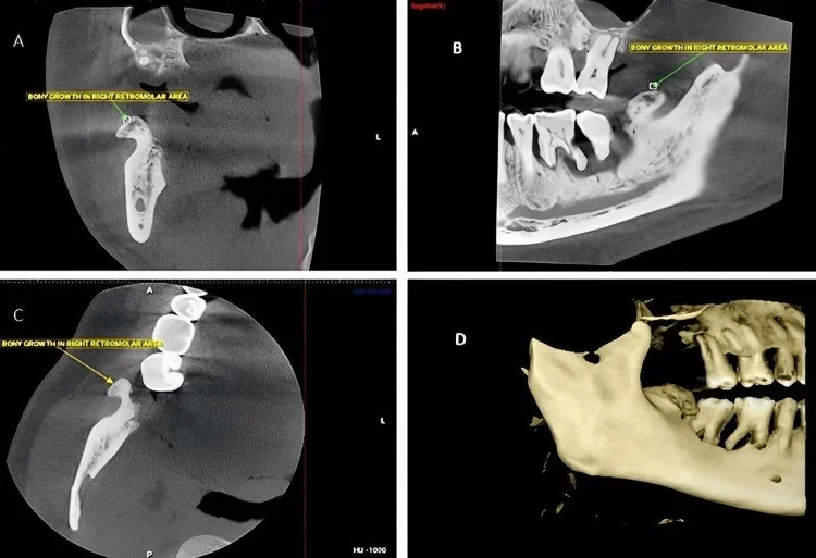
- Periferik osteom, mandibula dış korteksinden çıkıp yuvarlak, oval veya mantar biçimli iyi sınırlı kemik kitle olarak uzanabilir.
- Kompakt tip homojen yoğun kemik görünümündedir; spongiyöz tipte trabeküler yapı ve aralarda ilik alanları seçilebilir.
- Kortikal tabanla kesintisiz devamlılık, lezyonun çeneden kaynaklandığını destekler.
- Yavaş büyümeye eşlik eden düzgün kortikal remodeling görülebilir; agresif permeatif yıkım osteom için beklenen bir görünüm değildir.
- Lezyonun kondil, angulus, korpus veya maksiller sinüs duvarıyla ilişkisi çok düzlemli görüntülerde belirlenir.
- Çenede birden fazla osteom veya eşlik eden gömülü/süpernümerer dişler Gardner sendromu açısından dikkat çekici kraniyofasiyal örüntüdür.
Ölçütler ve sınıflamalar
- Anatomik kökene göre osteom
- Periferik osteom periosttan gelişen ve korteks dışına büyüyen kemik kitlesidir; santral osteom endosteal kökenlidir. BT’de korteksle devamlılık ve lezyonun dışa uzanımı multiplanar olarak doğrulanır.
Normalde
Normal mandibula ve maksillada kortikal kontur kesintisiz, düzgün ve diş alveolleriyle uyumludur; dış yüzeyden kemik yoğunluğunda ayrı bir kitle kabarmaz. Aynı kesit düzeyinde iki taraflı korteks kalınlığını ve konturunu karşılaştır; osteomda odak kortekse bağlı kalırken komşu kemik iliği ve çevre yumuşak doku planları çoğunlukla korunur.
Normal BT ile kıyasla
Aynı bölgenin, kaynak veri setinde patoloji içermediği belirtilen bir BT incelemesini kesit kesit kaydırın; organ sınırlarını açarak anatomiyi hasta görüntüsüyle karşılaştırın. Küçük rastlantısal bulgular tümüyle dışlanmamıştır.
Kaynak: TotalSegmentator v2.01 veri seti (Wasserthal ve ark., Radiology: Artificial Intelligence 2023), CC BY 4.0, vaka s0849; organ sınırları veri setinin otomatik segmentasyonundan, birleştirilerek sadeleştirildi. Görüntüler 2,5 mm kesit aralığına yeniden örneklendi.
Ayırıcı tanı
- Torus veya oral ekzostoz
- tipik yerleşimde düzgün kortikal çıkıntı, özellikle mandibula lingual yüzünde ya da alveoler sırttadır.
- Kondanse osteit, çene kemik yoğunluğu lezyonlarının ayırıcı tanısında dikkate alınmalıdır.
- İdiyopatik osteoskleroz
- medüller kemik içinde yoğun odak oluşturur, dış kortekse bağlı ekzofitik kitle görünümü taşımaz.
- Ossifiye fibrom
- kemik içinde genişleyen, çevresinde sınır oluşturan miks dansiteli lezyon olabilir; osteom gibi kortikal kemik kabartısı değildir.
- Osteosarkom
- kortikal yıkım, agresif periostal yanıt veya yumuşak doku bileşeni benign osteomdan kuşku uyandıracak biçimde ayrılır.
Tuzaklar
- Torus da olgun kemik yoğunluğundadır; mandibula lingual yüzündeki simetrik veya karşılıklı çıkıntıyı tekil periferik osteom olarak adlandırmadan yerleşimi ve karşı tarafı incele.
- Küçük osteom aksiyel kesitte kortikal kalınlaşma gibi görünebilir; koronal ve sagittal rekonstrüksiyonlarda dışa doğru gerçek kitleleşmeyi doğrula.
- Birden çok çene osteomu yalnız lokal bulgu olarak bırakılmamalı; görüntü alanındaki ek osteomları ve eşlik eden diş anomalilerini ara.
Kendini dene
Asemptomatik hastada mandibula angulusuna bağlı, homojen kemik yoğunluklu, düzgün sınırlı dışa büyüyen kitle görülüyor. En olası tanı nedir?
Cevabı göster
Periferik osteom. Korteksle devamlı, olgun kemik yoğunluklu ve iyi sınırlı dışa büyüme osteomu destekler. Ossifiye fibrom, kemik içinde genişleyen ve çevresinde az ossifiye doku hali bulunan ekspansil bir odaktır; osteosarkomda ise kortikal yıkım ve düzensiz sınırlar gibi agresif radyografik bulgular gözlenir.
Çok sayıda çene osteomuna gömülü dişler eşlik ediyor. Görüntüleme açısından hangi ilişki akla gelmelidir?
Cevabı göster
Gardner sendromu. Çoklu kraniyofasiyal osteomlar ve diş anomalileri Gardner sendromu örüntüsünü düşündürür. Diğer seçenekler osteom ve gömülü diş birlikteliğini bu şekilde karakterize etmez.
İlgili konular
Kaynaklar
Bu sayfadaki 38 cümle ve 2 quiz sorusu aşağıdaki kaynaklardan alıntılarla tek tek karşılaştırıldı; 2 cümle bu karşılaştırmada düzeltildi (2026-10-08).
- Management of Anterior Mandibular Exostosis With Cone‐Beam Computed Tomography: A Rare Case Reportpmc.ncbi.nlm.nih.gov
- Imaging Features of Mandibular Ramus Osteoma on Computed Tomography: A Case Report With Literature Analysispmc.ncbi.nlm.nih.gov
- Dilated Odontoma of the Mandible: A Rare Case Report with Discussion about the Differential Diagnosispmc.ncbi.nlm.nih.gov
- Concurrent resection and orthognathic surgery for the management of mandibular condyle osteoma: a case report and literature reviewpmc.ncbi.nlm.nih.gov
- Evaluation of Jaw Pathologies by Computed Tomographypmc.ncbi.nlm.nih.gov
- Multidetector Row Computed Tomography in Maxillofacial Imaging.pubmed.ncbi.nlm.nih.gov
- Orthognathic Surgery in Cleidocranial Dysplasia.pubmed.ncbi.nlm.nih.gov
- Ligature-induced peri-implantitis in mice.pubmed.ncbi.nlm.nih.gov
- Nuclear Medicine Imaging in the Dentomaxillofacial Region.pubmed.ncbi.nlm.nih.gov
- A review on computer-aided design and manufacturing of patient-specific maxillofacial implants.pubmed.ncbi.nlm.nih.gov
- The MB3 canal in maxillary molars: a micro-CT study.pubmed.ncbi.nlm.nih.gov
- External apical root resorption and vectors of orthodontic tooth movement.pubmed.ncbi.nlm.nih.gov
- A morphometric analysis of the immature human infraorbital canal.pubmed.ncbi.nlm.nih.gov
- 3D-cinematic rendering for dental and maxillofacial imaging.pubmed.ncbi.nlm.nih.gov
- Occlusal Angles of Equine Incisors.pubmed.ncbi.nlm.nih.gov
- Delayed Implants Outcome in Maxillary Molar Region.pubmed.ncbi.nlm.nih.gov
- The Specific Morphological Features of Alveolar Bone.pubmed.ncbi.nlm.nih.gov
- Dental Anomalies Associated with Craniometaphyseal Dysplasia.pubmed.ncbi.nlm.nih.gov
- A systematic approach to CT evaluation of orbital trauma.pubmed.ncbi.nlm.nih.gov
- Multiple Osteomas on the Skull: A Case Report and Literature Review.pubmed.ncbi.nlm.nih.gov
- Imaging Features of Mandibular Ramus Osteoma on Computed Tomography: A Case Report With Literature Analysis.pubmed.ncbi.nlm.nih.gov
- Peripheral Osteoma on the Medial Surface of the Mandibular Ramus: A Case Report.pubmed.ncbi.nlm.nih.gov
- Cone-beam computed tomography assessment of the prevalence and distribution of bone islands of the jaws in a group of chinese patients.pubmed.ncbi.nlm.nih.gov
- Giant cementoblastoma involving multiple teeth: A rare case report and review of differential diagnoses.pubmed.ncbi.nlm.nih.gov
- Poor condyle position after mandibular reconstruction and a classification system for patients with "VSCU" based on computed tomography: a cross-sectional study with retrospective data collection.pubmed.ncbi.nlm.nih.gov
- Craniomaxillofacial Peripheral Osteomas in Gardner Syndrome.pubmed.ncbi.nlm.nih.gov
- Diagnostic and Treatment Protocols for Peripheral Craniofacial Osteomas.pubmed.ncbi.nlm.nih.gov
- Gardner syndrome initially misdiagnosed as bilateral breast malignancy: a case report and literature review.pubmed.ncbi.nlm.nih.gov
- Proposed differential diagnosis and classification of non-syndromic and syndromic supernumerary teeth.pubmed.ncbi.nlm.nih.gov
- Imaging of Skull Base Tumors.pubmed.ncbi.nlm.nih.gov
- Bone-related disorders of the jaw: A clinico-radiological diagnostic algorithm.pubmed.ncbi.nlm.nih.gov
- A practical overview of CT and MRI features of developmental, inflammatory, and neoplastic lesions of the sphenoid body and clivus.pubmed.ncbi.nlm.nih.gov
- Giant Peripheral Osteoma of the Mandibular Antegonial Notch in an Elderly Patient: A Case Report.pubmed.ncbi.nlm.nih.gov
- "Genetic tumor syndromes of the head and neck: Update in the genomic era".pubmed.ncbi.nlm.nih.gov
- Ossifying Fibroma of Non-odontogenic Origin: A Fibro-osseous Lesion in the Craniofacial Skeleton to be (Re-)considered.pubmed.ncbi.nlm.nih.gov
- A Rare Case of Peripheral Exophytic Osteoma of the Mandible Arising From an Extraction Site: Cone-Beam Computed Tomography (CBCT) Findings.pubmed.ncbi.nlm.nih.gov
- A Rare Case of Peripheral Osteoma of the Alveolar Bone of the Maxilla in a 13-Year-Old Boy.pubmed.ncbi.nlm.nih.gov
- The Curious Case of a 'Maverick' Cementoblastoma!pubmed.ncbi.nlm.nih.gov
- Non-odontogenic tumors of the facial bones in children and adolescents: role of multiparametric imaging.pubmed.ncbi.nlm.nih.gov
- Management of Anterior Mandibular Exostosis With Cone-Beam Computed Tomography: A Rare Case Report.pubmed.ncbi.nlm.nih.gov
- Case Report: Ossifying parosteal lipoma of the mandible with cortical continuity: surgical considerations from a rare case and literature review.pubmed.ncbi.nlm.nih.gov
- Condylar Osteochondroma in an Older Adult With Facial Asymmetry: Case Report and Differentiation From Condylar Hyperplasia.pubmed.ncbi.nlm.nih.gov
- Intramedullary osteosclerosis of the tibia with inflammatory features: Multimodality imaging findings.pubmed.ncbi.nlm.nih.gov
- Giant intraosseous (ectopic) meningioma of the temporoparietal calvarium with rapid expansion over 5 years: A case report.pubmed.ncbi.nlm.nih.gov
- Masses of developmental and genetic origin affecting the paediatric craniofacial skeleton.pubmed.ncbi.nlm.nih.gov
- International Harmonization of Nomenclature and Diagnostic Criteria (INHAND): Nonproliferative and Proliferative Lesions of the Rabbit.pubmed.ncbi.nlm.nih.gov
- Impaction of Deciduous and Permanent Teeth Related to Local Obstacles: A Retrospective Study of 10 Years of Institutional Experience.pubmed.ncbi.nlm.nih.gov
- Gardner syndrome: When cervical-facial osteomas reveal the tip of the iceberg: A case report and literature review.pubmed.ncbi.nlm.nih.gov
- Pediatric Intraosseous Cavernous Hemangioma of the Mandible: A Case Series Highlighting Diagnostic Challenges and Surgical Management.pubmed.ncbi.nlm.nih.gov
- Fibrous dysplasia of the jaws: four cases with conservative and resective surgical management.pubmed.ncbi.nlm.nih.gov
- Mandibular Osseous Changes in Children and Adolescents With Beta Thalassemia Major: A Panoramic Radiomorphometric Study.pubmed.ncbi.nlm.nih.gov
- CBCT-Based Assessment of Mandibular Canal Position and Bone Morphology at Critical Osteotomy Sites for Sagittal Split Ramus Osteotomy.pubmed.ncbi.nlm.nih.gov
- Osteoporosis Diagnostics in Dental Radiology (Panoramic Radiograph): The Mini Osteoporosis Pre-Screening (MOPS).pubmed.ncbi.nlm.nih.gov
- Osteomyelitis of the Jaw Bones and Its Mimics: Resolving the Diagnostic Enigma.pubmed.ncbi.nlm.nih.gov
- Benign and malignant lesions of the mandible and maxilla: imaging findings and systematic approach.pubmed.ncbi.nlm.nih.gov
- Efficiency and Safety of Dental Implantation in the Area of Hyperdense Jaw Lesions: A Narrative Review.pubmed.ncbi.nlm.nih.gov
- The quiet flame: whole-body MRI and the underestimated burden of pediatric chronic non-bacterial osteomyelitis (CNO).pubmed.ncbi.nlm.nih.gov
- Osteoblastoma of the Mandible: A Case Report.pubmed.ncbi.nlm.nih.gov
- The Occurrence and Outcomes of Cemento-Osseous Dysplasias (COD) in the Jaw Bones of the Population of Lower Silesia, Poland.pubmed.ncbi.nlm.nih.gov
- The World Health Organization Classification of Odontogenic Lesions: A Summary of the Changes of the 2022 (5th) Edition.pubmed.ncbi.nlm.nih.gov
- Multimodal Imaging of Osteosarcoma: From First Diagnosis to Radiomics.pubmed.ncbi.nlm.nih.gov
- Cone-Beam Computed Tomography Imaging of Primary Non-Hodgkin Lymphoma of the Jaws: A 6-Case Series.pubmed.ncbi.nlm.nih.gov
- Maxillofacial tubercular osteomyelitis: diagnostic and therapeutic perspectives from a case series.pubmed.ncbi.nlm.nih.gov
- Sarcomatoid squamous cell carcinoma of the mandible: Radiologic-pathologic correlation and diagnostic pitfalls of an aggressive osteolytic lesion.pubmed.ncbi.nlm.nih.gov
- Imaging of pediatric calvarial and skull base tumors: A COG Diagnostic Imaging Committee/SPR Oncology Committee/ASPNR White Paper.pubmed.ncbi.nlm.nih.gov
- Craniofacial Osteomas: From Diagnosis to Therapy.pubmed.ncbi.nlm.nih.gov
- Novel Mutation in APC Gene Associated with Multiple Osteomas in a Family and Review of Genotype-Phenotype Correlations of Extracolonic Manifestations in Gardner Syndrome.pubmed.ncbi.nlm.nih.gov
- Developmental disorders of the dentition: an update.pubmed.ncbi.nlm.nih.gov
- Intraoral Scanning Enables Virtual-Splint-Based Non-Invasive Registration Protocol for Maxillofacial Surgical Navigation.pubmed.ncbi.nlm.nih.gov
- Cranio-Maxillo-Facial Reconstruction with Polyetheretherketone Patient-Specific Implants: Aesthetic and Functional Outcomes.pubmed.ncbi.nlm.nih.gov
- Utility of optical see-through head mounted displays in augmented reality-assisted surgery: A systematic review.pubmed.ncbi.nlm.nih.gov
- Large Peripheral Osteomas and Dental Implants: A Case Report.pubmed.ncbi.nlm.nih.gov
- Bone marrow edema in children: chronic nonbacterial osteomyelitis and its mimickers.pubmed.ncbi.nlm.nih.gov
- Familial multiple impacted supernumerary teeth: Case report.pubmed.ncbi.nlm.nih.gov
- Gardner's Syndrome: Vigilance Better than Negligence - A Case Report.pubmed.ncbi.nlm.nih.gov
- Rare Presentation of Cemento-Ossifying Fibroma in the Anterior Maxilla: A Case Report.pubmed.ncbi.nlm.nih.gov
- Respiratory Distress Arising from a Peripheral Osteoma: A Rare Case Report.pubmed.ncbi.nlm.nih.gov
- Performance of AI Chatbots in Preliminary Diagnosis of Maxillofacial Pathologies.pubmed.ncbi.nlm.nih.gov
- Clinicopathological and molecular insights into odontogenic tumors associated with syndromes: A comprehensive review.pubmed.ncbi.nlm.nih.gov
- Rare Germline Variants in the Adenomatous Polyposis Coli Gene Associated with Dental and Osseous Anomalies.pubmed.ncbi.nlm.nih.gov
- A novel mutation of adenomatous polyposis coli (APC) gene results in the formation of supernumerary teeth.pubmed.ncbi.nlm.nih.gov
- One of the rarest syndromes in dentistry: gardner syndrome.pubmed.ncbi.nlm.nih.gov
- Concurrent resection and orthognathic surgery for the management of mandibular condyle osteoma: a case report and literature review.pubmed.ncbi.nlm.nih.gov
- How Clinical and Radiological Findings in Chronic Mandibular Osteomyelitis Do Not Always Correlate: Diagnostic Dilemmas in Dental-Related Bone Inflammations.pubmed.ncbi.nlm.nih.gov
- 2025 FelineVMA feline oral health and dental care guidelines.pubmed.ncbi.nlm.nih.gov
- Idiopathic Osteosclerosis in Orthodontic Patients: A Report of Two Cases.pubmed.ncbi.nlm.nih.gov
- Decompression Tube Fixation on Adjacent Teeth for Cystic Lesions in the Jaws: A Long-Term Follow-Up Analysis of Predictive Factors.pubmed.ncbi.nlm.nih.gov
- Parosteal lipoma of the mandible: A case report and review of the literature.pubmed.ncbi.nlm.nih.gov
- Solitary submandibular soft tissue osteochondroma: A rare case report.pubmed.ncbi.nlm.nih.gov
Eğitim amaçlıdır; hasta başında tanı ve tedavi kararının yerine geçmez. İçerik yapay zekâ destekli bir süreçle güncel literatüre göre cümle cümle kaynaklarla karşılaştırıldı, ancak uzman radyolog incelemesinden geçmedi. Klinik kararlarda güncel kılavuzları, kurum protokollerini ve radyoloji raporunu esas alın.