Özet
Çölyak enteropatisi, ince barsak mukozasında villöz hasara ve malabsorpsiyon bulgularına yol açan immün aracılı bir hastalıktır. Refrakter çölyak hastalarında katlantı örüntüsü anormalliği, ince bağırsak duvar kalınlaşması ve dilatasyon iyileşmiş çölyak grubuna göre daha sık bildirilmiştir; bu bulgular kesitsel görüntülemede görülebilir. Dirençli çölyak hastalarında kesitsel görüntülemede genel olarak ince bağırsak katlantı örüntüsünde bozulma, direnç olmayan hastalara kıyasla daha yüksek oranda saptanır. Kesitsel görüntüleme bulguları, dirençli çölyak tipinin ayrımı ve mukozal iyileşme durumunun ayırt edilmesinde klinik ve histopatolojik verilerle birlikte kullanılır.
Faz ve pencere
- Portal
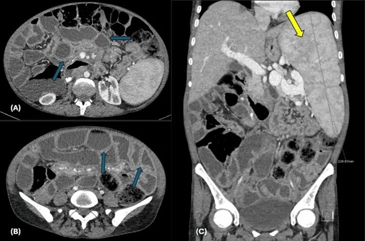
- Enterografi teknikleri, yeterli barsak distansiyonu sağlandığında ince bağırsak duvarı, lümen ve mezenter yapısını detaylı olarak değerlendirmeyi mümkün kılar. Dirençli çölyakta kesitsel görüntülemede ince bağırsak duvar kalınlaşması ve dilatasyon sık görülür; ayrıca mezenterik lenf nodu büyümesi ve kavitasyon sendromu gibi özel komplikasyonlar izlenebilir.
Önerilen pencereler: Batın (G 400 / M 50), Yumuşak doku (G 400 / M 40).
BT bulguları
- Dirençli çölyak hastalarında ince bağırsak katlantı örüntüsünde bozulma, sağlıklı mukozaya sahip hastalara kıyasla anlamlı olarak daha yaygındır.
- Dirençli olmayan çölyak hastalarında ince bağırsak katlantı örüntüsündeki anormallikler dirençli alt tiplere göre çok daha seyrek izlenir.
- İleal jejunizasyon — normalde daha seyrek görünmesi beklenen ileal katlantılar belirginleşerek jejunal örüntüye yaklaşır.
- İnce barsak dilatasyonu — lümen genişlemesi ve sıvı içerikli anslar malabsorptif enteropatiye eşlik edebilir, ancak özgül değildir.
- Duodenum ve jejunum duvar değişikliği — mukozal katlantıların düzensizliği veya duvar kalınlaşması hastalık yüküyle birlikte görülebilir; inflamatuvar nedenlerle örtüşür.
- Geçici enteroenterik invajinasyon — iç içe geçmiş barsak duvarı ve mezenter yağının oluşturduğu hedef görünümü olabilir; kalıcı geçiş engeli veya kurşun nokta olup olmadığı araştırılır.
- Mezenterik lenf nodlarında merkezi nekrotik veya yağlı kavitasyon izlenmesi, çölyak hastalığına özgü nadir bir komplikasyon olan kaviter mezenterik lenf nodu sendromunu düşündürür.
- Refrakter çölyakta katlantı örüntüsü anormallikleri iyileşmiş çölyaka göre daha sık bildirilmiştir; ileal jejunizasyon, ileum katlantılarının normal jejunumdakine benzer görünmesidir.
- Tip II refrakter çölyak hastalarında ince bağırsak duvar kalınlaşması ve dilatasyonu, tip I refrakter ve iyileşmiş çölyak gruplarına göre daha sık bildirilmiştir.
Normalde
Yeterli barsak distansiyonu, MR enterografi görüntü kalitesini etkileyen temel parametrelerden biridir. Normal jejunum görünümü, ileal katlantı kalınlaşmasının “jejunizasyon” olarak adlandırılmasında karşılaştırma ölçütüdür. Kesitsel görüntüleme refrakter çölyak tipini tek başına sınıflandırmaz; S10 çalışmasında sınıflandırma, ince bağırsak biyopsisinde T hücre reseptör yeniden düzenlenmesi veya T hücre immünofenotiplemesiyle yapılmıştır.
Normal BT ile kıyasla
Aynı bölgenin, kaynak veri setinde patoloji içermediği belirtilen bir BT incelemesini kesit kesit kaydırın; organ sınırlarını açarak anatomiyi hasta görüntüsüyle karşılaştırın. Küçük rastlantısal bulgular tümüyle dışlanmamıştır.
Kaynak: TotalSegmentator v2.01 veri seti (Wasserthal ve ark., Radiology: Artificial Intelligence 2023), CC BY 4.0, vaka s0119; organ sınırları veri setinin otomatik segmentasyonundan, birleştirilerek sadeleştirildi. Görüntüler 2,5 mm kesit aralığına yeniden örneklendi.
Ayırıcı tanı
- Crohn hastalığında kesitsel görüntülemede duvar kalınlaşması, abses, darlık, fistül ve mezenterik değişiklikler gibi inflamatuvar ve penetran bulgular öne çıkar.
- Giardiyaziste duodenal villöz atrofi ve BT-enterografide ince barsak anslarında dilatasyon görülebilir.
- İnce barsak lenfoması
- fokal veya uzun segmentli kitle, belirgin duvar kalınlaşması, anevrizmal lümen genişlemesi ya da hacimli nodlar görülebilir.
- İntestinal tüberküloz
- terminal ileoçekal tutulum ve merkezî nekrozlu mezenterik nodlar çölyak katlantı örüntüsünden farklıdır.
- Geçici invajinasyon için patolojik kurşun nokta
- kalıcı kitle, belirgin proksimal dilatasyon veya devam eden invajinasyon varsa çölyakla ilişkili geçici bulgudan ayrılmalıdır.
- Katlantı örüntüsü anormallikleri refrakter çölyakta iyileşmiş çölyaka göre daha sık bildirilmiştir; refrakter çölyak tipinin sınıflandırılması için ince bağırsak biyopsisinde T hücre reseptör yeniden düzenlenmesi veya T hücre immünofenotiplemesi kullanılmıştır.
Tuzaklar
- Yetersiz oral kontrast veya çökmüş anslar katlantı azalması izlenimi verir; jejunoileal örüntüyü yalnız iyi distandü segmentlerde kıyaslayın.
- S10 çalışmasında katlantı örüntüsü anormalliği iyileşmiş çölyak grubunun %16’sında görülmüştür.
- Geçici kısa invajinasyonu otomatik olarak tümör kabul etmeyin; devamlılık, kurşun nokta, proksimal obstrüksiyon ve çevresel sıvı bulgularını arayın.
- Katlantı örüntüsü çölyak için ipucu olsa da BT ile kesin tanı konmaz; görüntüyü klinik, seroloji ve biyopsiyle ilişkilendirin.
- Yetersiz distansiyon MR enterografi görüntü kalitesini etkileyebilir; ince bağırsak distansiyonunu artırmaya yönelik stratejiler kullanılabilir.
Kendini dene
S10 çalışmasında tip II refrakter çölyak hastalarında katlantı örüntüsü anormalliği hangi oranda bildirilmiştir?
Cevabı göster
%53. S10’da katlantı örüntüsü anormalliği tip II refrakter çölyakta %53, tip I refrakter çölyakta %43 ve iyileşmiş çölyakta %16 oranında bildirilmiştir.
Erişkinlerde BT'de tesadüfen saptanan invajinasyonların izlem görüntülemesindeki seyri için aşağıdakilerden hangisi doğrudur?
Cevabı göster
Çoğu kendiliğinden gerileyen benign, geçici süreçlerdir; invajinasyona yol açan patolojik bir lezyon seyrek saptanır.. S39 kohortunda izlem görüntülemesi yapılan hastaların %74,6'sında invajinasyon kendiliğinden gerilemiş; tüm kohortta patolojik bir öncü lezyon %11,3, ileus ise %4,7 oranında bildirilmiştir.
İlgili konular
- Enfeksiyöz enterit
- Crohn hastalığında mezenterik yağ proliferasyonu
- Crohn ile ilişkili ince barsak adenokarsinomu
Kaynaklar
Bu sayfadaki 38 cümle ve 2 quiz sorusu aşağıdaki kaynaklardan alıntılarla otomatik olarak karşılaştırıldı; 26 cümle bu karşılaştırmada düzeltildi (2026-10-09).
- Celiac Disease Emerging After ASCT in Multiple Myeloma: A Rare Diagnostic Intersection and Literature Reviewpmc.ncbi.nlm.nih.gov
- Olmesartan-Induced Enteritis Mimicking Small Bowel Crohn's Diseasepmc.ncbi.nlm.nih.gov
- Nutritional remission without mucosal healing in type II refractory celiac disease: a case reportpmc.ncbi.nlm.nih.gov
- Usual Interstitial Pneumonia in a Patient With Celiac Disease: Expanding the Spectrum of Pulmonary Manifestationspmc.ncbi.nlm.nih.gov
- Recurrent Transpyloric Reflux of a Video Capsule: A Rare Kinetic Sign of Superior Mesenteric Artery Syndromepmc.ncbi.nlm.nih.gov
- Headache Associated with Coeliac Disease: A Systematic Review and Meta-Analysis.pubmed.ncbi.nlm.nih.gov
- Interdisciplinary approach in emergency revascularization and treatment for acute mesenteric ischemia.pubmed.ncbi.nlm.nih.gov
- Gastric ischaemia: a rare cause of gastric ulcers.pubmed.ncbi.nlm.nih.gov
- Lactase genetic polymorphisms and coeliac disease in children: a cohort study.pubmed.ncbi.nlm.nih.gov
- Cross-sectional imaging in refractory celiac disease.pubmed.ncbi.nlm.nih.gov
- Case report of a gallbladder agenesis, a diagnostic challenge.pubmed.ncbi.nlm.nih.gov
- Management of chronic atherosclerotic mesenteric ischemia.pubmed.ncbi.nlm.nih.gov
- Intestinal Spirocheteosis in a Patient with Celiac Disease.pubmed.ncbi.nlm.nih.gov
- Intestinal pseudo-obstruction caused by Giardia lamblia infection.pubmed.ncbi.nlm.nih.gov
- Incidental Diagnosis of Monomorphic Epitheliotropic Intestinal T-Cell Lymphoma: A Case Report.pubmed.ncbi.nlm.nih.gov
- Diagnosis and staging in gastrointestinal lymphoma.pubmed.ncbi.nlm.nih.gov
- Immunomodulatory activity of recombinant α-gliadin conjugated to cholera toxin in DQ8 transgenic mice.pubmed.ncbi.nlm.nih.gov
- Celiac Disease on FDG PET/CT.pubmed.ncbi.nlm.nih.gov
- Nutritional remission without mucosal healing in type II refractory celiac disease: a case report.pubmed.ncbi.nlm.nih.gov
- Pancytopenia as the Initial Presentation of Celiac Disease.pubmed.ncbi.nlm.nih.gov
- Celiac Diseasepubmed.ncbi.nlm.nih.gov
- Epidemiological and clinical trends of malignant small-bowel tumors in Asia.pubmed.ncbi.nlm.nih.gov
- Eosinophilic Duodenitis and Jejunitis with Ascites and Peripheral Eosinophilia: A Diagnostic Challenge-Case Report.pubmed.ncbi.nlm.nih.gov
- Role of magnetic resonance in bowel pathologies: Beyond inflammatory bowel disease.pubmed.ncbi.nlm.nih.gov
- Malignancies in Celiac Disease-A Hidden Threat with Diagnostic Pitfalls.pubmed.ncbi.nlm.nih.gov
- A clinician's perspective on the new organ mesentery and non-vascular mesenteropathies.pubmed.ncbi.nlm.nih.gov
- POCUS for acute abdominal pain: practical scan protocols on gastrointestinal diseases and an evidence review.pubmed.ncbi.nlm.nih.gov
- Small Bowel Imaging from Stepchild of Roentgenology to MR Enterography: Part I: Guidance in Performing and Observing Normal and Abnormal Imaging Findings.pubmed.ncbi.nlm.nih.gov
- Duodenal imaging on the spotlight: from A to Z.pubmed.ncbi.nlm.nih.gov
- The Many Faces of Intestinal Tumors in Adults, Including the Primary Role of CT Imaging in Emergencies and the Important Role of Cross-Sectional Imaging: A Pictorial Review.pubmed.ncbi.nlm.nih.gov
- Intestinal tuberculosis versus crohn's disease: Clinical and radiological recommendations.pubmed.ncbi.nlm.nih.gov
- Gastrointestinal imaging-practical magnetic resonance imaging approach.pubmed.ncbi.nlm.nih.gov
- Jejunal amyloidosis: a rare case presented with sudden weight loss and altered bowel habits.pubmed.ncbi.nlm.nih.gov
- Nephrocalcinosis Following Roux-en-Y Gastric Bypass Secondary to Suspected Enteric Hyperoxaluria: A Case Report.pubmed.ncbi.nlm.nih.gov
- Analysis of Diagnosis and Treatment of Active Lupus Enteritis Accompanied by Intestinal Pseudo-Obstruction as the Initial Manifestation: A Case Report and Literature Review.pubmed.ncbi.nlm.nih.gov
- Chronic intestinal pseudo-obstruction secondary to intestinal leiomyositis in a ragdoll cat.pubmed.ncbi.nlm.nih.gov
- Case report of distal gastrectomy applied in primary duodenal bulb adenocarcinoma.pubmed.ncbi.nlm.nih.gov
- Imaging findings of gastrointestinal tract tumors in children and adolescents.pubmed.ncbi.nlm.nih.gov
- Clinical course and predictive factors of incidentally detected adult intussusceptions on CT: a retrospective cohort study.pubmed.ncbi.nlm.nih.gov
- A Rare Presentation of Adult Intussusception.pubmed.ncbi.nlm.nih.gov
- Idiopathic double-site jejunoileal intussusception with volvulus and transmural necrosis in a young adult: case report.pubmed.ncbi.nlm.nih.gov
- Intestinal Lymphangioma With Extensive Secondary Lymphangiectasia Causing Life-Threatening Hemorrhage: First Case Requiring Small Bowel Transplantation.pubmed.ncbi.nlm.nih.gov
- Cavitating mesenteric lymph node syndrome: a rare complication of celiac disease-case report and literature review.pubmed.ncbi.nlm.nih.gov
- Mosunetuzumab Successfully Treated Rituximab-Refractory Lymphoma-Associated Protein-Losing Enteropathy With Concomitant Immune Thrombocytopenia in Follicular Lymphoma: A Case Report.pubmed.ncbi.nlm.nih.gov
- Transabdominal ultrasonography of the small bowel.pubmed.ncbi.nlm.nih.gov
- The Overlap Between Crohn's Disease and Intestinal Tuberculosis: A Never-Ending Story.pubmed.ncbi.nlm.nih.gov
- Uncommon ileal perforation due to intestinal tuberculosis: A case report and literature review.pubmed.ncbi.nlm.nih.gov
- Crohn's disease: review and standardization of nomenclature.pubmed.ncbi.nlm.nih.gov
- Unmasking the Parasite: When Giardiasis Looks Like Gastroenteritis.pubmed.ncbi.nlm.nih.gov
- A Case Report and Literature Review of an Uncommon Manifestation of Giardiasis: Protein-Losing Enteropathy in an Immunocompetent Host.pubmed.ncbi.nlm.nih.gov
- CVID Enteropathy Associated With Chronic Norovirus Infection: Background, Clinical Features, and Therapeutic Aspects.pubmed.ncbi.nlm.nih.gov
- When Histology Is Unknown: Surgical Decision-Making in Small Bowel Tumors-From Preoperative Imaging to Intraoperative Findings: A Narrative Review.pubmed.ncbi.nlm.nih.gov
- Ileal tuberculosis presenting with severe unexplained anemia and unusual pseudo-tumoral imaging features: A case report.pubmed.ncbi.nlm.nih.gov
- Monomorphic Epitheliotropic Intestinal T-Cell Lymphoma: A Case of Extranodal Non-Hodgkin Lymphoma in the Jejunum with Invasion of the Abdominal Wall and Transverse Colon.pubmed.ncbi.nlm.nih.gov
- Incidental mass found on screening colonoscopy.pubmed.ncbi.nlm.nih.gov
- Hiding in Plain Sight: A Case of Intestinal Tuberculosis Masquerading as Benign Lower GI Bleeding.pubmed.ncbi.nlm.nih.gov
- Early postoperative ileoileal intussusception following adhesiolysis and enterouterine fistula resection in an adult: a rare case report.pubmed.ncbi.nlm.nih.gov
- A Collision Tumour Phenomenon: Dual Metastatic Deposits From Melanoma and Adrenal Carcinoma Causing Small Bowel Intussusception.pubmed.ncbi.nlm.nih.gov
- Ileocecal Lipoma as a Lead Point for Adult Ileocolic Intussusception: A Diagnostic Challenge.pubmed.ncbi.nlm.nih.gov
- Imaging of Strictures in Crohn's Disease.pubmed.ncbi.nlm.nih.gov
- Challenges and Strategies to Optimising the Quality of Small Bowel Magnetic Resonance Imaging in Crohn's Disease.pubmed.ncbi.nlm.nih.gov
- Imaging Modalities for Evaluation of Intestinal Obstruction.pubmed.ncbi.nlm.nih.gov
- Artificial Intelligence in Gastrointestinal Wireless Capsule Endoscopy: A Systematic Literature Review and Meta-Analysis.pubmed.ncbi.nlm.nih.gov
- Real-time microscopic visualisation: current applications and challenges of confocal laser endomicroscopy in the diagnosis and treatment of small bowel disorders.pubmed.ncbi.nlm.nih.gov
- Atypical presentation for a gastrointestinal basidiobolomycosis and rare side effect of Voriconazole: A case report and literature review.pubmed.ncbi.nlm.nih.gov
- The role of enteroscopy in the diagnosis and interventional management of small intestine diseases.pubmed.ncbi.nlm.nih.gov
- Cancer Risk in Clinically Recognized Celiac Disease: A Nationwide Propensity-Matched Cohort Study.pubmed.ncbi.nlm.nih.gov
- Challenges in the diagnosis of marginal zone lymphoma with symptoms of small intestinal disease: a case report and scoping review of the literature.pubmed.ncbi.nlm.nih.gov
- A Twisting Diagnosis: A Case Report of Double Intussusception Uncovering an Unexpected Malignant Melanoma.pubmed.ncbi.nlm.nih.gov
- Chronic Intestinal Failure and Short Bowel Syndrome in Adults: The State of the Art.pubmed.ncbi.nlm.nih.gov
- The Importance of Magnetic Resonance Enterography in Monitoring Inflammatory Bowel Disease: A Review of Clinical Significance and Current Challenges.pubmed.ncbi.nlm.nih.gov
- Celiac disease - a pluripathological model in pediatric practice.pubmed.ncbi.nlm.nih.gov
- Celiac Disease Emerging After ASCT in Multiple Myeloma: A Rare Diagnostic Intersection and Literature Review.pubmed.ncbi.nlm.nih.gov
- Olmesartan-Induced Enteritis Mimicking Small Bowel Crohn's Disease.pubmed.ncbi.nlm.nih.gov
- Imaging More than Skin-Deep: Radiologic and Dermatologic Presentations of Systemic Disorders.pubmed.ncbi.nlm.nih.gov
- Atypical presentation of an impending thoracoabdominal aortic aneurysm rupture: The hyperattenuating crescent sign as a life-saving imaging feature.pubmed.ncbi.nlm.nih.gov
- Median Arcuate Ligament Syndrome: Diagnostic Challenges and Outcomes Following Laparoscopic Decompression.pubmed.ncbi.nlm.nih.gov
- Risk of malignancy and surveillance of premalignant gastrointestinal lesions in adult celiac disease.pubmed.ncbi.nlm.nih.gov
- Mesenteric lymph node cavitation syndrome revealing celiac disease: Diagnostic and follow-up imaging findings.pubmed.ncbi.nlm.nih.gov
- Progressive Symptoms in an Older Patient Recently Diagnosed With Coeliac Disease and Rapid Progression to Enteropathy-Associated T-cell Lymphoma (EATL).pubmed.ncbi.nlm.nih.gov
- Adult Intussusception and Ischemic Bowel Potentially Associated with CurQD Supplementation: A Case Report of A Diagnosis Driven by Point-of-care Ultrasound.pubmed.ncbi.nlm.nih.gov
- Adult Colonic Intussusception: A Focused Review of Diagnostic and Management Strategies.pubmed.ncbi.nlm.nih.gov
- Upper Gastrointestinal Crohn's Disease: Shedding Light on the Obscure L4 Classification Meaning.pubmed.ncbi.nlm.nih.gov
- Diagnostic and Clinical Impact of Double-Balloon Enteroscopy in Small-Bowel Inflammatory Lesions: A Retrospective Cohort Study in a Turkish Population.pubmed.ncbi.nlm.nih.gov
- Diagnosis of Pediatric Inflammatory Bowel Disease (PIBD)-Implications for the Asia-Pacific Region: A Position Paper from the Asian Pan-Pacific Society for Pediatric Gastroenterology, Hepatology, and Nutrition PIBD Working Group.pubmed.ncbi.nlm.nih.gov
- Non-celiac Enteropathies.pubmed.ncbi.nlm.nih.gov
- Eosinophilic Ascites: An Underrecognized Entity in Peritoneal Disorders.pubmed.ncbi.nlm.nih.gov
- Small Intestine Tumors: Diagnostic Role of Multiparametric Ultrasound.pubmed.ncbi.nlm.nih.gov
- Diverting ileostomy for treatment of ileoanal pouch dysfunction: a technical note.pubmed.ncbi.nlm.nih.gov
- Multiple Transient Idiopathic Small Bowel Intussusceptions in an Adult: A Rare Intraoperative Finding.pubmed.ncbi.nlm.nih.gov
- Inverted Meckel's Diverticulum: A Rare Cause of Intussusception.pubmed.ncbi.nlm.nih.gov
- Primary Hyperparathyroidism in Celiac Disease: Diagnostic Challenges and Therapeutic Strategies.pubmed.ncbi.nlm.nih.gov
- Delayed diagnosis of vascular compression syndrome mimicking chronic gastritis: a case report.pubmed.ncbi.nlm.nih.gov
- Delayed Diagnosis of Type 1 Gaucher Disease at Age 15 After Years of Mild Cytopenias and Splenomegaly: A Case Report and Long-Term Follow-Up.pubmed.ncbi.nlm.nih.gov
- Could Capsule Endoscopy Be Useful in Detection of Suspected Small Bowel Bleeding and IBD-10 Years of Single Center Experience.pubmed.ncbi.nlm.nih.gov
- Imaging of Gastrointestinal Tract Ailments.pubmed.ncbi.nlm.nih.gov
- Endoscopic Bariatric Therapies in Inflammatory Bowel Disease: Patient Selection, Nutritional Risk, and Periprocedural Management.pubmed.ncbi.nlm.nih.gov
- European Society for the Study of Coeliac Disease (ESsCD) 2025 Updated Guidelines on the Diagnosis and Management of Coeliac Disease in Adults. Part 2: Management, Follow-Up, and Complex Disease Courses.pubmed.ncbi.nlm.nih.gov
- Gaps in the diagnosis of small bowel adenocarcinoma among patients with Crohn's disease: a tertiary care perspective.pubmed.ncbi.nlm.nih.gov
- Small Bowel Imaging from Stepchild of Roentgenology to MR Enterography, Part II: The Reliable Disclosure of Crohn's Disease and Non-Inflammatory Small Bowel Disorder Plot through MRI Findings.pubmed.ncbi.nlm.nih.gov
- The role of multidetector computed tomography enterography in the evaluation of localized malignant small intestinal lesions: retrospective radiological and pathological experience.pubmed.ncbi.nlm.nih.gov
- Gut and liver involvement in pediatric hematolymphoid malignancies.pubmed.ncbi.nlm.nih.gov
- Management Strategy for Non-Responsive and Refractory Celiac Disease in Adults: A Review Article.pubmed.ncbi.nlm.nih.gov
- Inflammatory bowel disease imaging: Current practice and future directions.pubmed.ncbi.nlm.nih.gov
- Small bowel diaphragm disease from long-term non-steroidal anti-inflammatory use.pubmed.ncbi.nlm.nih.gov
- Impact of deep learning-based reconstruction and anti-peristaltic agent on the image quality and diagnostic performance of magnetic resonance enterography comparing single breath-hold single-shot fast spin echo with and without anti-peristaltic agent.pubmed.ncbi.nlm.nih.gov
- Granulomatous lymphocytic interstitial lung disease in common variable immune deficiency: an in-depth clinical, immunological, functional and radiological exploration with a focus on its management, challenged by chronic CMV infection.pubmed.ncbi.nlm.nih.gov
- European Society for the Study of Coeliac Disease 2025 Updated Guidelines on the Diagnosis and Management of Coeliac Disease in Adults. Part 1: Diagnostic Approach.pubmed.ncbi.nlm.nih.gov
- Increased Fecal Bile Acid Excretion in a Significant Subset of Patients with Other Inflammatory Diarrheal Diseases.pubmed.ncbi.nlm.nih.gov
- Severe celiac trunk stenosis following blunt trauma in a patient with diabetic ketoacidosis: a diagnostic dilemma of traumatic injury versus median arcuate ligament syndrome.pubmed.ncbi.nlm.nih.gov
- Case Report: Rapid altitude transition as an environmental trigger for acute sigmoid volvulus following oxaliplatin-based chemotherapy.pubmed.ncbi.nlm.nih.gov
- Eosinophilic Ascites: A Rare Diagnosis With an Even Rarer Etiology.pubmed.ncbi.nlm.nih.gov
- Molecular Mechanisms of Intestinal Adaptation in Short Bowel Syndrome: A Comprehensive Review.pubmed.ncbi.nlm.nih.gov
- IVMT-Rx-3 Microemulsion as Low-Dose Metronomic Chemotherapy for Melanoma Metastasis.pubmed.ncbi.nlm.nih.gov
- Non-nutritive sweeteners improve growth, reduce diarrhea, and modulate intestinal and systemic metabolism in weaned pigs.pubmed.ncbi.nlm.nih.gov
Eğitim amaçlıdır; hasta başında tanı ve tedavi kararının yerine geçmez, tıbbi tavsiye değildir. Metinler yapay zekâ desteğiyle yazıldı ve otomatik bir adımda kaynak alıntılarıyla karşılaştırıldı; bu bir tıbbi doğrulama değildir ve içerik uzman radyolog incelemesinden geçmedi. Klinik kararlarda güncel kılavuzları, kurum protokollerini ve radyoloji raporunu esas alın.