Özet
Uzun süreli transmural Crohn hastalığında mezenterik yağ, en sık kronik tutulan ileumun mezenterik kenarı boyunca ansı kısmen ya da çevresel olarak saracak biçimde çoğalabilir. Kontrastlı BT-enterografi bu yağın hangi ansla ilişkili olduğunu, komşu ansları ayırıp ayırmadığını ve vasa recta belirginliği gibi eşlik eden mezenterik değişiklikleri gösterir. Bu görünüm kronik hastalık yükünü destekler; tek başına aktif alevlenme veya Crohn tanısı anlamına gelmez. Gözden kaçması kronik fibrofatty dönüşümün ve buna eşlik eden darlık, fistül ya da apse gibi komplikasyonların eksik raporlanmasına yol açabilir.
Faz ve pencere
- Portal tanısal
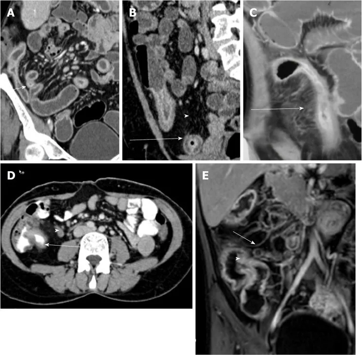
- Enterografi distansiyonu sağlanmış kontrastlı incelemede, terminal ileumun hastalıklı segmentine komşu mezenterik yağın normal yağdan daha hacimli biçimde ansın mezenterik kenarını sarması ve komşu ansları birbirinden itmesi aranır. Aynı alanda vasa recta uzamış ve belirgin olabilir; ayrı bir kontrastsız faz bu yağ proliferasyonunu tanımak için gerekli değildir.
Önerilen pencereler: Batın (G 400 / M 50), Yumuşak doku (G 400 / M 40).
BT bulguları
- Yağ sarmalanması (creeping fat) — hipertrofik mezenterik yağ, kronik tutulan terminal ileumun mezenterik yüzünden duvara doğru ilerler ve ansı kısmen çevreler.
- Anslar arası ayrışma — artmış yağ hacmi komşu ileal ansları birbirinden uzaklaştırabilir; bu görünüm yalnızca genel mezenterik yağ fazlalığı olarak yorumlanmamalıdır.
- Tutulan ansla mekânsal eşleşme — yağ proliferasyonu, duvar kalınlaşması veya kronik darlık gösteren ileal segmentle aynı mezenterik pediküle bağlanmalıdır.
- Mezenterik damar belirginliği (comb sign) — tutulan segmente uzanan vasa recta paralel, tarak dişleri benzeri çizgilenme oluşturabilir; bu bulgu eşlik eden hiperemiyi gösterir, yağ proliferasyonunun kendisi değildir.
- Kronik darlık eşliği — fibrofatty mezenter, lümen daralması ve proksimal ans genişlemesiyle birlikte olabilir; geçiş noktasını koronal planda takip edin.
- Transmural komplikasyon komşuluğu — mezenterik yağ içinde fistül hattı, ekstraluminal gaz veya çevresel kontrastlanan koleksiyon varsa bunları yağ sarmalanmasından ayrı tanımlayın.
Normalde
Normal terminal ileum mezenterinde yağ dokusu ansı çevreleyen belirgin bir kılıf oluşturmaz; ince barsak ansları mezenterik köke doğal aralıklarla uzanır. Aynı düzeyde hedef barsak çevresindeki mezenterik yağın sarma derecesini ve yağ içindeki damarların uzanımını değerlendirin.
Normal BT ile kıyasla
Aynı bölgenin, kaynak veri setinde patoloji içermediği belirtilen bir BT incelemesini kesit kesit kaydırın; organ sınırlarını açarak anatomiyi hasta görüntüsüyle karşılaştırın. Küçük rastlantısal bulgular tümüyle dışlanmamıştır.
Kaynak: TotalSegmentator v2.01 veri seti (Wasserthal ve ark., Radiology: Artificial Intelligence 2023), CC BY 4.0, vaka s0119; organ sınırları veri setinin otomatik segmentasyonundan, birleştirilerek sadeleştirildi. Görüntüler 2,5 mm kesit aralığına yeniden örneklendi.
Ayırıcı tanı
- Akut mezenterik inflamasyon
- yağ içinde bulanıklık ve sıvı çizgilenmesi ön plandadır; kronik Crohn yağ sarmalanmasındaki hacim artışı ve ansı örtme daha az belirgindir.
- Mezenterik lipomatozis
- yaygın yağ artışı belirli bir hastalıklı barsak segmentine sarılmadan görülür; Crohn ile ilişkili yağ segmental ve tutulan ansa bağlıdır.
- Mezenterik pannükülit
- mezenterik kökte yağ dansitesi artışı, küçük nodüller ve damar çevresi korunma görülebilir; dağılım terminal ileum duvarındaki kronik hastalıkla sınırlı değildir.
- Perienterik ödem
- sıvı ve çizgilenme şeklinde artmış dansite yapar; mezenterik yağın ans çevresinde gerçek hacimsel proliferasyonundan farklıdır.
Tuzaklar
- Tek başına kalın mezenterik yağ Crohn'a özgü değildir; kronik yağ sarmalanmasını aynı ansın duvar bulgusu ve mezenterik bağlantısıyla birlikte gösterin.
- Comb sign aktif hiperemiyi işaret eder, creeping fat ile eş anlamlı değildir; damarları ve yağ hacmini ayrı ayrı tarif edin.
- Yağ sarmalanmasının bulunmaması Crohn hastalığını dışlamaz; özellikle erken hastalıkta ya da teknik olarak yetersiz distansiyonda bu bulgu seçilmeyebilir.
Kendini dene
Kronik Crohn ileumu çevresinde hacimli mezenterik yağ ansları ayırıyor. Bu görünümün adı nedir?
Cevabı göster
Yağ sarmalanması. Tutulan barsak ansının mezenterik kenarını saran fibrofatty proliferasyon creeping fat olarak adlandırılır. Pannükülit ve lipomatozis genellikle bu şekilde tek bir Crohn segmentini sarmaz; iskemi ise damar ve duvar perfüzyon bulgularıyla değerlendirilir.
Crohn segmentine dik uzanan paralel, belirgin mezenterik damarlar hangi bulgudur?
Cevabı göster
Comb sign. Mezenterik hipervaskülarite (comb sign), Crohn hastalığında aktif inflamasyonu yansıtır; bu bulgu creeping fat ile aynı değildir.
İlgili konular
Kaynaklar
Bu sayfadaki 32 cümle ve 2 quiz sorusu aşağıdaki kaynaklardan alıntılarla tek tek karşılaştırıldı; 3 cümle bu karşılaştırmada düzeltildi (2026-10-09).
- Semi‐Automatic Assessment of Crohn's Disease Activity by Combined Analysis of Bowel Lesions and Creeping Fatpmc.ncbi.nlm.nih.gov
- Can Creeping Fat Be a Prognostic Factor in Crohn’s Disease?pmc.ncbi.nlm.nih.gov
- Comparing the Diagnostic Accuracy of Magnetic Resonance Enterography and Computed Tomography Enterography in Inflammatory Bowel Diseases: A Prospective Studypmc.ncbi.nlm.nih.gov
- Mesenteric and antimesenteric border subregionalization using MR enterography: advancing fibrosis evaluation in Crohn diseasepmc.ncbi.nlm.nih.gov
- Bildgebende Diagnostik bei Erkrankungen des Dünndarmspmc.ncbi.nlm.nih.gov
- Can Creeping Fat Be a Prognostic Factor in Crohn's Disease?pubmed.ncbi.nlm.nih.gov
- The role of adipose tissue in inflammatory bowel diseases.pubmed.ncbi.nlm.nih.gov
- Creeping Fat in Crohn's Disease-Surgical, Histological, and Radiological Approaches.pubmed.ncbi.nlm.nih.gov
- Creeping fat is associated with transmural healing in patients with Crohn's disease receiving ustekinumab.pubmed.ncbi.nlm.nih.gov
- Detecting and characterizing creeping fat in Crohn's disease: agreement between intestinal ultrasound and computed tomography enterography.pubmed.ncbi.nlm.nih.gov
- CT energy spectral parameters of creeping fat in Crohn's disease and correlation with inflammatory activity.pubmed.ncbi.nlm.nih.gov
- Dual-energy computed tomography quantification of creeping fat in Crohn's disease: A new radiologic biomarker for transmural disease monitoring.pubmed.ncbi.nlm.nih.gov
- A novel computed tomography enterography radiomics combining intestinal and creeping fat features could predict surgery risk in patients with Crohn's disease.pubmed.ncbi.nlm.nih.gov
- Creeping fat in patients with ileo-colonic Crohn's disease correlates with disease activity and severity of inflammation: A preliminary study using energy spectral computed tomography.pubmed.ncbi.nlm.nih.gov
- Correlation between radiomic features of Crohn's disease and secondary loss of response to infliximab.pubmed.ncbi.nlm.nih.gov
- Degree of Creeping Fat Assessed by Computed Tomography Enterography is Associated with Intestinal Fibrotic Stricture in Patients with Crohn's Disease: A Potentially Novel Mesenteric Creeping Fat Index.pubmed.ncbi.nlm.nih.gov
- Correlation between computed tomography imaging features of mesenteric fat and inflammatory activity in Crohn's disease.pubmed.ncbi.nlm.nih.gov
- Main Imaging Features of Crohn's Disease: Agreement between MR-Enterography and CT-Enterography.pubmed.ncbi.nlm.nih.gov
- Bowel inflammatory presentations on computed tomography in adult patients with severe aplastic anemia during flared inflammatory episodes.pubmed.ncbi.nlm.nih.gov
- Mesenteric and antimesenteric border subregionalization using MR enterography: advancing fibrosis evaluation in Crohn disease.pubmed.ncbi.nlm.nih.gov
- Update on intestinal ultrasound in pediatric inflammatory bowel disease.pubmed.ncbi.nlm.nih.gov
- Mesenteric adipose tissue: from protective gatekeeper to driver of inflammatory bowel disease.pubmed.ncbi.nlm.nih.gov
- Comparing the Diagnostic Accuracy of Magnetic Resonance Enterography and Computed Tomography Enterography in Inflammatory Bowel Diseases: A Prospective Study.pubmed.ncbi.nlm.nih.gov
- SAR Consensus Recommendations for Defining Small Bowel Crohn Disease Strictures at CT and MR Enterography.pubmed.ncbi.nlm.nih.gov
- [Imaging diagnostics for diseases of the small bowel].pubmed.ncbi.nlm.nih.gov
- The role of lipid metabolism and creeping fat in disease assessment and prediction for Crohn's disease.pubmed.ncbi.nlm.nih.gov
- Semi-Automatic Assessment of Crohn's Disease Activity by Combined Analysis of Bowel Lesions and Creeping Fat.pubmed.ncbi.nlm.nih.gov
- Characterization of mesocolic adipose hyperplasia in a rat 2,4,6-trinitrobenzenesulfonic acid colitis model and comparison to creeping fat in Crohn disease.pubmed.ncbi.nlm.nih.gov
- Mesenteric fat proliferation on magnetic resonance enterography is a predictor of long-term disease complications in crohn's disease.pubmed.ncbi.nlm.nih.gov
- How to use intestinal ultrasonography in patients with Crohn disease: its role in the assessment of disease activity and disease monitoring in the era of the treat-to-target strategy.pubmed.ncbi.nlm.nih.gov
- Radiology plus ileocolonoscopy versus radiology alone in Crohn's disease: prognosis prediction and mutual agreement.pubmed.ncbi.nlm.nih.gov
- Magnetic resonance enterography (MRE) and ultrasonography (US) in the study of the small bowel in Crohn's disease: state of the art and review of the literature.pubmed.ncbi.nlm.nih.gov
- Mesenteric Adipose Tissue as a Modulator of Intestinal Inflammation: Mechanistic Insights and Model Systems in Crohn's Disease.pubmed.ncbi.nlm.nih.gov
- Elastography as a Discriminator Between Fibrotic and Inflammatory Strictures in Crohn's Disease: A Dead End or Bright Future in Clinical Decision-Making? Critical Review.pubmed.ncbi.nlm.nih.gov
- Imaging in inflammatory bowel disease: current and future perspectives.pubmed.ncbi.nlm.nih.gov
- Automated quantification of mesenteric hyperaemia in Crohn's disease using time-of-flight magnetic resonance angiography.pubmed.ncbi.nlm.nih.gov
- Pediatric Small Bowel Crohn Disease: Correlation of US and MR Enterography.pubmed.ncbi.nlm.nih.gov
- Abdominal ultrasonography with color Doppler analysis in the assessment of ileal Crohn's disease: comparison with magnetic resonance enterography.pubmed.ncbi.nlm.nih.gov
- Intestinal Ultrasound for the Pediatric Gastroenterologist: A Guide for Inflammatory Bowel Disease Monitoring in Children: Expert Consensus on Behalf of the International Bowel Ultrasound Group (IBUS) Pediatric Committee.pubmed.ncbi.nlm.nih.gov
- Therapeutic strategies in Crohn's disease in an emergency surgical setting.pubmed.ncbi.nlm.nih.gov
- Assessment of Crohn's disease-associated small bowel strictures and fibrosis on cross-sectional imaging: a systematic review.pubmed.ncbi.nlm.nih.gov
- The Role of Intestinal Ultrasonography in Children with Inflammatory Bowel Disease.pubmed.ncbi.nlm.nih.gov
- POCUS for acute abdominal pain: practical scan protocols on gastrointestinal diseases and an evidence review.pubmed.ncbi.nlm.nih.gov
- Twist in the cecum: A case series of mobile cecum, cecal volvulus and cecal bascule.pubmed.ncbi.nlm.nih.gov
- Early-onset colorectal cancer in patients under 30: a case series from Tanzania highlighting an emerging epidemiological shift.pubmed.ncbi.nlm.nih.gov
- Concurrent Amyand and Littre Hernias With Incidental Hodgkin Lymphoma: A Diagnostic and Surgical Challenge.pubmed.ncbi.nlm.nih.gov
- Surgical Anatomy of the Gastrointestinal Tract in Cats.pubmed.ncbi.nlm.nih.gov
- Computed Tomography and Magnetic Resonance Enterography: From Protocols to Diagnosis.pubmed.ncbi.nlm.nih.gov
- Acute Ileal Diverticulitis: Computed Tomography and Ultrasound Findings.pubmed.ncbi.nlm.nih.gov
- Pneumatosis intestinalis revisited: associated imaging findings, clinical correlation, management, risk stratification and future perspectives.pubmed.ncbi.nlm.nih.gov
- Relative colon wall attenuation - distinguishing ischemia from other colon wall changes.pubmed.ncbi.nlm.nih.gov
- Imaging Characteristics of Pylorogastric, Duodenogastric, and True-Gastrogastric Intussusception in 25 Dogs and One Cat.pubmed.ncbi.nlm.nih.gov
- Anatomical, Physiological, and Functional Diversity of Adipose Tissue.pubmed.ncbi.nlm.nih.gov
- Mesenteric Panniculitis in a Patient with Ulcerative Colitis in Remission on Vedolizumab Therapy: A Case Report and Literature Review.pubmed.ncbi.nlm.nih.gov
- Characteristic CT Imaging Features of Atraumatic Peritoneal and Subperitoneal Pathologies in Emergency Settings: A Pictorial Review.pubmed.ncbi.nlm.nih.gov
- Crohn's disease: research progress in decoding pathogenic multi-network and precision management of artificial intelligence radiomics.pubmed.ncbi.nlm.nih.gov
- Crohn Disease Strictures: Multimodality Imaging to Identify Imaging Biomarkers.pubmed.ncbi.nlm.nih.gov
- Protocol to interrogate the role of creeping fat and mechanical tension in Crohn's disease using a surgical mouse model of intestinal fibrosis.pubmed.ncbi.nlm.nih.gov
- Disturbance of Oxylipin Metabolism Mediated by Autophagy of Mesenteric Adipocytes Orchestrates Gut Inflammation in Crohn's Disease.pubmed.ncbi.nlm.nih.gov
- Gastrointestinal imaging-practical magnetic resonance imaging approach.pubmed.ncbi.nlm.nih.gov
- A Novel Chronic Enteropathy Associated with SLCO2A1 Gene Mutation: Enterography Findings in a Multicenter Korean Registry.pubmed.ncbi.nlm.nih.gov
- Radiological insights into diverticulitis: Clinical manifestations, complications, and differential diagnosis.pubmed.ncbi.nlm.nih.gov
- Primary Midgut Volvulus: An Unusual Case of a Young Adult Necessitating Extensive Bowel Resection.pubmed.ncbi.nlm.nih.gov
- Peritoneal disease: key imaging findings that help in the differential diagnosis.pubmed.ncbi.nlm.nih.gov
- Intriguing Role of the Mesentery in Ileocolic Crohn's Disease.pubmed.ncbi.nlm.nih.gov
- Imaging of Acute Abdominopelvic Pain in Pregnancy and Puerperium-Part II: Non-Obstetric Complications.pubmed.ncbi.nlm.nih.gov
- Crohn's disease at radiological imaging: focus on techniques and intestinal tract.pubmed.ncbi.nlm.nih.gov
- Radiology of fibrosis part II: abdominal organs.pubmed.ncbi.nlm.nih.gov
- Bleeding per rectum in pediatric population: A pictorial review.pubmed.ncbi.nlm.nih.gov
- Comparison of US Strain Elastography and Entero-MRI to Typify the Mesenteric and Bowel Wall Changes during Crohn's Disease: A Pilot Study.pubmed.ncbi.nlm.nih.gov
- Expanding the role of radiomics and artificial intelligence in the management of inflammatory bowel disease: Insights, opportunities, and challenges.pubmed.ncbi.nlm.nih.gov
- Spectrum of bowel wall thickening on ultrasound with pathological correlation in children.pubmed.ncbi.nlm.nih.gov
- Serum proteomics in paediatric inflammatory bowel disease from a case-control study: biomarker discovery and ulcerative colitis-Crohn's disease differentiation.pubmed.ncbi.nlm.nih.gov
- Unravelling mesenteric fibrosis in small intestinal neuroendocrine tumours and implications for clinical management.pubmed.ncbi.nlm.nih.gov
- Two sides of the same coin: protective versus pathogenic mesentery in inflammatory bowel diseases - a comprehensive review.pubmed.ncbi.nlm.nih.gov
- Small Bowel Imaging from Stepchild of Roentgenology to MR Enterography, Part II: The Reliable Disclosure of Crohn's Disease and Non-Inflammatory Small Bowel Disorder Plot through MRI Findings.pubmed.ncbi.nlm.nih.gov
- CT of acute abdomen in the elderly.pubmed.ncbi.nlm.nih.gov
- Actionable imaging findings in the daily PET/CT scenario.pubmed.ncbi.nlm.nih.gov
- Duodenal imaging on the spotlight: from A to Z.pubmed.ncbi.nlm.nih.gov
- Small bowel imaging in Crohn's disease patients.pubmed.ncbi.nlm.nih.gov
- How to perform gastrointestinal ultrasound: Anatomy and normal findings.pubmed.ncbi.nlm.nih.gov
- Magnetic resonance enterography: A stepwise interpretation approach and role of imaging in management of adult Crohn's disease.pubmed.ncbi.nlm.nih.gov
- The Importance of Magnetic Resonance Enterography in Monitoring Inflammatory Bowel Disease: A Review of Clinical Significance and Current Challenges.pubmed.ncbi.nlm.nih.gov
- The Role of Magnetic Resonance Enterography in Crohn's Disease: A Review of Recent Literature.pubmed.ncbi.nlm.nih.gov
- Imaging Approach to Diverticular Disease of the Small Bowel.pubmed.ncbi.nlm.nih.gov
Eğitim amaçlıdır; hasta başında tanı ve tedavi kararının yerine geçmez. İçerik yapay zekâ destekli bir süreçle güncel literatüre göre cümle cümle kaynaklarla karşılaştırıldı, ancak uzman radyolog incelemesinden geçmedi. Klinik kararlarda güncel kılavuzları, kurum protokollerini ve radyoloji raporunu esas alın.