Özet
Sınıflandırılamayan hepatoselüler adenom, tanımlı moleküler veya histopatolojik adenom alt tiplerine uymayan benign hepatosit tümörüdür. Çok fazlı BT lezyonun kontrastlanmasını ve kanama, yağ ya da nekroz gibi iç bileşenlerini gösterir; sınıflandırılamayan alt tipi görüntü görünümünden teşhis edemez. Bu nedenle görüntü, klinik bağlam ve gerektiğinde doku tanısı birlikte ele alınmalıdır. Lezyonun HCC ile örtüşen görünümü varsa onu sırf adenom olarak adlandırmak maligniteyi geciktirebilir.
Faz ve pencere
- Kontrastsız
- Adenom karaciğerle izodens olabilir; yağ ve nekroz düşük, akut kanama yüksek atenüasyonlu alanlar oluşturabilir. Bu seri iç bileşenleri tanımlar, sınıflandırılamayan alt tipi göstermez.
- Arteriyel tanısal
- Adenom belirgin ya da heterojen kontrastlanabilir; bu boyanma hepatoselüler tümörlerde ortak olduğundan özgül alt tip bulgusu değildir.
- Portal tanısal
- Lezyon karaciğerle izodensleşebilir veya göreli hipodens hale gelebilir. Kanama ve nekroz iç yapıyı heterojen kılar; görünüm β-katenin aktive adenom ya da HCC ile örtüşebilir.
- Gecikmiş
- Sınıflandırılamayan HCA için özgül radyolojik özellik tanımlanmamıştır; bir olguda tümör gecikmiş fazda normal karaciğere benzer görünmüştür.
Önerilen pencereler: Batın (G 400 / M 50), Karaciğer (G 150 / M 30).
BT bulguları
- Karaciğer parankiminden ayrılan, düzgün sınırlı veya hafif lobüle solid hepatoselüler kitle
- Arteriyel fazda belirgin kontrastlanma; boyanmanın homojen ya da heterojen olması moleküler sınıflandırma sağlamaz
- Kontrastsız seride yağ, akut kan, eski kan ürünü veya nekrozla ilişkili atenüasyon farklılıkları
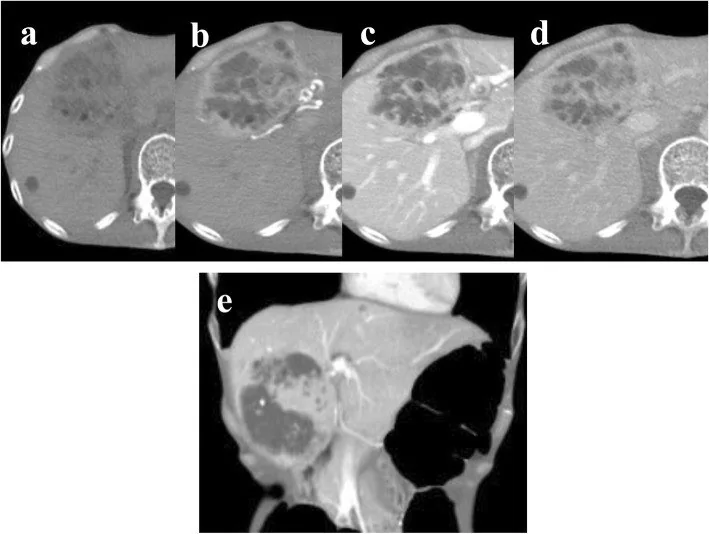
- Bir sınıflandırılamayan HCA olgusunda portal fazda kontrastlanma sürmüş, gecikmiş fazda tümör normal karaciğere benzer görünmüştür; HCA ile HCC ayrımı görüntülemeyle güç olabilir.
- Santral arterli yıldızsı skarın yokluğu FNH’yi daha az olası kılar, ancak tek başına adenom tanısı koydurmaz
- Kitle büyümesiyle karaciğer kapsülünde bombeleşme ve çevre organlarda yer değiştirme
- Kanama şüphesinde lezyon çevresinde perihepatik sıvı veya hematom; kontrast dışa kaçışı varsa aktif kanama olasılığı
Ölçütler ve sınıflamalar
- Hepatoselüler adenom moleküler alt tipleri
- Güncel moleküler sınıflama sekiz grubu içerir: HNF1A-inaktive; inflamatuvar; CTNNB1 exon 3 β-katenin aktive; CTNNB1 exon 7/8 β-katenin aktive; inflamatuvar/CTNNB1 exon 3 mikst; inflamatuvar/CTNNB1 exon 7/8 mikst; sonic hedgehog; sınıflandırılamayan. BT bu genetik alt tipleri tek başına güvenilir biçimde belirleyemez; referans doku temelli immünohistokimya veya moleküler incelemedir.
Normalde
Sınıflandırılamayan HCA tanısı alan bir olguda BT'de lezyon arteriyel fazda hafif-orta kontrastlanmış ve bu kontrastlanma portal ve denge fazlarında sürmüştür.
Normal BT ile kıyasla
Aynı bölgenin, kaynak veri setinde patoloji içermediği belirtilen bir BT incelemesini kesit kesit kaydırın; organ sınırlarını açarak anatomiyi hasta görüntüsüyle karşılaştırın. Küçük rastlantısal bulgular tümüyle dışlanmamıştır.
Kaynak: TotalSegmentator v2.01 veri seti (Wasserthal ve ark., Radiology: Artificial Intelligence 2023), CC BY 4.0, vaka s0541; organ sınırları veri setinin otomatik segmentasyonundan, birleştirilerek sadeleştirildi. Görüntüler 2,5 mm kesit aralığına yeniden örneklendi.
Ayırıcı tanı
- Hepatoselüler karsinom
- riskli zeminde çevresel halka biçiminde olmayan arteriyel hiper-kontrastlanma, portal/gecikmiş fazda yıkanma ve kapsül HCC lehinedir; adenomla örtüşme olabilir.
- Fokal nodüler hiperplazi
- santral yıldızsı skar ve merkezden çevreye dallanan arteriyel damarlar FNH lehinedir.
- Hemanjiom
- çevresel kesintili nodüler boyanmanın sonraki fazlarda santrale ilerlemesi vasküler tümörü düşündürür.
- Hipervasküler metastaz
- eşlik eden primer tümör ve çoklu arteriyel boyanan odaklar metastazı destekler.
- Fokal yağ değişikliği
- segmental veya coğrafi atenüasyon farkı damarları ve karaciğer konturunu bozmaz, kitle etkisi yapmaz.
Tuzaklar
- ‘Sınıflandırılamayan’ tanısı BT’de özel bir patern anlamına gelmez; bu adenom grubu görüntüleme ile β-katenin aktive adenomdan ayrılamaz.
- Sınıflandırılamayan HCA'lı bir olguda portal faz kontrastlanması sürmüş ve tümör gecikmiş fazda normal karaciğere benzer görünmüştür; portal fazdaki bulguyu diğer fazlarla birlikte değerlendirin.
- Santral skar yokluğu FNH olasılığını azaltabilir, ancak skar bulunmaması adenomun alt tipini belirlemez.
- Çevre dokuda hematom ve lezyon içi yüksek atenüasyon varken kitleyi yalnız benign insidental bulgu olarak raporlama.
Kendini dene
Histolojide belirgin bir moleküler değişiklik saptanmayan hepatoselüler adenom BT’de arteriyel boyanıyor. Hangi çıkarım doğrudur?
Cevabı göster
Alt tip BT ile konamaz. Sınıflandırılamayan HCA tanısı, tanımlı moleküler ve histopatolojik gruplara girmemeye dayanır. Arteriyel boyanma bu alt tip için özgül değildir; BT HCC’yi kesin dışlayamaz veya β-katenin aktivasyonunu gösteremez.
HCC açısından riskli zemini olan hastada portal fazda hipodens ve kapsül benzeri sınırı bulunan hepatik kitle saptanıyor. En uygun görüntüleme yaklaşımı hangisidir?
Cevabı göster
Kontrastsız ve tüm kontrastlı fazları, klinik risk bağlamıyla birlikte değerlendir. HCC açısından riskli zeminde kontrastsız seri ile tüm uygun kontrast fazları ve hastanın klinik riski birlikte değerlendirilmelidir. Portal faz hipodensliği tek başına yıkanma ya da tanı ölçütü değildir; tek bir özellik adenom, FNH veya hemanjiyom sonucunu vermez.
İlgili konular
Kaynaklar
Bu sayfadaki 44 cümle ve 2 quiz sorusu aşağıdaki kaynaklardan alıntılarla tek tek karşılaştırıldı; 5 cümle bu karşılaştırmada düzeltildi (2026-10-09).
- Unclassified hepatocellular adenoma with histological brown pigment deposition and serum PIVKA-II level elevation: a case reportpmc.ncbi.nlm.nih.gov
- Unclassified hepatocellular adenoma with beta-catenin mutation: a case reportpmc.ncbi.nlm.nih.gov
- Mesenchymal hamartoma of the liver in a young adult female: a diagnostic challenge mimicking hepatic adenomapmc.ncbi.nlm.nih.gov
- Giant Atypical Hepatocellular Adenoma of Unknown Etiology in a Young Male: A Case Reportpmc.ncbi.nlm.nih.gov
- Multifocal hemorrhagic hepatocellular adenomas following vigorous physical exertion: Radiologic–pathologic correlation and endovascular managementpmc.ncbi.nlm.nih.gov
- Hepatic adenoma: evolution of a more individualized treatment approach.pubmed.ncbi.nlm.nih.gov
- Unclassified hepatocellular adenoma in a middle-aged woman with glucose intolerance.pubmed.ncbi.nlm.nih.gov
- Unclassified hepatocellular adenoma with histological brown pigment deposition and serum PIVKA-II level elevation: a case report.pubmed.ncbi.nlm.nih.gov
- Contrast-enhanced ultrasound patterns of hepatocellular adenoma: an Italian multicenter experience.pubmed.ncbi.nlm.nih.gov
- Case of resected multiple hepatocellular adenomas in a young man with severe obesity.pubmed.ncbi.nlm.nih.gov
- A case of hepatocellular adenoma with pedunculated development and difficulty in diagnosis.pubmed.ncbi.nlm.nih.gov
- Mesenchymal hamartoma of the liver in a young adult female: a diagnostic challenge mimicking hepatic adenoma.pubmed.ncbi.nlm.nih.gov
- Giant Atypical Hepatocellular Adenoma of Unknown Etiology in a Young Male: A Case Report.pubmed.ncbi.nlm.nih.gov
- Hepatocellular Carcinoma Precursor Lesions: From Pathological Basis to Risk Stratification and Precision Intervention.pubmed.ncbi.nlm.nih.gov
- Diagnosis and Management of Benign Liver Tumors.pubmed.ncbi.nlm.nih.gov
- Hepatocellular adenoma update: diagnosis, molecular classification, and clinical course.pubmed.ncbi.nlm.nih.gov
- Surgical Management of a Giant Inflammatory Hepatocellular Adenoma in a Young Female.pubmed.ncbi.nlm.nih.gov
- Case Report: Focal nodular hyperplasia of the liver in an adolescent-a case report and literature review.pubmed.ncbi.nlm.nih.gov
- Case Report: Hypervascular lipid-rich intrahepatic adrenocortical adenoma in the right posterior liver: a diagnostic challenge for hepatobiliary surgeons.pubmed.ncbi.nlm.nih.gov
- Resolution of Recurrent Severe Hypoglycemia After Surgical Excision of a Primary Hepatic Neoplasm of Uncertain Histogenesis in a Dog: A Case Report.pubmed.ncbi.nlm.nih.gov
- Ectopic adrenal gland in the liver leading to a misdiagnosis of hepatocellular carcinoma: A case report.pubmed.ncbi.nlm.nih.gov
- Case Report: Contrast-enhanced US and CT imaging features of nodule-in-nodule hepatocellular carcinoma in a dog.pubmed.ncbi.nlm.nih.gov
- Use of the Term "Atypical Hepatic Hemangioma" in Radiology Reports: An Analysis of Diagnostic Precision.pubmed.ncbi.nlm.nih.gov
- Washout on Contrast-Enhanced Ultrasound of Benign Focal Liver Lesions-A Review on Its Frequency and Possible Causes.pubmed.ncbi.nlm.nih.gov
- Primary Hepatic Perivascular Epithelioid Cell Tumors: From Diagnosis to Treatment.pubmed.ncbi.nlm.nih.gov
- TERT drives liver tumorigenesis beyond telomere elongation.pubmed.ncbi.nlm.nih.gov
- Benign Biliary Tumors and Precursor Neoplasms: An Updated Clinicopathological and Molecular Review Based on the 2026 WHO Classification.pubmed.ncbi.nlm.nih.gov
- Special Issue: EACR 2026 Congress: Innovative Cancer Science, 8-11 June 2026.pubmed.ncbi.nlm.nih.gov
- Hepatic mucinous cystic neoplasm with hematoma: A case report.pubmed.ncbi.nlm.nih.gov
- Coexistence of Small Duct Type and Biliary Adenofibroma-Associated Large Duct Type Intrahepatic Cholangiocarcinoma within a Single Lesion: A Case Report.pubmed.ncbi.nlm.nih.gov
- Ancillary Diagnostic Utility of Glycoprotein Nonmetastatic Melanoma Protein B (GPNMB) in Diagnostically Challenging Hepatic Angiomyolipoma.pubmed.ncbi.nlm.nih.gov
- Insights into hepatocellular adenomas in Asia: molecular subtypes, clinical characteristics, imaging features, and hepatocellular carcinoma risks.pubmed.ncbi.nlm.nih.gov
- Primary hepatic neuroendocrine neoplasms: imaging characteristics and misdiagnosis analysis.pubmed.ncbi.nlm.nih.gov
- Are the washout values currently accepted for lesion characterization in dedicated adrenal CT adequate for diagnosis?pubmed.ncbi.nlm.nih.gov
- Percutaneous ablation of the small renal mass-techniques and outcomes.pubmed.ncbi.nlm.nih.gov
- Splenoses and Other Ectopic and Heterotopic Splenic Tissue: The Use of Long-Lasting Enhancement in Contrast-Enhanced Ultrasound to Avoid Unnecessary Intervention.pubmed.ncbi.nlm.nih.gov
- Benign focal liver lesions: The role of magnetic resonance imaging.pubmed.ncbi.nlm.nih.gov
- Hepatic tumors: pitfall in diagnostic imaging.pubmed.ncbi.nlm.nih.gov
- Metastatic Fibrolamellar Hepatocellular Carcinoma in a Young Adult: A Case Report and Narrative Review.pubmed.ncbi.nlm.nih.gov
- Screening Sonography Detection of Liver Masses: Assessing the Non-emergent Nature of Focal Nodular Hyperplasia.pubmed.ncbi.nlm.nih.gov
- Giant Cavernous Hepatic Hemangioma Masking a Separate Pleural Empyema: A Diagnostic Pitfall and Anchoring Bias.pubmed.ncbi.nlm.nih.gov
- Non-infectious Hepatic Cystic Lesions: A Narrative Review.pubmed.ncbi.nlm.nih.gov
- International Harmonization of Nomenclature and Diagnostic Criteria (INHAND): Nonproliferative and Proliferative Lesions of the Rabbit.pubmed.ncbi.nlm.nih.gov
- International Harmonization of Nomenclature and Diagnostic Criteria (INHAND): Non-proliferative and Proliferative Lesions of the Non-human Primate ( M. fascicularis ).pubmed.ncbi.nlm.nih.gov
- Multifocal hemorrhagic hepatocellular adenomas following vigorous physical exertion: Radiologic-pathologic correlation and endovascular management.pubmed.ncbi.nlm.nih.gov
- Case Report: Rare spontaneous hepatic rupture hemorrhage complicated by autoimmune liver disease.pubmed.ncbi.nlm.nih.gov
- Managing Life-Threatening Spontaneous Liver Rupture in Pregnancy: A Case Study.pubmed.ncbi.nlm.nih.gov
- Case Report: Multimodality imaging features and serial metabolic changes on <sup>18</sup> F-FDG PET/CT in a dog with granulomatous hepatitis.pubmed.ncbi.nlm.nih.gov
- Contrast-Enhanced CT of the Coelom in a Female Bald Eagle (<i>Haliaeetus leucocephalus</i>): Development of a Preliminary CT-Based Anatomical Segmentation Workflow.pubmed.ncbi.nlm.nih.gov
- Spontaneous subcapsular hepatic hematoma with hepatic rupture and hemorrhage in the postpartum period: a case report and literature review.pubmed.ncbi.nlm.nih.gov
- Liver Imaging Reporting and Data System (LI-RADS): A Comprehensive Review.pubmed.ncbi.nlm.nih.gov
- The diagnostic value of virtual unenhanced spectral CT images in acute pancreatitis: a comparison with true unenhanced images.pubmed.ncbi.nlm.nih.gov
- Quantitative assessment of multi-phase contrast-enhanced CT features in hepatic epithelioid hemangioendothelioma.pubmed.ncbi.nlm.nih.gov
- Pathology and diagnostic approaches to well-differentiated hepatocellular lesions: a narrative review.pubmed.ncbi.nlm.nih.gov
- Molecular diagnostics of hepatobiliary and pancreatic neoplasias.pubmed.ncbi.nlm.nih.gov
- Artificial Intelligence in Oncology: A Comprehensive Cross-Cancer Translational Readiness Analysis Across 18 Malignancies.pubmed.ncbi.nlm.nih.gov
- Human biomarker navigator.pubmed.ncbi.nlm.nih.gov
- Immunohistochemistry in intrahepatic cholangiocarcinoma: histological subtyping and drug selection.pubmed.ncbi.nlm.nih.gov
- Hepatic perivascular epithelioid cell tumor: A rare mesenchymal tumor with epithelioid features: A case report.pubmed.ncbi.nlm.nih.gov
- Current approach to diagnosis and treatment of focal nodular hyperplasia in children with a management algorithm.pubmed.ncbi.nlm.nih.gov
- Separation of liver focal nodular hyperplasia (FNH) and liver malignant tumors by a combination of T2-weighted imaging signal and three diffusion magnetic resonance metrics of diffusion-derived vessel density, slow diffusion coefficient, and apparent diffusion coefficient.pubmed.ncbi.nlm.nih.gov
- Diagnosis of a Liver Lymphangioma Using Contrast-Enhanced Ultrasonography (CEUS): Single Case Report.pubmed.ncbi.nlm.nih.gov
- Lower Abdominal and Periumbilical Pain in a Patient With Focal Nodular Hyperplasia of the Liver: A Case Report and Literature Review.pubmed.ncbi.nlm.nih.gov
- Calcified liver metastases from medullary thyroid carcinoma in a Multiple Endocrine Neoplasia Type 2A patient.pubmed.ncbi.nlm.nih.gov
- Contrast-Enhanced CT Finding of Hot Spot Sign Secondary to Supra Hilar Mass Causing Superior Vena Cava Syndrome: A Case Report.pubmed.ncbi.nlm.nih.gov
- Primary intraosseous anastomosing hemangioma of the sacrum: illustrative case.pubmed.ncbi.nlm.nih.gov
- Photoacoustic and ultrasound Imaging of placental functions in gestational diabetes: From diagnosis to precision interventions.pubmed.ncbi.nlm.nih.gov
- Steatotic liver disease trajectory: A holistic EASL guidance on the multidisciplinary approach to screening and patient management.pubmed.ncbi.nlm.nih.gov
- The tumor microbiome in cancer progression: mechanisms and therapeutic potential.pubmed.ncbi.nlm.nih.gov
- Scoping review of clinical practice guidelines on the management of benign liver tumours.pubmed.ncbi.nlm.nih.gov
- Benign Liver Tumors.pubmed.ncbi.nlm.nih.gov
- Application of indocyanine green fluorescence imaging in hepatobiliary surgery.pubmed.ncbi.nlm.nih.gov
- Imaging findings of primary hepatic leiomyosarcoma: a case report and literature review.pubmed.ncbi.nlm.nih.gov
- A narrative review of foundation models for medical image segmentation: zero-shot performance evaluation on diverse modalities.pubmed.ncbi.nlm.nih.gov
- Integrating Radiomics and Artificial Intelligence (AI) in Stereotactic Body Radiotherapy (SBRT)/Stereotactic Radiosurgery (SRS): Predictive Tools for Tailored Cancer Care.pubmed.ncbi.nlm.nih.gov
- Incidental Adrenal Nodules and Masses: The Imaging Approach.pubmed.ncbi.nlm.nih.gov
- Congenital focal nodular hyperplasia-like lesion mimicking hepatoblastoma: anecdotal but plausible.pubmed.ncbi.nlm.nih.gov
- Hepatocellular carcinoma (HCC) and focal nodular hyperplasia (FNH) showing iso- or hyperintensity in the hepatobiliary phase: differentiation using Gd-EOB-DTPA enhanced MRI radiomics and deep learning features.pubmed.ncbi.nlm.nih.gov
- Features of Nodules in Explants of Children Undergoing Liver Transplantation for Biliary Atresia.pubmed.ncbi.nlm.nih.gov
- Primary Hepatic Masses in Ten Dogs: A Retrospective Surgical Case Series.pubmed.ncbi.nlm.nih.gov
- Ectopic liver tissue on the gallbladder wall with independent vascular supply: a case report.pubmed.ncbi.nlm.nih.gov
- Imaging insights into pediatric liver masses: A comprehensive minireview for hepatology practice.pubmed.ncbi.nlm.nih.gov
- Focal Liver Lesions other than Hepatocellular Carcinoma in Cirrhosis: Diagnostic Challenges.pubmed.ncbi.nlm.nih.gov
- Update on the Pathology of Pediatric Liver Tumors: A Pictorial Review.pubmed.ncbi.nlm.nih.gov
- Liver masses: a clinical, radiologic, and pathologic perspective.pubmed.ncbi.nlm.nih.gov
- Multimodal imaging features to diagnose abdominal complications of sickle cell disease.pubmed.ncbi.nlm.nih.gov
- Rare Solid Pancreatic Lesions on Cross-Sectional Imaging.pubmed.ncbi.nlm.nih.gov
- Intraindividual comparison of photon-counting detector CT and third-generation dual-source energy-integrating detector CT in multiphasic contrast-enhanced abdominal CT.pubmed.ncbi.nlm.nih.gov
- Quantitative Conspicuity of Pancreatic Canine Insulinoma: A Comparison of Dynamic 4D CT and Dual-Source, Dual-Energy Bolus-Triggered Multiphase CT Imaging.pubmed.ncbi.nlm.nih.gov
- Deep learning-based combined noise reduction and contrast enhancement for post-neoadjuvant pancreatic cancer CT: does improved image quality translate to better resectability assessment?pubmed.ncbi.nlm.nih.gov
- Ectopic β-hCG-Expressing Hepatic Neoplasm with Cutaneous Metastasis in a Young Postpartum Woman: A Case Report and Literature Review.pubmed.ncbi.nlm.nih.gov
- Portal vein aneurysm mimicking a pancreatic mass: A case report with an emphasis on imaging pitfalls.pubmed.ncbi.nlm.nih.gov
- Triple-Phase Computed Tomography for Differentiating Canine Cutaneous and Subcutaneous Mast Cell Tumors From Benign Lesions: A Size-Based Evaluation.pubmed.ncbi.nlm.nih.gov
- Giant Hepatobiliary Cystadenoma Requiring Biliary Reconstruction in a Cat: A Case Report.pubmed.ncbi.nlm.nih.gov
- Clinical and CT/MRI features of hepatic AL amyloidosis: preliminary experience in 10 cases.pubmed.ncbi.nlm.nih.gov
- Photon-counting CT for pancreatic cancer: advancing precision imaging and virtual surgical navigation.pubmed.ncbi.nlm.nih.gov
- Imaging Considerations before and after Liver-Directed Locoregional Treatments for Metastatic Colorectal Cancer.pubmed.ncbi.nlm.nih.gov
- Imaging features of lacrimal gland disease.pubmed.ncbi.nlm.nih.gov
- Pediatric Primary Hepatic Tumors: Diagnostic Considerations.pubmed.ncbi.nlm.nih.gov
- I thought it was a hemangioma! A pictorial essay about common and uncommon liver hemangiomas' mimickers.pubmed.ncbi.nlm.nih.gov
- Guidelines on the Use of Therapeutic Apheresis in Clinical Practice-Evidence-Based Approach From the Writing Committee of the American Society for Apheresis: The Tenth Special Issue.pubmed.ncbi.nlm.nih.gov
- Patterns of injury and violence in Yaoundé Cameroon: an analysis of hospital data.pubmed.ncbi.nlm.nih.gov
- Pitfalls of Adrenal Washout on CT in the Adult Population: A Pictorial Review.pubmed.ncbi.nlm.nih.gov
- Case Report: A rare case of primary hepatic paraganglioma: a mimicker of hepatocellular carcinoma.pubmed.ncbi.nlm.nih.gov
- Case Report: Indocyanine green fluorescence imaging in complex focal nodular hyperplasia resection: report of two cases.pubmed.ncbi.nlm.nih.gov
- Clinical significance and outcomes of left-sided gallbladder for cholecystectomy: A retrospective cohort study based on 12-year single-center experience.pubmed.ncbi.nlm.nih.gov
- Oncocytic adrenal adenoma: Diagnostic challenge of an incidentaloma; a case report.pubmed.ncbi.nlm.nih.gov
Eğitim amaçlıdır; hasta başında tanı ve tedavi kararının yerine geçmez. İçerik yapay zekâ destekli bir süreçle güncel literatüre göre cümle cümle kaynaklarla karşılaştırıldı, ancak uzman radyolog incelemesinden geçmedi. Klinik kararlarda güncel kılavuzları, kurum protokollerini ve radyoloji raporunu esas alın.