
Özet
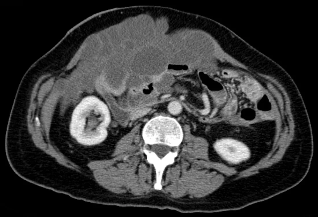
Psödomiksoma peritonei (PMP), çoğunlukla rüptüre apendiks müsinöz neoplazisinden kaynaklanan, periton içinde ilerleyici müsinöz asit ve implant birikimiyle tanımlanan klinik tablodur. Over/adneks tutulumu çoğu kez gastrointestinal kaynaklı sekonder yayılımdır; kaynak araştırmasında apendiks ve adneksler birlikte değerlendirilir. Portal venöz kontrastlı BT; loküle, kitle etkili müsin havuzlarını, serozal bası çentiklerini, peritoneal dağılımı ve olası apendiks odağını gösterir. BT müsin ile basit asit ayrımını desteklese de hücreselliği ve histolojik dereceyi tek başına belirleyemez.
Faz ve pencere
- Portal tanısal
- Loküle, çoğu kez suya yakın düşük dansiteli fakat kitle etkisi yapan müsinöz sıvı; septalar, yumuşak doku implantları, kalsifikasyonlar ve karaciğer/dalak yüzeyinde basıya bağlı scalloping aranır.
Önerilen pencereler G genişlik, M merkez; değerler Hounsfield birimi (HU)
- BatınG 400 / M +50
- Yumuşak dokuG 400 / M +40
BT bulguları
- Kitle etkili düşük dansiteli müsin — sıvı yoğunluklu içerik serbestçe dağılmak yerine kompartımanları doldurup ince barsakları merkeze veya bir yana iter.
- Karaciğer ve dalak yüzeylerinde scalloping (çentiklenme), dıştan bası yapan müsinöz birikimlerin oluşturduğu kontur değişikliğidir.
- Lokülasyon ve septalar — müsin havuzları peritoneal bölmelere uyar ve ince iç bölmeler içerebilir.
- Omental birikim — yoğun müsin ve tümör dokusu büyük omentumda plak veya kitle etkili kek görünümü oluşturabilir.
- Peritoneal yumuşak doku implantları — müsin gölcükleri içinde ya da çevresinde kontrastlanan nodüler bileşenler bulunabilir.
- Kalsifikasyon — amorf veya kıvrımlı kalsifik odaklar peritoneal müsinöz depozitlerde izlenebilir.
- Apendiks kaynak şüphesi — genişlemiş, mukosel benzeri apendiks veya kalsifiye duvarlı apendiks primer odağa işaret edebilir; görünür olmaması kaynağı dışlamaz.
- Adneks değerlendirmesi — overlerde eşlik eden müsinöz kitle ya da metastatik görünüm olup olmadığı kaynak araştırmasında kaydedilir.
- Peritoneal yaygınlığı tarif etmek için PCI kullanılabilir; 13 abdominopelvik bölgenin her birinde en büyük lezyon boyutu 0–3 arasında skorlanır. BT ile belirlenen PCI hastalık yükünü olduğundan az gösterebilir; bu durum özellikle ince bağırsakların sınırlı görüntülenmesiyle ilişkilidir.
- Psödomiksoma peritoneide müsinöz asit düşük dansiteli ve kitle etkili olabilir; septalar ile karaciğer ve dalak yüzeylerinde scalloping tanıyı destekler.
Ölçütler ve sınıflamalar
- WHO/PSOGI peritoneal hastalık sınıflaması
- PSOGI sınıflaması peritoneal hastalığı aselüler müsin, düşük dereceli müsinöz karsinom peritonei ve yüksek dereceli müsinöz karsinom peritonei olarak üç grupta sınıflandırır.
- AJCC TNM Version 9
- AJCC Version 9, apendiks kanserleri için bir evreleme sistemidir. AJCC Version 9 evrelemesinde apendiks kanserlerinde non-müsinöz, müsinöz ve taşlı yüzük hücreli histolojiler prognostik farklılıkları nedeniyle ayrı ele alınır. BT'de apendiks müsinöz lezyonlarının histolojik alt tiplerini ayırt etmek güç olabilir.
Normalde
Müsinöz birikimde lokülasyon veya septalar, kitle etkisi ve karaciğer ya da dalak yüzeylerinde scalloping olup olmadığını değerlendirin.
Normal BT ile kıyasla
Aynı bölgenin, kaynak veri setinde patoloji içermediği belirtilen bir BT incelemesini kesit kesit kaydırın; organ sınırlarını açarak anatomiyi hasta görüntüsüyle karşılaştırın. Küçük rastlantısal bulgular tümüyle dışlanmamıştır.
Kaynak: TotalSegmentator v2.01 veri seti (Wasserthal ve ark., Radiology: Artificial Intelligence 2023), CC BY 4.0, vaka s0119; organ sınırları veri setinin otomatik segmentasyonundan, birleştirilerek sadeleştirildi. Görüntüler 2,5 mm kesit aralığına yeniden örneklendi.
Ayırıcı tanı
- Basit asit
- homojen sıvı bağımlı boşluklarda yayılır, visseral konturları scallop etmez ve belirgin kitle etkisiyle barsakları toplamaz.
- Peritoneal karsinomatozis
- çoğunlukla kontrastlanan solid nodüller ve plaklar baskındır; yaygın müsinöz materyal ve visseral scalloping psödomiksomayı destekler.
- Kistik lenfanjiektazi (lenfatik malformasyon) nadir görülebilir; kistik lezyonlar değerlendirilirken içerik, bölütlenme, duvar kalınlığı, septalar ve kontrastlanma incelenir.
- Over müsinöz tümörü
- büyük adneksiyel kitle primer tümör olabilir; eşzamanlı peritoneal müsin yayılımı varsa görüntüleme tek başına primer odağı kesinleştirmeyebilir.
- Tüberküloz peritoniti
- loküle asit yapabilir fakat düzgün kontrastlanan periton, omental smudging ve nekrotik nodlar müsinöz bası örüntüsünden ayrılır.
Tuzaklar
- Müsin dansitesi basit sıvıya yakın olabilir; organ yüzeylerinde çentiklenme ve ansların yer değiştirmesi gibi kitle etkilerini özellikle arayın.
- Apendiks BT'de seçilemeyebilir veya önceki apendektomi kaydı olabilir; apendiksin görünmemesi psödomiksoma olasılığını ortadan kaldırmaz.
- Scalloping, karaciğer veya dalak yüzeyindeki peritoneal implantların parankime dıştan basısıyla oluşabilir; bu kontur bulgusunu parankim içi metastaz olarak değerlendirmeyin.
- Adneksiyel müsinöz kitleyi otomatik olarak primer over tümörü saymayın; apendiks ve peritoneal dağılımla beraber değerlendirin.
Kendini dene
BT'de düşük dansiteli, kitle etkisi yapan müsinöz asit karaciğer ve dalak yüzeylerinde scalloping oluşturuyor. Bu örüntü en çok hangi tanıyı destekler?
Cevabı göster
Psödomiksoma peritonei. Kitle etkili müsinöz asit ve karaciğer-dalak yüzeylerinde scalloping, psödomiksoma peritonei için tanımlanan BT bulgularıdır.
Peritoneal müsinöz hastalık düşünülüyor. Görüntülemede hangi primer kaynak özellikle araştırılmalıdır?
Cevabı göster
Apendiks müsinöz neoplazisi. PMP çoğunlukla rüptüre apendiks müsinöz neoplazisiyle ilişkilidir; bu nedenle apendiks ve apendektomi yatağı özellikle değerlendirilir. Over/adneks tutulumu sıklıkla sekonderdir ve görüntüleme tek başına primer kaynağı kesinleştirmez.
İlgili konular
Kaynaklar
Bu sayfadaki 46 cümle ve 2 quiz sorusu aşağıdaki kaynaklardan alıntılarla otomatik olarak karşılaştırıldı; 11 cümle bu karşılaştırmada düzeltildi (2026-10-09).
- CT imaging of peritoneal carcinomatosis with surgical correlation: a pictorial reviewpmc.ncbi.nlm.nih.gov
- Pseudomyxoma Peritonei.pubmed.ncbi.nlm.nih.gov
- Mucocoele and mucinous tumours of the appendix: A review of the literature.pubmed.ncbi.nlm.nih.gov
- Pathologic diagnosis, origin, and natural history of pseudomyxoma peritonei.pubmed.ncbi.nlm.nih.gov
- Multimodality imaging of common and uncommon peritoneal diseases: a review for radiologists.pubmed.ncbi.nlm.nih.gov
- CT imaging of peritoneal carcinomatosis and its mimics.pubmed.ncbi.nlm.nih.gov
- A general overview of mucocele of appendix.pubmed.ncbi.nlm.nih.gov
- Management of pseudomyxoma peritonei.pubmed.ncbi.nlm.nih.gov
- Borderline epithelial ovarian tumors: what the radiologist should know.pubmed.ncbi.nlm.nih.gov
- Appendicular Mucocele: Possibilities and Limits of Laparoscopy. Brief Series and Review of the Literature.pubmed.ncbi.nlm.nih.gov
- [Incidental finding of mucinous neoplasia of the appendix : Treatment strategies].pubmed.ncbi.nlm.nih.gov
- Spectrum of computed tomography manifestations of appendiceal neoplasms: acute appendicitis and beyond.pubmed.ncbi.nlm.nih.gov
- Mucinous appendiceal neoplasms: pathologic classification, clinical implications, imaging spectrum and mimics.pubmed.ncbi.nlm.nih.gov
- Low Grade Appendiceal Mucinous Neoplasm - A Case Series A Silent Threat with Deadly Consequences.pubmed.ncbi.nlm.nih.gov
- Diagnostic uncertainty and management of low-grade appendiceal mucinous neoplasm-a case report and review of the literature.pubmed.ncbi.nlm.nih.gov
- Pseudomyxoma Peritonei: An Incidental Diagnosis With Atypical Intraoperative Findings Discovered During a Laparoscopic Cholecystectomy.pubmed.ncbi.nlm.nih.gov
- Umbilical hernia as the initial manifestation of pseudomyxoma peritonei: a case report.pubmed.ncbi.nlm.nih.gov
- Bilateral ovarian masses, massive ascites, and prior appendectomy: must it be ovarian cancer? --a case report of pseudomyxoma peritonei 24 years after appendectomy.pubmed.ncbi.nlm.nih.gov
- High-volume mucinous ascites secondary to low-grade mucinous neoplasm arising in a mature cystic teratoma: a case report.pubmed.ncbi.nlm.nih.gov
- Appendiceal Mucinous Adenocarcinoma Presenting as Flank and Thigh Abscess: An Unusual Extra-abdominal Manifestation in a Rural Setting.pubmed.ncbi.nlm.nih.gov
- CT and MR in peritoneal malignancies: pearls and pitfalls at preoperative examination.pubmed.ncbi.nlm.nih.gov
- Peritoneal disease: key imaging findings that help in the differential diagnosis.pubmed.ncbi.nlm.nih.gov
- Pseudomyxoma peritonei originating from transverse colon mucinous adenocarcinoma: CT and MRI insights from a rare case.pubmed.ncbi.nlm.nih.gov
- Frequency and clinicopathological features of somatic neoplasms arising in ovarian mature teratomas: a single-center experience.pubmed.ncbi.nlm.nih.gov
- Exceptional response to chemo-immunotherapy in a patient with HER2-negative, TMB-high metastatic gastric mucinous adenocarcinoma: a case report and literature review.pubmed.ncbi.nlm.nih.gov
- Cytoreductive surgery and hyperthermic intraperitoneal chemotherapy for appendiceal mucocele tumors: Five case reports and review of literature.pubmed.ncbi.nlm.nih.gov
- Characteristic CT Imaging Features of Atraumatic Peritoneal and Subperitoneal Pathologies in Emergency Settings: A Pictorial Review.pubmed.ncbi.nlm.nih.gov
- Low-Grade Appendiceal Mucinous Neoplasm Mimicking Pelvic Inflammatory Disease: A Case Report.pubmed.ncbi.nlm.nih.gov
- Peritoneal Carcinosis: What the Radiologist Needs to Know.pubmed.ncbi.nlm.nih.gov
- Approach to Cystic Lesions in the Abdomen and Pelvis, with Radiologic-Pathologic Correlation.pubmed.ncbi.nlm.nih.gov
- CT semantic features of primary gastric cancer: a preoperative predictive model for peritoneal metastasis with superior efficacy to conventional direct CT assessment.pubmed.ncbi.nlm.nih.gov
- Magnetic Resonance Imaging Features of Hepatic Hydatid Disease: A Pictorial Review with Emphasis on Atypical Presentations and Differential Diagnosis.pubmed.ncbi.nlm.nih.gov
- Manifestations of Ovarian Cancer in Relation to Other Pelvic Diseases by MRI.pubmed.ncbi.nlm.nih.gov
- Appendix Mucinous Neoplasm.pubmed.ncbi.nlm.nih.gov
- Low-Grade Appendiceal Mucinous Neoplasm: A Case Highlighting Diagnostic and Management Considerations.pubmed.ncbi.nlm.nih.gov
- Mechanical Small Bowel Obstruction Secondary to Low-Grade Appendiceal Mucinous Neoplasm: A Case Report.pubmed.ncbi.nlm.nih.gov
- Proliferative Sparganosis Due to Spirometra mansoni in a Dog: A Detailed Diagnostic Picture.pubmed.ncbi.nlm.nih.gov
- Splenoses and Other Ectopic and Heterotopic Splenic Tissue: The Use of Long-Lasting Enhancement in Contrast-Enhanced Ultrasound to Avoid Unnecessary Intervention.pubmed.ncbi.nlm.nih.gov
- Splenic Flexure Colon Cancer Presenting as a Perisplenic Abscess With Subsequent Intrasplenic Abscess Formation and Portal Venous Gas: A Case Report.pubmed.ncbi.nlm.nih.gov
- Low-grade appendiceal mucinous neoplasm vs. high-grade appendiceal mucinous neoplasm: similarities, differences, and current management challenges.pubmed.ncbi.nlm.nih.gov
- Peritoneal metastasis histological grade independently predicts outcome in pseudomyxoma peritonei treated with curative surgery.pubmed.ncbi.nlm.nih.gov
- Development of a prognostic model for postoperative survival in appendiceal pseudomyxoma peritonei based on clinical indicators.pubmed.ncbi.nlm.nih.gov
- Circulating tumor DNA in appendiceal malignancies: a comprehensive review of clinical applications and limitations.pubmed.ncbi.nlm.nih.gov
- Outcomes of Patients With Appendiceal Neoplasms Based on Clinicopathological Features and Surgical Intervention: A Retrospective Observational Study at a Tertiary Cancer Centre in India.pubmed.ncbi.nlm.nih.gov
- Intratumoral spatial heterogeneity at non-contrast CT predicts histological grading of invasive pulmonary adenocarcinoma: a multicenter retrospective study.pubmed.ncbi.nlm.nih.gov
- Prognostic Factors in Pseudomyxoma Peritonei with Emphasis on the Predictive Role of Peritoneal Cancer Index and Tumor Markers.pubmed.ncbi.nlm.nih.gov
- DNA methylation profiling of a lipomatous meningioma: illustrative case.pubmed.ncbi.nlm.nih.gov
- Appendiceal Mucinous Neoplasms: From Clinic to Pathology and Prognosis.pubmed.ncbi.nlm.nih.gov
- Defining a role for systemic chemotherapy in local and advanced appendix adenocarcinoma.pubmed.ncbi.nlm.nih.gov
- Signet-ring cell carcinoma of the appendix presenting as diffuse uterine involvement: a case report and literature review.pubmed.ncbi.nlm.nih.gov
- CT features with histopathological correlation in inflammatory versus benign & malignant neoplastic appendiceal mucoceles: a retrospective cross-sectional study.pubmed.ncbi.nlm.nih.gov
- Retrospective analysis of Claudin 18.2 expression in 64 patients with advanced gastrointestinal peritoneal disease: a potential therapeutic target for high-grade appendiceal mucinous carcinoma.pubmed.ncbi.nlm.nih.gov
- Collaborative Diagnostic Pathways for Unilateral Orbital Disease: A Clinicopathological Series.pubmed.ncbi.nlm.nih.gov
- Mucinous neoplasms of the appendix: a current comprehensive clinicopathologic and imaging review.pubmed.ncbi.nlm.nih.gov
- Patterns of injury and violence in Yaoundé Cameroon: an analysis of hospital data.pubmed.ncbi.nlm.nih.gov
- Total laparoscopic radical hysterectomy for cervical cancer in prolapsed uterus.pubmed.ncbi.nlm.nih.gov
- Pseudomyxoma Peritonei: A Case Report.pubmed.ncbi.nlm.nih.gov
- Primary and secondary tumors of the peritoneum: key imaging features and differential diagnosis with surgical and pathological correlation.pubmed.ncbi.nlm.nih.gov
- Computed tomography imaging of ovarian peritoneal carcinomatosis: a pictorial review.pubmed.ncbi.nlm.nih.gov
- A rare case of primary retroperitoneal mucinous cystadenocarcinoma presenting with recurrent infected psoas abscess.pubmed.ncbi.nlm.nih.gov
- Pseudomyxoma peritonei leading to "jelly belly" abdomen: a case report and review of the literature.pubmed.ncbi.nlm.nih.gov
- Evaluating the Diagnostic Performance of PET/MR Versus CECT in Determining Resectability in Ovarian Cancer.pubmed.ncbi.nlm.nih.gov
- Mucinous adenocarcinoma with peritoneal carcinomatosis presenting as acute abdomen: a case report.pubmed.ncbi.nlm.nih.gov
- Sclerosing encapsulating peritonitis (Abdominal Cocoon) associated with Non-Hodgkin's lymphoma of bowel wall: a rare case report.pubmed.ncbi.nlm.nih.gov
- Mimics and Pitfalls of Primary Ovarian Malignancy Imaging.pubmed.ncbi.nlm.nih.gov
- Synchronous Low-Grade Appendiceal Mucinous Neoplasm and Neuroendocrine Tumor Presenting as Pseudomyxoma Peritonei: A Case Report.pubmed.ncbi.nlm.nih.gov
- Clinical Outcomes of Low-Grade Appendiceal Mucinous Neoplasms in a Single Institution.pubmed.ncbi.nlm.nih.gov
- Tips and Tricks in Thoracic Radiology for Beginners: A Findings-Based Approach.pubmed.ncbi.nlm.nih.gov
- Abdominal Lymphatic Malformations Presenting as Protein-Losing Enteropathy.pubmed.ncbi.nlm.nih.gov
- Giant filarial retroperitoneal cyst: a diagnostic dilemma.pubmed.ncbi.nlm.nih.gov
- Case report: Filariasis presenting as an intra-abdominal cyst.pubmed.ncbi.nlm.nih.gov
- Pseudomyxoma Peritonei Presenting With Progressive Abdominal Distension and Abdominal Hernia: A Case Report.pubmed.ncbi.nlm.nih.gov
- Classic multivisceral transplant for advanced nonresectable pseudomyxoma peritonei: a case report.pubmed.ncbi.nlm.nih.gov
- Peritoneal Tuberculosis Mimicking Pseudomyxoma Peritonei: A Diagnostic Challenge.pubmed.ncbi.nlm.nih.gov
- Appendiceal-origin pseudomyxoma peritonei masked by culture-positive neutrocytic ascites: a case report.pubmed.ncbi.nlm.nih.gov
- Unusual giant low-grade appendiceal mucinous neoplasm: A case report and literature review.pubmed.ncbi.nlm.nih.gov
- A Unique Case of Intra-Abdominal Diffuse Lymphangiomatosis Mimicking a Pseudomyxoma Peritonei.pubmed.ncbi.nlm.nih.gov
- Appendiceal Mucinous Neoplasms and Pseudomyxoma Peritonei: Current Classification and the Role of Intraperitoneal Chemotherapy.pubmed.ncbi.nlm.nih.gov
- Head-to-head comparison of [<sup>18</sup>F]AlF-NOTA-FAPI-04 PET/CT and CE-MRI for diagnosing biliary tract cancer: a retrospective study.pubmed.ncbi.nlm.nih.gov
- Large-Cell Neuroendocrine Carcinoma of the Cervix: Case Report and Literature Review.pubmed.ncbi.nlm.nih.gov
- Giant Mesenteric Lymphangioma in a Young Adult: A Rare Clinical Entity.pubmed.ncbi.nlm.nih.gov
- Low-grade appendiceal mucinous neoplasm with heterotopic ossification and pseudomyxoma peritonei: a case report.pubmed.ncbi.nlm.nih.gov
- Diagnostic Pitfalls in Peritoneal Carcinomatosis: A Case of Pseudomyxoma Peritonei.pubmed.ncbi.nlm.nih.gov
- Comprehensive Understanding and Evolutional Therapeutic Schemes for Pseudomyxoma Peritonei: A Literature Review.pubmed.ncbi.nlm.nih.gov
- Mucocele of the appendix: what to expect.pubmed.ncbi.nlm.nih.gov
- Beyond embryological remnants: imaging of ligamentum teres hepatis and falciform ligament pathologies.pubmed.ncbi.nlm.nih.gov
- A Rare Presentation of Advanced-Stage Low-Grade Serous Carcinoma in a 27-Year-Old Patient. Navigating a Complex Postoperative Course: Submassive Pulmonary Embolism and a Large Retroperitoneal Haematoma Following Radical Ovarian Cytoreductive Surgery.pubmed.ncbi.nlm.nih.gov
- Successful surgical removal of a retroperitoneal paraganglioma in the celiac artery trifurcation in a dog.pubmed.ncbi.nlm.nih.gov
- Appendiceal Tumors: A Narrative Review.pubmed.ncbi.nlm.nih.gov
- Beyond the Inflamed Appendix: Low-Grade Appendiceal Mucinous Neoplasm-Emphasis on Macroscopic Findings.pubmed.ncbi.nlm.nih.gov
- KRAS inhibition is an effective therapy for appendiceal adenocarcinoma.pubmed.ncbi.nlm.nih.gov
- The Many Faces of the Angry Peritoneum.pubmed.ncbi.nlm.nih.gov
- Extraovarian fibrothecomas: Two case reports and comprehensive review of ovarian sex cord-stromal fibroma-thecoma tumors.pubmed.ncbi.nlm.nih.gov
- Defining a 'cells to society' research framework for appendiceal tumours.pubmed.ncbi.nlm.nih.gov
- Survival outcomes used to validate version 9 of the American Joint Committee on Cancer staging system for appendiceal cancer.pubmed.ncbi.nlm.nih.gov
- Left-sided omental torsion secondary to recurrent inguinal hernia in a 24-year-old male: A case report and review of literature.pubmed.ncbi.nlm.nih.gov
- Role of diagnostic laparoscopy in preoperative staging and resectability assessment for cytoreductive surgery combined with hyperthermic intraperitoneal chemotherapy in peritoneal metastasis.pubmed.ncbi.nlm.nih.gov
- Research progress in the diagnosis and treatment of pseudomyxoma peritonei.pubmed.ncbi.nlm.nih.gov
- The clinical and pathological features of low-grade appendiceal mucinous neoplasm (LAMN).pubmed.ncbi.nlm.nih.gov
- New insights in the management of pseudomyxoma peritonei.pubmed.ncbi.nlm.nih.gov
- Pseudomyxoma peritonei presenting with pyloric obstruction: a case report.pubmed.ncbi.nlm.nih.gov
- Ten-year recurrence-free survival in stage IV perforated appendiceal mucinous adenocarcinoma with high-risk features: a case report.pubmed.ncbi.nlm.nih.gov
- Late Pulmonary and Lymph Node Recurrence 14 Years after Curative Resection of Incidentally Diagnosed Appendiceal Adenocarcinoma with Hepatic Metastasis.pubmed.ncbi.nlm.nih.gov
- Appendiceal Neuroendocrine Tumor Presenting as Acute Appendicitis: A Case Report.pubmed.ncbi.nlm.nih.gov
- Clinicopathologic and Molecular Characteristics of High-Grade Appendiceal Mucinous Neoplasms.pubmed.ncbi.nlm.nih.gov
- Advanced Computational Modeling and Machine Learning for Risk Stratification, Treatment Optimization, and Prognostic Forecasting in Appendiceal Neoplasms.pubmed.ncbi.nlm.nih.gov
- Essentials for Pathological Evaluation of Peritoneal Surface Malignancies and Synoptic Reporting of Cytoreductive Surgery Specimens-A review and evidence-based guide.pubmed.ncbi.nlm.nih.gov
- Multicystic peritoneal mesothelioma mimicking recurrent pseudomyxoma peritonei.pubmed.ncbi.nlm.nih.gov
- A 6.3 kg uterine tumor resembling an ovarian sex cord-stromal tumor (UTROSCT): A case report.pubmed.ncbi.nlm.nih.gov
- Rare Appendicular Pathologies: Diagnostic Challenges, Surgical Management, and Outcomes in a Retrospective Tertiary-Center Cohort with Literature Review.pubmed.ncbi.nlm.nih.gov
- The Varied Presentation of Appendiceal Mucinous Neoplasms: A Rural Case Series.pubmed.ncbi.nlm.nih.gov
Eğitim amaçlıdır; hasta başında tanı ve tedavi kararının yerine geçmez, tıbbi tavsiye değildir. Metinler yapay zekâ desteğiyle yazıldı ve otomatik bir adımda kaynak alıntılarıyla karşılaştırıldı; bu bir tıbbi doğrulama değildir ve içerik uzman radyolog incelemesinden geçmedi. Klinik kararlarda güncel kılavuzları, kurum protokollerini ve radyoloji raporunu esas alın.