Özet
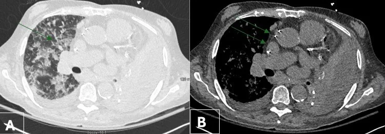
Yetişkinde aynı lobda yineleyen veya beklenenden geç gerileyen konsolidasyon, bronş tıkanıklığını ve özellikle malign bir odağı düşündürebilir; tıkanmanın distalinde enfeksiyon ya da sekresyon birikimi eşlik edebilir. BT'de opak alanla ilişkili bronşun lümeninde darlık, oklüzyon veya endobronşiyal gölge olup olmadığını değerlendirin. BT'de bronş darlığı veya oklüzyonu ve mukus impaksiyonu görülebilir; endobronşiyal tümör şüphesinde bronkoskopi değerlendirmeyi tamamlar. Tıkayıcı odağın gözden kaçması altta yatan malignitenin tanısını geciktirebilir ve apse, ampiyem ya da fistül gibi komplikasyonların fark edilmemesine yol açabilir. Tıkanma distalindeki konsolidasyonun görüntüsü toplum kökenli pnömoniyle örtüşebilir; postobstrüktif pnömoni ile toplum kökenli pnömoniyi görüntüleme bulgularıyla ayırt etmek güç olabilir.
Faz ve pencere
- Kontrastsız tanısal
- İnce kesitli toraks BT'de hava ile dolu bronşun fokal daralması veya kesilmesi, lümendeki yumuşak doku ya da sekresyon, tıkanmanın distalindeki konsolidasyon ve eşlik eden hacim kaybı birlikte değerlendirilebilir. Toraks BT'de bronş duvarı kalınlaşması, darlık veya oklüzyon değerlendirilebilir; kontrastlı BT hiler-mediastinal kitlenin damarları çevrelemesini ve bronşa basısını gösterebilir.
Önerilen pencereler G genişlik, M merkez; değerler Hounsfield birimi (HU)
- AkciğerG 1500 / M −600
- MediastenG 350 / M +50
BT bulguları
- Bronşta fokal daralma — BT'de bronş stenozu veya oklüzyonu postobstrüktif pnömonide daha sık görülebilir.
- Endobronşiyal yumuşak doku — MSCT'de endobronşiyal gölgeler postobstrüktif pnömoni grubunda daha sık görülür.
- Dıştan bası — hiler kitle ya da büyümüş lenf düğümleri bronşu çevreleyerek daraltabilir; bronş duvarı ve komşu yapılarla ilişkisini gösterin.
- Tıkanma distalinde hava boşluğu opasitesi — aynı bronş ağacına uyan segmentte veya lobda konsolidasyon ve sekresyon birikimi bulunabilir.
- Eşlik eden obstrüktif atelektazi — lob hacmi azalabilir; ataletikside hacim kaybına bağlı olarak mediyastinum yönünde sapma gözlenebilir (S38, S40).
- Distal bronşlarda mukus — bronş lümenini dolduran dallanan tübüler opasiteler mukus retansiyonunu destekleyebilir; bu görünüm tek başına nedeni belirlemez.
- Parankim veya plevra uzanımı — konsolidasyon içinde nekrotik boşluk, plevral sıvı/kalınlaşma ya da bronş ile plevra arasında fistül şüphesi aranır.
- Kontrastlı toraks BT, hiler veya mediastinal kitlenin damarları çevrelemesini ve bronşa bası yapmasını gösterebilir; BT'de bronş darlığı ya da oklüzyonu ve eşlik eden konsolidasyon da değerlendirilebilir.
Normalde
Bronş açıklığını değerlendirirken BT'de darlık, oklüzyon ve lümen içi gölge olup olmadığını araştırın. Atelektazide hacim kaybını ve etkilenen bronşta daralma ya da oklüzyon bulgularını araştırın.
Normal BT ile kıyasla
Aynı bölgenin, kaynak veri setinde patoloji içermediği belirtilen bir BT incelemesini kesit kesit kaydırın; organ sınırlarını açarak anatomiyi hasta görüntüsüyle karşılaştırın. Küçük rastlantısal bulgular tümüyle dışlanmamıştır.
Kaynak: TotalSegmentator v2.01 veri seti (Wasserthal ve ark., Radiology: Artificial Intelligence 2023), CC BY 4.0, vaka s1278; organ sınırları veri setinin otomatik segmentasyonundan, birleştirilerek sadeleştirildi. Görüntüler 2,5 mm kesit aralığına yeniden örneklendi.
Ayırıcı tanı
- Primer hava yolu tümörü
- bronş lümenine uzanan veya bronş duvarını düzensiz kalınlaştıran odak tıkanma noktasında görünür; histolojik ayrım BT ile kesinleşmez.
- Mukus tıkacı
- lümeni dolduran materyal bronş boyunca tübüler biçimde uzanabilir; bronş duvarına tutunan ayrı bir kitle eşlik etmeyebilir.
- Yabancı cisim aspirasyonu
- BT, yabancı cismin yerini ve özelliklerini göstererek tanıya yardımcı olabilir; klinik öykü de değerlendirmeye katkı sağlar.
- Komplike olmayan lober pnömoni
- hava boşluğu konsolidasyonu görülür; eşlik eden fokal bronş kesilmesi ya da kitle gösterilmesi alternatif bir obstrüktif neden düşündürür.
- Pulmoner infarkt
- plevraya taban veren periferik kama biçimli opasite beklenir; merkezde tıkayıcı bronş odağı bulunmaz.
Tuzaklar
- Yoğun distal konsolidasyon hilusa uzanan küçük bir endobronşiyal odağı örtebilir; bronşu opasitenin içinden değil, proksimal hava yolundan başlayarak takip edin.
- Mukus tıkacı kitle benzeri görünüm oluşturabilir ve endobronşiyal tümörle görüntü özellikleri örtüşebilir; kontrastlanma ve komşu hiler yapılarla ilişki ayırıcı tanıya katkı sağlar ancak BT tek başına histolojik tanı koydurmaz.
- Tıkanma distalindeki mukus, konsolide ya da kollabe akciğerde dallanan tübüler dolumlar oluşturabilir; distal mukus dolumları proksimal bronşun açık olduğunu tek başına göstermez.
- Aynı lobdaki yinelenen enfeksiyonu yalnız yeni pnömoni olarak adlandırmak altta yatan tıkanmayı saklayabilir; önceki incelemelerle bronş açıklığının değişimini kıyaslayın.
Kendini dene
Postobstrüktif pnömoni ile toplum kökenli pnömoninin BT bulguları karşılaştırıldığında, hangi bulgular postobstrüktif pnömonide daha sık görülür?
Cevabı göster
Bronş duvarı kalınlaşması, darlık veya oklüzyon, mukus impaksiyonu ve endobronşiyal gölgeler. Karşılaştırmalı çalışmada bronş duvarı kalınlaşması, bronş darlığı veya oklüzyonu, mukus impaksiyonu ve endobronşiyal gölgeler postobstrüktif pnömonide daha sık bildirilmiştir.
BT'de lob bronşında lümen içi gölge saptanan ve endobronşiyal tümör şüphesi bulunan hastada lezyonun doğrudan değerlendirilmesi için hangi yöntem öne çıkar?
Cevabı göster
Bronkoskopi. BT'de endobronşiyal gölgeler görülebilir; endobronşiyal tümör şüphesinde erken bronkoskopi önerilir.
İlgili konular
Kaynaklar
Bu sayfadaki 37 cümle ve 2 quiz sorusu aşağıdaki kaynaklardan alıntılarla otomatik olarak karşılaştırıldı; 18 cümle bu karşılaştırmada düzeltildi (2026-10-09).
- [Pulmonary carcinoid tumors].pubmed.ncbi.nlm.nih.gov
- Mediastinum Teratoma Rupture: A Rare Manifestation.pubmed.ncbi.nlm.nih.gov
- Erroneous bronchial transection after video assisted thoracoscopic surgery (VATS) pulmonary resection diagnosed with bronchoscopy: Case report.pubmed.ncbi.nlm.nih.gov
- Endobronchial deposits of chronic lymphocytic leukemia - an unusual cause of central airway obstruction.pubmed.ncbi.nlm.nih.gov
- Endobronchial hamartoma as a cause of pneumonia.pubmed.ncbi.nlm.nih.gov
- Liquefied Lung Cancer: An Uncommon Form of Squamous Cell Carcinoma of the Lung.pubmed.ncbi.nlm.nih.gov
- Rigid-Flexible Bronchoscopy With Holmium Laser Cauterization for Pine Nut Shell Removal and Balloon Dilation for Subsequent Bronchial Stenosis in a 16-Month-Old Girl.pubmed.ncbi.nlm.nih.gov
- Comparison between community-acquired pneumonia and post-obstructive pneumonia associated with endobronchial tumors.pubmed.ncbi.nlm.nih.gov
- Chili pepper aspiration in elderly patients: a case series highlighting diagnostic challenges and the role of cryoextraction.pubmed.ncbi.nlm.nih.gov
- Disseminated <i>Mycobacterium kansasii</i> infection with osseous involvement in anti-interferon-γ autoantibody-associated adult-onset immunodeficiency: a case report and literature review.pubmed.ncbi.nlm.nih.gov
- Lipoid Pneumonia: HRCT and MRI Spectrum, Diagnostic Pitfalls, and Imaging-Based Diagnostic Workflow.pubmed.ncbi.nlm.nih.gov
- Beyond the Usual Suspects: Rare Causes of Hemoptysis.pubmed.ncbi.nlm.nih.gov
- Unilateral "white lung" in a child revealing plastic bronchitis: A reversible cause of airway obstruction.pubmed.ncbi.nlm.nih.gov
- Fibrosing Mediastinitis in Pregnancy: A Rare Case With Antepartum Hemoptysis and Airway Obstruction.pubmed.ncbi.nlm.nih.gov
- The first CT-based classification system for pulmonary actinomycosis: correlating imaging patterns with therapeutic strategies and prognosis in a highly misdiagnosed disease.pubmed.ncbi.nlm.nih.gov
- Post-Obstructive Pneumonia in Patients with Cancer: A Review.pubmed.ncbi.nlm.nih.gov
- Bronchoscopic management of airway foreign bodies in adults: a narrative educational review.pubmed.ncbi.nlm.nih.gov
- Pulmonary Pathology of COVID-19 Following 8 Weeks to 4 Months of Severe Disease: A Report of Three Cases, Including One With Bilateral Lung Transplantation.pubmed.ncbi.nlm.nih.gov
- Postobstructive pneumonia in lung cancer.pubmed.ncbi.nlm.nih.gov
- Pneumonia in normal and immunocompromised children: an overview and update.pubmed.ncbi.nlm.nih.gov
- Bronchoscopic local instillation of amphotericin B deoxycholate as adjunctive therapy for pulmonary mucormycosis with inadequate response to posaconazole: a retrospective case series.pubmed.ncbi.nlm.nih.gov
- Imaging appearances, CT evolution patterns, and surgical prognosis of stage I lung squamous cell carcinoma.pubmed.ncbi.nlm.nih.gov
- Endobronchial lipoma presenting as a fat-attenuation lesion causing left upper lobar bronchial obstruction: A case report.pubmed.ncbi.nlm.nih.gov
- An unusual presentation of tracheobronchial tuberculosis mimicking airway tumor: a case report and literature review.pubmed.ncbi.nlm.nih.gov
- Diagnostic and surgical challenges of pulmonary mucoepidermoid carcinoma: a case report.pubmed.ncbi.nlm.nih.gov
- Giant Right Sphenoid Wing Meningioma as a Reversible Frontal Network Lesion: A Pseudo-bvFTD Case with Venous-Sparing Skull-Base Resection.pubmed.ncbi.nlm.nih.gov
- Anatomically Precise Microsurgical Resection of a Posterior Fossa Cerebellar Metastasis in an Elderly Patient with Preservation of Venous Outflow, Dentate Nucleus, and Cerebrospinal Fluid Pathways.pubmed.ncbi.nlm.nih.gov
- Fleischner Society: Glossary of Terms for Thoracic Imaging.pubmed.ncbi.nlm.nih.gov
- Congenital Bronchial Atresia Mimicking Pulmonary Sequestration in the Lower Lobe: A Case Report.pubmed.ncbi.nlm.nih.gov
- Anatomical structure of pulmonary vessels and bronchi in the right upper lobe based on three-dimensional reconstruction.pubmed.ncbi.nlm.nih.gov
- Congenital Absence of the Left Pulmonary Artery Associated With Right-Sided Aortic Arch Presenting as Hemoptysis in an Adult: A Case Report.pubmed.ncbi.nlm.nih.gov
- Quantitative perfusion imaging from non-contrast micro-ct for pulmonary embolism evaluation in preclinical models.pubmed.ncbi.nlm.nih.gov
- CXCR4-targeted PET/CT in early systemic sclerosis-associated interstitial lung disease: a prospective proof-of-concept study of in vivo inflammatory activity.pubmed.ncbi.nlm.nih.gov
- Monitoring pneumonia onset and severe respiratory infectious diseases using quantitative computed tomography, validation amidst the coronavirus disease 2019 outbreak at the end of 2022 in the Chinese mainland.pubmed.ncbi.nlm.nih.gov
- Hampton's Hump-A Rare Radiological Feature in Patients with Pulmonary Embolism in a Single-Center Study.pubmed.ncbi.nlm.nih.gov
- Exploring varied radiologic appearance in pulmonary embolism with CT pulmonary angiography: Case series with literature review.pubmed.ncbi.nlm.nih.gov
- Multimodality imaging of acute and chronic pulmonary thromboembolic disease.pubmed.ncbi.nlm.nih.gov
- Disproportionate Mediastinal Shift From an Isolated Right Middle Lobe Collapse: A Case Report.pubmed.ncbi.nlm.nih.gov
- Preoperative localization of pulmonary nodules near the fissures: electromagnetic navigation bronchoscopy vs. hook-wire percutaneous localization.pubmed.ncbi.nlm.nih.gov
- Deep-learning based quantitative evaluation of postoperative atelectasis following right upper lobectomy.pubmed.ncbi.nlm.nih.gov
- Endobronchial epithelioid leiomyoma (leiomyoblastoma) causing complete right-lung atelectasis: A case report.pubmed.ncbi.nlm.nih.gov
- Early Structural Lung Changes in Primary Ciliary Dyskinesia (PCD).pubmed.ncbi.nlm.nih.gov
- Use of Extracorporeal Membrane Oxygenation for Lung Cancer-Related Massive Hemoptysis: A Case Report and Literature Review.pubmed.ncbi.nlm.nih.gov
- The cavernous sinus: An anatomic study with clinical implication.pubmed.ncbi.nlm.nih.gov
- Task force of the Brazilian Society of Otology - evaluation and management of peripheral facial palsy.pubmed.ncbi.nlm.nih.gov
- Tuberculosis revisted: classic imaging findings in childhood.pubmed.ncbi.nlm.nih.gov
- Chest imaging using signs, symbols, and naturalistic images: a practical guide for radiologists and non-radiologists.pubmed.ncbi.nlm.nih.gov
- Multi-detector computed tomography imaging of large airway pathology: A pictorial review.pubmed.ncbi.nlm.nih.gov
- International Society of Paediatric Surgical Oncology (IPSO) Minimally Invasive Surgery (MIS) Guidelines.pubmed.ncbi.nlm.nih.gov
- PET-CT for assessing mediastinal lymph node involvement in patients with suspected resectable non-small cell lung cancer.pubmed.ncbi.nlm.nih.gov
- Long-term safety and effectiveness of the "OptEase" vena cava filter.pubmed.ncbi.nlm.nih.gov
- Typical endobronchial carcinoid tumor in a 15-year-old female: A case report.pubmed.ncbi.nlm.nih.gov
- Tracheobronchial Tumors: Radiologic-Pathologic Correlation of Tumors and Mimics.pubmed.ncbi.nlm.nih.gov
- Foreign body aspiration in adult airways: therapeutic approach.pubmed.ncbi.nlm.nih.gov
- Flexible Bronchoscopic En Bloc Cryoextraction of Endobronchial Leiomyoma Using a 1.7-mm Cryoprobe: A Case Report with One-Year Follow-Up.pubmed.ncbi.nlm.nih.gov
- Lung and lobe torsion: a narrative review.pubmed.ncbi.nlm.nih.gov
- Atypical type D extended sleeve resection after neoadjuvant chemoimmunotherapy in a 69-year-old man with locally advanced lung cancer and a tracheobronchial branching anomaly: a case report.pubmed.ncbi.nlm.nih.gov
- Chest Imaging for Pulmonary TB-An Update.pubmed.ncbi.nlm.nih.gov
- Surgery for lung cancer: postoperative changes and complications-what the Radiologist needs to know.pubmed.ncbi.nlm.nih.gov
- Back to Basics - 'Must Know' Classical Signs in Thoracic Radiology.pubmed.ncbi.nlm.nih.gov
- Pictorial essay of radiological features of benign intrathoracic masses.pubmed.ncbi.nlm.nih.gov
- Pulmonary angiography with 64-multidetector-row computed tomography in normal dogs.pubmed.ncbi.nlm.nih.gov
- EACR 2025 Congress: Innovative Cancer Science, 16-19 June 2025.pubmed.ncbi.nlm.nih.gov
- Case Report: Severe bronchiectasis in three kittens with detection of <i>Mycoplasma felis</i>: radiographic and computed tomography findings.pubmed.ncbi.nlm.nih.gov
- Intracavitary Drainage Followed by Lobectomy for a Giant Bulla Causing Respiratory Failure due to Severe Mediastinal Compression: A Case Report.pubmed.ncbi.nlm.nih.gov
- Mobile Cone-Beam CT-Guided Transbronchial Cryobiopsy of a <i>Penicillium</i> Granuloma via the Right Post-Eparterial Bronchus.pubmed.ncbi.nlm.nih.gov
- Sleeve resection of a typical carcinoid tumour in a case of aberrant anatomy.pubmed.ncbi.nlm.nih.gov
- Anatomic reference measures for central airway anatomy in Indian adults: implications for precision airway management in surgical patient safety.pubmed.ncbi.nlm.nih.gov
- Morphological variations of fissures, lobes, and hilar pattern of the lung in a select South African sample.pubmed.ncbi.nlm.nih.gov
- Pulmonary Leiomyoma in a Post-hysterectomy Patient: A Diagnostic Challenge.pubmed.ncbi.nlm.nih.gov
- Communicating bronchopulmonary foregut malformations: Revisiting a rare entity.pubmed.ncbi.nlm.nih.gov
- Complications of Lung Transplantation: Update on Imaging Manifestations and Management.pubmed.ncbi.nlm.nih.gov
- Bronchoscopy and the Pulmonologist in Contemporary Lung Cancer Care.pubmed.ncbi.nlm.nih.gov
- Current trends in image-guided chest interventions.pubmed.ncbi.nlm.nih.gov
- Narrative review of congenital lung lesions.pubmed.ncbi.nlm.nih.gov
- Spectral CT-derived peripheral iodine concentration in acute pulmonary embolism: association with subsegmental emboli and pulmonary infarction.pubmed.ncbi.nlm.nih.gov
- Host, Pathogen, and Pulmonary Anatomy in Pneumonia: An Integrated Imaging and Clinical Perspective.pubmed.ncbi.nlm.nih.gov
- Case Report: Adult fibrinous bronchitis associated with lymphatic reflux: a probable diagnosis based on convergent evidence.pubmed.ncbi.nlm.nih.gov
- Challenges in the Early Diagnosis, Screening and Management of Heart Failure in Patients with Chronic Obstructive Pulmonary Disease.pubmed.ncbi.nlm.nih.gov
- CT Imaging Features of Pulmonary Sarcoidosis: Typical and Atypical Radiological Features and Their Differential Diagnosis.pubmed.ncbi.nlm.nih.gov
- Precision Medicine in Asthma: The Role of Biomarkers.pubmed.ncbi.nlm.nih.gov
- Non-Small-Cell Lung Cancer: From Histopathological Classification to Precision Oncology-A Narrative Review.pubmed.ncbi.nlm.nih.gov
- The fate of the Fontan circulation.pubmed.ncbi.nlm.nih.gov
- Evaluation of a caudal midline glossectomy on tongue volume and upper airway cross-sectional areas in brachycephalic dogs: a cadaveric study.pubmed.ncbi.nlm.nih.gov
- Transformative biomechanics and mechanobiology breakthroughs shaping the future of health and medicine.pubmed.ncbi.nlm.nih.gov
- Research hotspots and frontiers of application of mass spectrometry breath test in respiratory diseases.pubmed.ncbi.nlm.nih.gov
- A Frequently Missed Pulmonary Infarction: Clinical and 18F-FDG PET/CT Manifestation of Hilar Tumor-Induced Pulmonary Infarction.pubmed.ncbi.nlm.nih.gov
- Active pulmonary tuberculosis: something old, something new, something borrowed, something blue.pubmed.ncbi.nlm.nih.gov
- A nursing perspective on bronchoscopic intervention strategies for children with macrolide-unresponsive <i>Mycoplasma pneumoniae</i> pneumonia.pubmed.ncbi.nlm.nih.gov
- Chlamydia trachomatis Pneumonia in a 50-Day-Old Full-Term Male Infant Associated With Mucus Plugging of the Left Upper Lobe Bronchus and Left Upper Lobe Atelectasis.pubmed.ncbi.nlm.nih.gov
- Carinal Squamous Cell Carcinoma Causing Progressive Central Airway Obstruction: CT, FDG PET/CT, Bronchoscopic Pre- and Post-Debulking, and Pathologic Correlation.pubmed.ncbi.nlm.nih.gov
- Management of pneumonia by the emergency radiologist.pubmed.ncbi.nlm.nih.gov
- Clinical classification of chronic obstructive pulmonary disease with invasive pulmonary aspergillosis: a retrospective study.pubmed.ncbi.nlm.nih.gov
- Diagnostic Dilemma of Rounded Atelectasis in the Left Lower Lobe Showing High Uptake of 18F-Fluorodeoxyglucose: A Surgical Conundrum.pubmed.ncbi.nlm.nih.gov
- Diffuse cystic lung diseases-a primer for radiologists.pubmed.ncbi.nlm.nih.gov
- CT features of feline cystic bronchiectasis forming mass lesions.pubmed.ncbi.nlm.nih.gov
- Fibrotic Pulmonary Sarcoidosis-From Pathogenesis to Management.pubmed.ncbi.nlm.nih.gov
- A sheep in wolf's clothing? Herpes-simplex-virus endobronchial pseudotumor.pubmed.ncbi.nlm.nih.gov
- Mimicking Metastasis: Endobronchial Tuberculosis Presenting as a Mass in a Pediatric Patient With Yolk Sac Tumor.pubmed.ncbi.nlm.nih.gov
- Short-term prognosis, mid- and long-term clinical follow-up of bronchoscopic intervention therapy combined with bronchial artery embolization on massive hemoptysis.pubmed.ncbi.nlm.nih.gov
- Overview of malignant central airway obstruction.pubmed.ncbi.nlm.nih.gov
- Bacterial Granulomatous Lung Diseases: Radiological Findings and Differential Diagnosis.pubmed.ncbi.nlm.nih.gov
- Endobronchial fibroepithelial polyp presenting as persistent wheezing in a 9-year-old girl: a case report.pubmed.ncbi.nlm.nih.gov
- Multimodal interventional bronchoscopy for bronchial myelolipoma complicated with airway obstruction: a case report and literature review.pubmed.ncbi.nlm.nih.gov
- Primary pulmonary mucosa-associated lymphoid tissue lymphoma presenting as a right hilar mass: A case report.pubmed.ncbi.nlm.nih.gov
- Advances in diagnosis and treatment of lung cancer-associated obstructive pneumonia and lung abscess: synergistic strategies for infection control and antitumor therapy.pubmed.ncbi.nlm.nih.gov
- Stepwise diagnostic algorithm for high-attenuation pulmonary abnormalities on CT.pubmed.ncbi.nlm.nih.gov
- CT Findings of Central Airway Lesions Causing Airway Stenosis-Visualization and Quantification: A Pictorial Essay.pubmed.ncbi.nlm.nih.gov
- Case Report: Pediatric mediastinal actinomycosis mimicking lymphoma diagnosed by tissue metagenomic next-generation sequencing.pubmed.ncbi.nlm.nih.gov
- Exploring the key clinical and computed tomography features for distinguishing high-grade lung cancers from morphologically similar benign tumors: a two-center case-control study.pubmed.ncbi.nlm.nih.gov
Eğitim amaçlıdır; hasta başında tanı ve tedavi kararının yerine geçmez, tıbbi tavsiye değildir. Metinler yapay zekâ desteğiyle yazıldı ve otomatik bir adımda kaynak alıntılarıyla karşılaştırıldı; bu bir tıbbi doğrulama değildir ve içerik uzman radyolog incelemesinden geçmedi. Klinik kararlarda güncel kılavuzları, kurum protokollerini ve radyoloji raporunu esas alın.