Özet
Atelektazi, akciğerin bir bölümünün kısmi ya da tam kollapsıdır; anatomik yaygınlığı subsegmental, segmental, lober veya tüm akciğer düzeyinde tarif edilebilir. BT’de opasitenin fissürlerle ilişkisi, lob hacmindeki azalma ve bronşların açıklığı birlikte değerlendirilerek tutulan anatomik birim adlandırılır. Bu yaklaşım, eşlik edebilen plevral sıvı ve santral bronş tıkanıklığının göz önünde bulundurulmasına yardım eder. Hacim kaybını yalnızca opasite artışı olarak yorumlamak, kollapsın yaygınlığının ve olası obstrüktif nedenin yanlış değerlendirilmesine yol açabilir.
Okuma sırası
- Sağ akciğer lobları. Koronal kesitte sağ akciğeri hilustan diyaframa kadar tarayın; opasitenin tek segmentte mi, bir fissür çifti arasındaki lobda mı, yoksa tüm hemitoraksta mı uzandığını belirleyin.
- İnterlobar fissürler. Opasitenin tuttuğu lobun hacmini değerlendirin; lobar hacim kaybı atelektazi derecesiyle paralel artar ve BT'de gösterilebilir.
- Sağ üst lob bronşu. Hilustan çıkan sağ üst lob bronşunu opak bölgeye kadar izleyin; lümen açık mı, sekresyonla dolu mu, yoksa ani biçimde kesiliyor mu kaydedin.
- Sağ orta lob ve karşı akciğer. Çöken lobun dışındaki sağ orta lobu ve karşı akciğeri gözden geçirin; genişlemiş kalan parankimin kollapsa eşlik eden telafi edici genişlemeyi değerlendirin.
- Hilus ve mediastinal yapılar. Aynı düzeyde hilus, trakea, kalp kenarı ve diyaframı kıyaslayın; yapılar kollaps tarafına çekilmiş mi, efüzyon varsa ters yöne itilmiş mi ayırın.
Faz ve pencere
- Kontrastsız tanısal
- Kollabe segment veya lob, komşu havalı akciğere göre daha yoğun ve küçülmüş görünür; BT'de bronş lümenindeki tıkanıklık ve hacim kaybının kapsamı değerlendirilebilir. Hacim kaybının kapsamı BT’de değerlendirilebilir.
Önerilen pencereler: Akciğer (G 1500 / M -600), Mediasten (G 350 / M 50).
BT bulguları
- Lineer atelektazi BT’de görülebilir.
- Segmental kollaps — tek bir bronkopulmoner segmenti tutan hacim kaybıdır; mukus tıkaçları gibi nedenlerle gelişebilir.
- Lober kollaps — bir lobun küçülmesiyle tanınır; hilar siluet kaybı, hemidiyafram elevasyonu ve mediastinal kayma gibi dolaylı bulgular eşlik edebilir.
- Fissür yer değiştirmesi diğer hacim kaybı bulgularıyla birlikte değerlendirilmelidir.
- Hilus ve mediasten çekilmesi — belirgin tek taraflı hacim kaybında aynı taraftaki hilus, trakea veya kalp konturu kollabe alana yaklaşabilir.
- Diyafram kubbesi yükselmesi — alt lob veya tüm akciğerde hacim azalması ipsilateral hemidiyaframı yukarı çekebilir.
- Telafi edici genişleme — kollabe loba komşu loblar veya karşı akciğer genişleyebilir.
- Bronş açıklığı — BT'de bronş lümeninde endobronkiyal kitle ya da tıkaç saptanması, atelektazinin obstrüktif bir nedenle uyumlu olduğunu destekler.
- BT’de kronik alt lob kollapsı, lobun omurga boyunca retraksiyonu ile görülebilir.
- Yuvarlak atelektazi farklı etiyolojilerle ilişkili olabilir.
Ölçütler ve sınıflamalar
- Anatomik yaygınlığa göre atelektazi
- Subsegmental: alt segment düzeyinde tutulum; segmental: bir bronkopulmoner segment; lober: bir lob; total: bir akciğerin tamamı. Bu adlandırma tutulan anatomik hacmi belirtir, etiyolojiyi tek başına sınıflamaz.
Normalde
Normal BT’de izlenebilen fissürler ince ve düzenli seyreder; bronşlar açık, damar dalları düzenli dağılmıştır. İlgili bronş lümenini ve tutulan alanın hacmini BT'de inceleyin; hacim kaybının varlığı ve yaygınlığı bu değerlendirmeyle saptanabilir.
Normal BT ile kıyasla
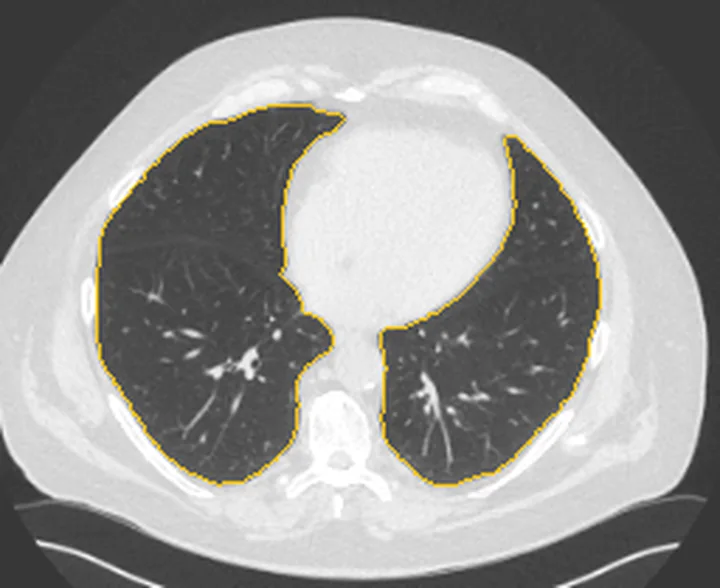
Aynı bölgenin, kaynak veri setinde patoloji içermediği belirtilen bir BT incelemesini kesit kesit kaydırın; organ sınırlarını açarak anatomiyi hasta görüntüsüyle karşılaştırın. Küçük rastlantısal bulgular tümüyle dışlanmamıştır.
Kaynak: TotalSegmentator v2.01 veri seti (Wasserthal ve ark., Radiology: Artificial Intelligence 2023), CC BY 4.0, vaka s1278; organ sınırları veri setinin otomatik segmentasyonundan, birleştirilerek sadeleştirildi. Görüntüler 2,5 mm kesit aralığına yeniden örneklendi.
Ayırıcı tanı
- Lober pnömoni
- BT'de tanınan, bir lobu tutan konsolidasyon paternidir ve toplum kökenli pnömonide en sık izlenen paternlerden biridir; kollapstan ayırmada tutulan alanın hacim kaybı değerlendirilir.
- Plevral efüzyon
- plevral aralıkta sıvı birikimidir; büyük efüzyon komşu akciğeri basıyla çökertebilir ve kompresif atelektaziye eşlik edebilir.
- Kitle benzeri yuvarlak atelektazi
- plevra tabanlı yuvarlak opasiteye komşu plevral kalınlaşma ve hilusa dönen bronkovasküler yapılar eşlik eder.
- Pulmoner infarkt
- BT’de plevra tabanlı kama biçimli opasite görülebilir. Akciğer grafisinde plevra tabanlı kama biçimli opasite Hampton hump olarak adlandırılır.
- Telafi edici genişleme
- kollapsa eşlik edebilen bir bulgudur; kollabe alanın komşusundaki loblar veya karşı akciğer hacim kazanabilir.
Tuzaklar
- BT’de lober pnömoni ve atelektazi ayrı paternler olarak tanımlanır; atelektazide hacim kaybı değerlendirilir.
- Büyük plevral efüzyon mediasteni karşı tarafa itebilir ve aynı tarafta bası atelektazisi oluşturabilir; kayma yönü tek başına kollapsı dışlamaz.
- Sağ orta lob kollapsında, lobun toplam akciğer hacmine katkısı görece küçük olduğundan mediastinal kayma genellikle minimaldir; BT izole kollapsı doğrulayabilir.
- Mediastinal ve hilar yer değiştirme, lober kollapsın dolaylı bulgusudur; tanı ve yaygınlık, bu kaymanın diğer ikincil bulgularla birlikte değerlendirilmesiyle önerilmelidir.
Kendini dene
BT’de bir lobun hacmi azalmış; aynı taraftaki hilus siluetinde azalma ve mediastinal yer değiştirme görülüyor. Tutulumun en doğru adı nedir?
Cevabı göster
Lober kollaps. Bir lobun hacim kaybı ve aynı taraftaki hilar yapıların yer değiştirmesi lober kollapsı destekler.
Alt lob opasitesi mevcut; tutulan lobun boyutu korunmuş ve hacim kaybı saptanmıyor. Hangi olasılık daha uygundur?
Cevabı göster
Lober pnömoni. Hacim kaybı eşlik etmeyen opasite, lobar kollapstan çok pnömonik konsolidasyonu düşündürür; BT'de lobar pnömoni ve atelektazi ayrı paternler olarak tanımlanır. Total veya obstrüktif kollapsta hacim azalması beklenir.
İlgili konular
Kaynaklar
Bu sayfadaki 44 cümle ve 2 quiz sorusu aşağıdaki kaynaklardan alıntılarla otomatik olarak karşılaştırıldı; 18 cümle bu karşılaştırmada düzeltildi (2026-10-09).
- Multimodality imaging of acute and chronic pulmonary thromboembolic diseasepmc.ncbi.nlm.nih.gov
- [Post-infectious bronchiolitis obliterans].pubmed.ncbi.nlm.nih.gov
- Structural Lung Disease and Clinical Phenotype in Bronchiectasis Patients: The EMBARC CT Study.pubmed.ncbi.nlm.nih.gov
- A clinical guideline for structured assessment of CT-imaging in congenital lung abnormalities.pubmed.ncbi.nlm.nih.gov
- Tomographic aspects of penetrating thoracic trauma: injuries from firearms and other weapons.pubmed.ncbi.nlm.nih.gov
- Visual Assessment of CT Findings in Smokers With Nonobstructed Spirometric Abnormalities in The COPDGene ® Study.pubmed.ncbi.nlm.nih.gov
- Computed tomographic findings of pulmonary atelectasis in healthy anesthetized Beagles.pubmed.ncbi.nlm.nih.gov
- Reexpansion pulmonary edema: computed tomography findings in 22 patients.pubmed.ncbi.nlm.nih.gov
- Evaluation of pulmonary disease using static lung volumes in primary ciliary dyskinesia.pubmed.ncbi.nlm.nih.gov
- The Diaphragm Acts as a Brake during Expiration to Prevent Lung Collapse.pubmed.ncbi.nlm.nih.gov
- Optimizing prone CT use for suspected interstitial lung abnormalities.pubmed.ncbi.nlm.nih.gov
- COVID-19 on Chest CT: Translating Known Microscopic Findings to Imaging Observations.pubmed.ncbi.nlm.nih.gov
- CT scan features as predictors of patient outcome after bronchial intervention in endobronchial TB.pubmed.ncbi.nlm.nih.gov
- Frequency and characterization of ancillary chest CT findings in COVID-19 pneumonia.pubmed.ncbi.nlm.nih.gov
- Proton Magnetic Resonance Imaging for Initial Assessment of Isolated Mycobacterium avium Complex Pneumonia.pubmed.ncbi.nlm.nih.gov
- The empty azygos fissure.pubmed.ncbi.nlm.nih.gov
- Ten good reasons to practice ultrasound in critical care.pubmed.ncbi.nlm.nih.gov
- [Clinico-radiological characteristics of bronchial carcinoid].pubmed.ncbi.nlm.nih.gov
- Lung reexpansion of obstructive atelectasis caused by radiotherapy after continuous gefitinib treatment in nonsmall cell lung cancer.pubmed.ncbi.nlm.nih.gov
- Thoracic Computed Tomography to Assess ARDS and COVID-19 Lungs.pubmed.ncbi.nlm.nih.gov
- Compact Arterial Monitoring Device Use in Resuscitative Endovascular Balloon Occlusion of the Aorta (REBOA): A Simple Validation Study in Swine.pubmed.ncbi.nlm.nih.gov
- Rapid identification of primary atopic disorders (PAD) by a clinical landmark-guided, upfront use of genomic sequencing.pubmed.ncbi.nlm.nih.gov
- Dapagliflozin for heart failure according to body mass index: the DELIVER trial.pubmed.ncbi.nlm.nih.gov
- Allergic bronchopulmonary aspergillosis presenting as lobar or total lung collapse.pubmed.ncbi.nlm.nih.gov
- Middle lobe syndrome: a rare presentation of allergic bronchopulmonary aspergillosis.pubmed.ncbi.nlm.nih.gov
- Total unilateral pulmonary collapse secondary to allergic bronchopulmonary aspergillosis: a case series of an unusual cause of complete atelectasis.pubmed.ncbi.nlm.nih.gov
- Partial replacement of left hemidiaphragm in dogs by either cryopreserved or decellularized heterograft patch.pubmed.ncbi.nlm.nih.gov
- The missing hilum: Chronic lobar collapse.pubmed.ncbi.nlm.nih.gov
- Long-term results of diaphragmatic plication in adults with unilateral diaphragm paralysis.pubmed.ncbi.nlm.nih.gov
- Complications related to endoscopic lung volume reduction for emphysema with endobronchial valves: results of a multicenter study.pubmed.ncbi.nlm.nih.gov
- Usefulness of FDG PET/CT in determining benign from malignant endobronchial obstruction.pubmed.ncbi.nlm.nih.gov
- Interventions for ulceration and other skin changes caused by nerve damage in leprosy.pubmed.ncbi.nlm.nih.gov
- Automated subfield volumetric analysis of amygdala, hippocampus, and thalamic nuclei in mesial temporal lobe epilepsy.pubmed.ncbi.nlm.nih.gov
- Reproducibility of the Unaided Subjective Assessment of Orbital Computed X-Ray Tomographic Features in Thyroid Eye Disease.pubmed.ncbi.nlm.nih.gov
- Deep-learning based quantitative evaluation of postoperative atelectasis following right upper lobectomy.pubmed.ncbi.nlm.nih.gov
- Is autonomic dysreflexia a cause of respiratory dysfunction after spinal cord injury?pubmed.ncbi.nlm.nih.gov
- AARC clinical practice guideline: effectiveness of nonpharmacologic airway clearance therapies in hospitalized patients.pubmed.ncbi.nlm.nih.gov
- Predictors of Airspace Disease on Chest X-ray in Emergency Department Patients With Clinical Bronchiolitis: A Systematic Review and Meta-analysis.pubmed.ncbi.nlm.nih.gov
- CT features of community-acquired pneumonia at the emergency department.pubmed.ncbi.nlm.nih.gov
- [Sarcoid pleural effusion].pubmed.ncbi.nlm.nih.gov
- Clinical Characteristics of Lung Abscess Caused by Streptococcus Constellatus Infection.pubmed.ncbi.nlm.nih.gov
- Case report: Malignant chemodectoma with hepatic metastasis in a cat.pubmed.ncbi.nlm.nih.gov
- Causative factors, imaging findings, and CT course of round atelectasis.pubmed.ncbi.nlm.nih.gov
- CT features of rounded atelectasis in chronic inflammatory pleural effusions in cats and dogs.pubmed.ncbi.nlm.nih.gov
- Diagnostic imaging and histopathologic features of rounded atelectasis in four cats and one dog: A descriptive case series study.pubmed.ncbi.nlm.nih.gov
- [Pulmonary cavities with Takayasu arteritis: report of 3 cases and literature review].pubmed.ncbi.nlm.nih.gov
- Pulmonary Infarction Due to Direct-acting Oral Anticoagulant Malabsorption.pubmed.ncbi.nlm.nih.gov
- Preferential location of acute pulmonary thromboembolism induced consolidative opacities: assessment with respiratory gated perfusion SPECT-CT fusion images.pubmed.ncbi.nlm.nih.gov
- Severe Pulmonary Infection in a 20-Month-Old Female.pubmed.ncbi.nlm.nih.gov
- Primary Pulmonary Leiomyosarcoma: An Unusual Cause of Pleural Effusion.pubmed.ncbi.nlm.nih.gov
- Interventional embolization for hemorrhagic pleural effusion in an infant with kaposiform lymphangiomatosis: a case report.pubmed.ncbi.nlm.nih.gov
- [A case of bronchoscopic treatment for endobronchial chondroma].pubmed.ncbi.nlm.nih.gov
- Endobronchial Lipoma: A Rare Cause of Bronchial Stenosis or Obstruction.pubmed.ncbi.nlm.nih.gov
- Obstructive bronchitis and recurrent pneumonia in esophageal achalasia in a child: A CARE compliant case report.pubmed.ncbi.nlm.nih.gov
- Role of ultrasound in confirmation of endotracheal tube in neonates: a review.pubmed.ncbi.nlm.nih.gov
- Persistent cough: An unexpected diagnosis.pubmed.ncbi.nlm.nih.gov
- Endobronchial Tuberculosis Simulating Carcinoid Tumor.pubmed.ncbi.nlm.nih.gov
- Point-of-Care Thoracic Ultrasonography in Patients With Cirrhosis and Liver Failure.pubmed.ncbi.nlm.nih.gov
- 36th International Symposium on Intensive Care and Emergency Medicine : Brussels, Belgium. 15-18 March 2016.pubmed.ncbi.nlm.nih.gov
- Acute life-threatening cardiac tamponade in a mechanically ventilated patient with COVID-19 pneumonia.pubmed.ncbi.nlm.nih.gov
- Comparison of early and late time-controlled adaptive ventilation on pulmonary gas exchange in anesthetized horses.pubmed.ncbi.nlm.nih.gov
- [Clinical and pathological findings of patients with interstitial lung disease associated with antisynthetase].pubmed.ncbi.nlm.nih.gov
- Disproportionate Mediastinal Shift From an Isolated Right Middle Lobe Collapse: A Case Report.pubmed.ncbi.nlm.nih.gov
- SeparSEPAR (Sociedad Española de Neumologia y Cirugía Torácica) Clinical Protocol on Endoscopic Lung Volume Reduction for Severe Emphysema.pubmed.ncbi.nlm.nih.gov
- The first CT-based classification system for pulmonary actinomycosis: correlating imaging patterns with therapeutic strategies and prognosis in a highly misdiagnosed disease.pubmed.ncbi.nlm.nih.gov
- Comprehensive review of pulmonary embolism imaging: past, present and future innovations in computed tomography (CT) and other diagnostic techniques.pubmed.ncbi.nlm.nih.gov
- Reading chest radiographs in the critically ill (Part II): Radiography of lung pathologies common in the ICU patient.pubmed.ncbi.nlm.nih.gov
- Long-term safety and effectiveness of the "OptEase" vena cava filter.pubmed.ncbi.nlm.nih.gov
- Impact of inferior pulmonary ligament dissection versus preservation during thoracoscopic upper lobectomy: a retrospective comparative analysis.pubmed.ncbi.nlm.nih.gov
- Ultra-high spatial resolution at photon-counting computed tomography: technical insights and sustainable applications in cardiothoracic imaging.pubmed.ncbi.nlm.nih.gov
- International Society of Paediatric Surgical Oncology (IPSO) Minimally Invasive Surgery (MIS) Guidelines.pubmed.ncbi.nlm.nih.gov
- ESR Essentials: imaging in nasal obstruction and epistaxis-practice recommendations by the European Society of Head and Neck Radiology.pubmed.ncbi.nlm.nih.gov
- Communicating bronchopulmonary foregut malformations: Revisiting a rare entity.pubmed.ncbi.nlm.nih.gov
- A study of excessive dynamic airway collapse in chronic obstructive pulmonary disease patients.pubmed.ncbi.nlm.nih.gov
- A rare case of symptomatic congenital lobar emphysema in an adolescent female who underwent expectant management of a congenital pulmonary malformation: a case report.pubmed.ncbi.nlm.nih.gov
- Fleischner Society: Glossary of Terms for Thoracic Imaging.pubmed.ncbi.nlm.nih.gov
- Cardiothoracic manifestations of neuroendocrine tumours.pubmed.ncbi.nlm.nih.gov
- Surgery for lung cancer: postoperative changes and complications-what the Radiologist needs to know.pubmed.ncbi.nlm.nih.gov
- Coexistence of Autoantibodies against the Golgi Complex and Ro52 Antigen in a Patient with Nonspecific Interstitial Pneumonia.pubmed.ncbi.nlm.nih.gov
- Case Report: Severe bronchiectasis in three kittens with detection of <i>Mycoplasma felis</i>: radiographic and computed tomography findings.pubmed.ncbi.nlm.nih.gov
- Development and validation of a nomogram for predicting recurrent plastic bronchitis in children with Mycoplasma pneumoniae pneumonia.pubmed.ncbi.nlm.nih.gov
- Thoracic abnormal air collections in patients in the intensive care unit: radiograph findings correlated with CT.pubmed.ncbi.nlm.nih.gov
- Consensus statement on thoracic radiology terminology in Portuguese used in Brazil and in Portugal.pubmed.ncbi.nlm.nih.gov
- Diffuse cystic lung diseases-a primer for radiologists.pubmed.ncbi.nlm.nih.gov
- CT Imaging Features of Pulmonary Sarcoidosis: Typical and Atypical Radiological Features and Their Differential Diagnosis.pubmed.ncbi.nlm.nih.gov
- The value of interlobar fissure semilunar sign based on multifactor joint analysis in predicting the invasiveness of ground glass nodules with interlobar fissure attachment in the lungs.pubmed.ncbi.nlm.nih.gov
- LobePrior segments lung lobes on computed tomography images in the presence of severe abnormalities.pubmed.ncbi.nlm.nih.gov
- Tracheobronchial injury after blunt thoracic trauma-lessons to learn in diagnosis, treatment, and postoperative care: a case series.pubmed.ncbi.nlm.nih.gov
- Effect of Right Middle-to-Lower Lobe Fixation on Postoperative Lung Volume after Right Upper Lobectomy.pubmed.ncbi.nlm.nih.gov
- Losing vigilance in diagnosing pulmonary alveolar microlithiasis: A report on four cases.pubmed.ncbi.nlm.nih.gov
- Giant Right Sphenoid Wing Meningioma as a Reversible Frontal Network Lesion: A Pseudo-bvFTD Case with Venous-Sparing Skull-Base Resection.pubmed.ncbi.nlm.nih.gov
- ESR Essentials: imaging of common paediatric pulmonary diseases-practice recommendations by the European Society of Paediatric Radiology.pubmed.ncbi.nlm.nih.gov
- Quantitative Imaging Metrics for the Assessment of Pulmonary Pathophysiology: An Official American Thoracic Society and Fleischner Society Joint Workshop Report.pubmed.ncbi.nlm.nih.gov
- Endoluminal approaches to postoperative air leaks.pubmed.ncbi.nlm.nih.gov
- Endobronchial Valves for Bronchoscopic Lung Volume Reduction in Severe Emphysema: A Reversible and Non-Surgical Treatment for Patients Who May or May Not Be Candidates for Lung Transplantation.pubmed.ncbi.nlm.nih.gov
- Endobronchial epithelioid leiomyoma (leiomyoblastoma) causing complete right-lung atelectasis: A case report.pubmed.ncbi.nlm.nih.gov
- Middle lobe torsion after chest tube drainage for primary spontaneous pneumothorax: a case report.pubmed.ncbi.nlm.nih.gov
- Anatomically Precise Microsurgical Resection of a Posterior Fossa Cerebellar Metastasis in an Elderly Patient with Preservation of Venous Outflow, Dentate Nucleus, and Cerebrospinal Fluid Pathways.pubmed.ncbi.nlm.nih.gov
- The cavernous sinus: An anatomic study with clinical implication.pubmed.ncbi.nlm.nih.gov
- Collagenous endobronchial obliteration after chemoradiation followed by consolidation osimertinib: a case report.pubmed.ncbi.nlm.nih.gov
- Congenital Pulmonary Airway Malformation Presenting as Pneumothorax: Two Case Reports.pubmed.ncbi.nlm.nih.gov
- Thoracotomy for Urgent Repair of Lung Torsion Identified as an Incidental Finding.pubmed.ncbi.nlm.nih.gov
- Structured respiratory physiotherapy protocol for resolution of atelectasis in pediatric intensive care.pubmed.ncbi.nlm.nih.gov
- Consensus recommendations on perioperative mechanical ventilation strategies: an evidence-based guideline by the Brazilian Society of Anesthesiology.pubmed.ncbi.nlm.nih.gov
- Middle Lobe Torsion after Unilateral Lung Transplant.pubmed.ncbi.nlm.nih.gov
- Overview of treatment related complications in malignant pleural mesothelioma.pubmed.ncbi.nlm.nih.gov
- Patterns of injury and violence in Yaoundé Cameroon: an analysis of hospital data.pubmed.ncbi.nlm.nih.gov
- Spectral CT-derived peripheral iodine concentration in acute pulmonary embolism: association with subsegmental emboli and pulmonary infarction.pubmed.ncbi.nlm.nih.gov
- Clinical features, management, and outcomes of pulmonary mucormycosis: a decade-long retrospective study from a single center in central China.pubmed.ncbi.nlm.nih.gov
- When Virchow's Triad Goes on Overdrive: Catastrophic Multi-System Thrombosis in the Setting of New-Onset Heart Failure in a Previously Healthy Man.pubmed.ncbi.nlm.nih.gov
- Multimodality imaging of acute and chronic pulmonary thromboembolic disease.pubmed.ncbi.nlm.nih.gov
Eğitim amaçlıdır; hasta başında tanı ve tedavi kararının yerine geçmez, tıbbi tavsiye değildir. Metinler yapay zekâ desteğiyle yazıldı ve otomatik bir adımda kaynak alıntılarıyla karşılaştırıldı; bu bir tıbbi doğrulama değildir ve içerik uzman radyolog incelemesinden geçmedi. Klinik kararlarda güncel kılavuzları, kurum protokollerini ve radyoloji raporunu esas alın.