Özet
Asbestozis, asbest liflerinin solunmasına bağlı pulmoner fibrozistir ve belirtiler maruziyetten onlarca yıl sonra ortaya çıkabilir. Yüksek çözünürlüklü BT, asbestozisin erken parankimal değişikliklerini ve eşlik eden plevral anormallikleri göğüs radyografisinden daha duyarlı gösterir. Plevral plakların görülmesi maruziyeti destekler, fakat tek başına parankimal asbestozis tanısı koydurmaz. Erken evre asbestozis tanısında radyolog ve patolojistlerin kullandığı kriterlerin uyuşmaması hastalığın gözden kaçmasına yol açabilir.
Faz ve pencere
- HRCT tanısal
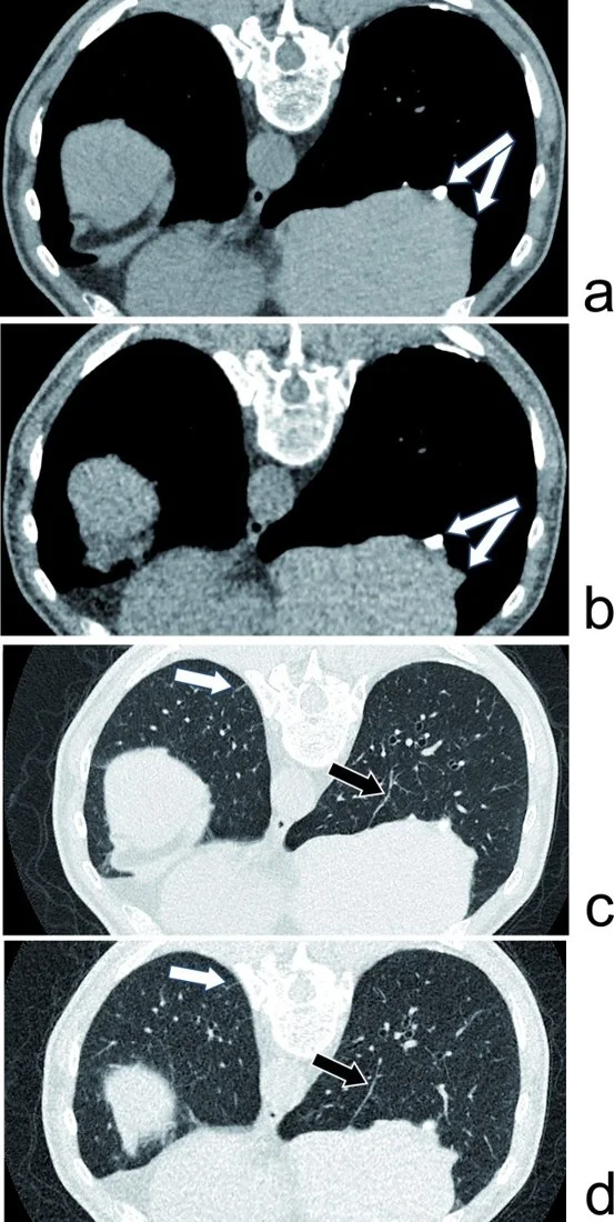
- Kontrastsız inspiryum ince kesitlerde bazal ve subplevral retikülasyon, subplevral çizgiler/noktalar, traksiyon bronşektazisi ve ileri olguda bal peteği görünümü aranır. Prone görüntüler sırtüstü bağımlı dansiteyi fibrozisten ayırmaya yardım eder; damar fazı asbestozis paternini saptamak için gerekli değildir.
Önerilen pencereler: Akciğer (G 1500 / M -600), Mediasten (G 350 / M 50).
BT bulguları
- Bazal ve subplevral retikülasyon — periferik interlobüler çizgiler ve düzensiz çizgisel opasiteler alt loblarda belirginleşir.
- Subplevral noktasal yapılar ve çizgiler — erken asbestoziste HRCT'de görülebilir.
- Traksiyon bronşektazisi — ileri asbestoziste konsolidasyon bulunan alanlarda görülebilir.
- Mimari distorsiyon ve alt lob hacim kaybı — fissür ve bronkovasküler demetler çekilir.
- Bal peteği — ileri fibrotik hastalıkta plevraya komşu kümelenmiş kistik hava boşlukları gelişebilir.
- Parankimal bantlar ve subplevral eğrisel çizgiler — asbest maruziyetli kişilerde BT'de görülebilir.
- Plevral plaklar veya diffüz plevral kalınlaşma — eşlik edebilir; parankim fibrozisinin yerine geçmez ve ayrı tanımlanmalıdır.
Normalde
Normal alt loblarda plevraya kadar uzanan havalı lobüller düzenlidir; bronşlar periferde incelir ve plevra düzgün, ince bir çizgi olarak izlenir. Aynı posterior bazal düzeyde iki akciğerin subplevral retikülasyonunu, bronşların çekilerek genişlemesini ve hacim kaybını karşılaştırın; şüpheli sırt bağımlı opasite prone seride kayboluyorsa fibrozis olmayabilir.
Normal BT ile kıyasla
Aynı bölgenin, kaynak veri setinde patoloji içermediği belirtilen bir BT incelemesini kesit kesit kaydırın; organ sınırlarını açarak anatomiyi hasta görüntüsüyle karşılaştırın. Küçük rastlantısal bulgular tümüyle dışlanmamıştır.
Kaynak: TotalSegmentator v2.01 veri seti (Wasserthal ve ark., Radiology: Artificial Intelligence 2023), CC BY 4.0, vaka s1278; organ sınırları veri setinin otomatik segmentasyonundan, birleştirilerek sadeleştirildi. Görüntüler 2,5 mm kesit aralığına yeniden örneklendi.
Ayırıcı tanı
- İdiyopatik pulmoner fibrozis
- UIP paterni asbestozisle örtüşür; plevral plak ve doğrulanmış maruziyet asbestozis lehine ek kanıttır.
- Bağ dokusu hastalığına bağlı UIP olasılığında sistemik bağ dokusu hastalığı bulguları değerlendirilmelidir.
- Asbestozisin HRCT paterni NSIP'den belirgin biçimde farklı, UIP paternine ise daha çok benzer.
- Bağımlı atelektazi
- posterior bazal opasite prone pozisyonda açılır; gerçek fibroziste distorsiyon ve traksiyon bronşektazisi kalır.
- Diffüz plevral kalınlaşma
- viseral plevrayı sarar, kostofrenik açıyı silebilir ve parankimal retikülasyondan ayrı bir plevral süreçtir.
Tuzaklar
- Sırtüstü çekimdeki bağımlı posterior yoğunluk erken fibrozis gibi görünebilir; prone seride açılan opasiteyi asbestozis retikülasyonu olarak işaretlemeyin.
- Plevral plağın hemen altındaki ince düzensizlik tek başına asbestozis kanıtı değildir; yaygın bazal retikülasyon, traksiyon ve hacim kaybı arayın.
- Plevral plak ve parankimal fibrozis farklı kompartımanlardır; plak varlığını parankim hastalığının şiddetiyle eş anlamlı kullanmayın.
- Plevral plaklar genellikle sınırlı kalınlaşma biçimindedir; plevral mezotelyomada çevresel nodüler kalınlaşma görülebilir ve benign-malign ayrımı görüntülemeyle her zaman yapılamaz.
Kendini dene
Asbest maruziyeti öyküsü olan bir kişide bazal-subplevral fibrotik değişiklikler görülüyor. Bu bulgular hangi hastalıkla uyumludur?
Cevabı göster
Asbestozis. Asbestozis, asbest maruziyetine bağlı pulmoner fibrozistir; bazal yerleşim bu hastalıkla uyumludur. İzole plevral plaklar asbestozis şüphesini artırabilir ancak tek başına tanı koydurmaz.
Sırtüstü HRCT'de posterior bazal çizgisel opasite görülüyor; prone seride tamamen kayboluyor. Bu değişiklik en çok neyi düşündürür?
Cevabı göster
Bağımlı atelektazi. Prone pozisyonda açılan posterior yoğunluk bağımlı atelektaziyi destekler. Bal peteği ve traksiyon bronşektazisi pozisyonla kaybolmaz; diffüz plevral kalınlaşma ise plevra boyunca görünür.
İlgili konular
Kaynaklar
Bu sayfadaki 38 cümle ve 2 quiz sorusu aşağıdaki kaynaklardan alıntılarla otomatik olarak karşılaştırıldı; 10 cümle bu karşılaştırmada düzeltildi (2026-10-09).
- Challenges in Identifying and Diagnosing Asbestos-Related Diseases in Emerging Economies: A Global Health Perspectivepmc.ncbi.nlm.nih.gov
- Asbestos exposure diagnosis in pulmonary tissuespmc.ncbi.nlm.nih.gov
- Lung cancer risk and occupational pulmonary fibrosis: systematic review and meta-analysispmc.ncbi.nlm.nih.gov
- Digital Pulmonology Practice with Phonopulmography Leveraging Artificial Intelligence: Future Perspectives Using Dual Microwave Acoustic Sensing and Imagingpmc.ncbi.nlm.nih.gov
- Imaging diagnosis of classical and new pneumoconiosis: predominant reticular HRCT patternpmc.ncbi.nlm.nih.gov
- Asbestos-associated pulmonary disease.pubmed.ncbi.nlm.nih.gov
- [Asbestosis and pleural plaques].pubmed.ncbi.nlm.nih.gov
- Occupational toxicology of asbestos-related malignancies.pubmed.ncbi.nlm.nih.gov
- Asbestos and the lung in the 21st century: an update.pubmed.ncbi.nlm.nih.gov
- Imaging diagnosis of classical and new pneumoconiosis: predominant reticular HRCT pattern.pubmed.ncbi.nlm.nih.gov
- Computed Tomography of Contemporary Occupational Lung Disease: A Pictorial Review.pubmed.ncbi.nlm.nih.gov
- A systematic review of the association between pleural plaques and changes in lung function.pubmed.ncbi.nlm.nih.gov
- High-resolution CT of the lungs: Indications and diagnosis.pubmed.ncbi.nlm.nih.gov
- [Occupational interstitial lung diseases].pubmed.ncbi.nlm.nih.gov
- Radiologic Diagnosis of Asbestosis in Korea.pubmed.ncbi.nlm.nih.gov
- [Effect of asbestos fibre dust exposures on lung function--a systematic review].pubmed.ncbi.nlm.nih.gov
- [Pneumoconiosis: high-resolution CT characteristics and patho-physiology].pubmed.ncbi.nlm.nih.gov
- Idiopathic Pulmonary Fibrosis: Epidemiology, Clinical Features, Prognosis, and Management.pubmed.ncbi.nlm.nih.gov
- Interstitial Lung Diseases in Developing Countries.pubmed.ncbi.nlm.nih.gov
- Genetic variability of immune checkpoints in asbestos-related diseases.pubmed.ncbi.nlm.nih.gov
- Neuropsychiatric disorders in pulmonary fibrosis: from brain network alterations to inflammatory mechanisms and therapeutic implications.pubmed.ncbi.nlm.nih.gov
- Mechanisms of Action of Herbal Preparations for the Rehabilitation of Dust-Related Diseases of the Bronchopulmonary System.pubmed.ncbi.nlm.nih.gov
- Advances in diagnosis of lung fibrosis: focus on present and future approaches.pubmed.ncbi.nlm.nih.gov
- Pleuroparenchymal Fibroelastosis in Connective Tissue Disease-Related Interstitial Lung Disease.pubmed.ncbi.nlm.nih.gov
- Comprehensive radiologic-pathologic correlation in systemic sclerosis-associated interstitial lung disease: identification of an early-stage CT findings.pubmed.ncbi.nlm.nih.gov
- Asbestos exposure diagnosis in pulmonary tissues.pubmed.ncbi.nlm.nih.gov
- Korean Guidelines for Diagnosis and Management of Interstitial Lung Diseases.pubmed.ncbi.nlm.nih.gov
- Fleischner Society: Glossary of Terms for Thoracic Imaging.pubmed.ncbi.nlm.nih.gov
- [Update in Diagnosis of Idiopathic Pulmonary Fibrosis and Interstitial Lung Abnormality].pubmed.ncbi.nlm.nih.gov
- Update in diagnosis and management of interstitial lung disease .pubmed.ncbi.nlm.nih.gov
- Occupational Lung Diseases: Spectrum of Common Imaging Manifestations.pubmed.ncbi.nlm.nih.gov
- Impact of coal mine dust exposure and cigarette smoking on lung disease in Appalachian coalminers.pubmed.ncbi.nlm.nih.gov
- Importance of chest HRCT in the diagnostic evaluation of fibrosing interstitial lung diseases.pubmed.ncbi.nlm.nih.gov
- Idiopathic pulmonary fibrosis: the radiologist's role in making the diagnosis.pubmed.ncbi.nlm.nih.gov
- The Clinical Significance and Translational Implications of Subclinical Interstitial Lung Abnormalities in Asymptomatic Adults: A Narrative Review.pubmed.ncbi.nlm.nih.gov
- ESR Essentials: imaging in fibrotic lung diseases-practice recommendations by the European Society of Thoracic Imaging.pubmed.ncbi.nlm.nih.gov
- Restrictions of VC and DLCO in relation to asbestos-related computed tomographic findings quantified by ICOERD-based parameters.pubmed.ncbi.nlm.nih.gov
- Asbestos-Related Respiratory Diseases in Spain: Insights From the Spanish Registry.pubmed.ncbi.nlm.nih.gov
- The usual Interstitial pneumonia pattern in autoimmune rheumatic diseases.pubmed.ncbi.nlm.nih.gov
- Potential asbestos exposure among patients with primary lung cancer in Japan.pubmed.ncbi.nlm.nih.gov
- Expert consensus on cancer treatment-related lung injury.pubmed.ncbi.nlm.nih.gov
- Consensus statement on thoracic radiology terminology in Portuguese used in Brazil and in Portugal.pubmed.ncbi.nlm.nih.gov
- Computed tomography of the chest in patients with COVID-19: what do radiologists want to know?pubmed.ncbi.nlm.nih.gov
- Interstitial Lung Abnormalities: What Radiologists Should Know.pubmed.ncbi.nlm.nih.gov
- Cigarette smoking decreases macrophage-dependent clearance to impact the biological effects of occupational and environmental particle exposures.pubmed.ncbi.nlm.nih.gov
- Secreted Phosphoprotein 1 in Lung Diseases.pubmed.ncbi.nlm.nih.gov
- Diagnostic Approaches for Idiopathic Pulmonary Fibrosis.pubmed.ncbi.nlm.nih.gov
- Occupational lung disease: when should I think of it and why is it important?pubmed.ncbi.nlm.nih.gov
- Diagnosis and Treatment of Hypersensitivity Pneumonitis: S2k Guideline of the German Respiratory Society and the German Society for Allergology and Clinical Immunology.pubmed.ncbi.nlm.nih.gov
- The role of vascularity and the fibrovascular interface in interstitial lung diseases.pubmed.ncbi.nlm.nih.gov
- Progressive Pulmonary Fibrosis: Where Are We Now?pubmed.ncbi.nlm.nih.gov
- Lipoid Pneumonia: HRCT and MRI Spectrum, Diagnostic Pitfalls, and Imaging-Based Diagnostic Workflow.pubmed.ncbi.nlm.nih.gov
- Computed tomography in ARDS, from morphological insights to AI-powered multi-modal analysis: a narrative review.pubmed.ncbi.nlm.nih.gov
- Comments and Illustrations of the European Federation of Societies for Ultrasound in Medicine Guidelines: Benign Pleura Lesions (Benign Pleura Thickening, Lesions and Masses)-What Can Be Seen on Transthoracic Ultrasound?pubmed.ncbi.nlm.nih.gov
- Tips and Tricks in Thoracic Radiology for Beginners: A Findings-Based Approach.pubmed.ncbi.nlm.nih.gov
- Integrating CT radiomics and clinical data with machine learning to predict fibrosis progression in coalworker pneumoconiosis.pubmed.ncbi.nlm.nih.gov
- Comparison of Asbestos Victim Relief Available Outside of Conventional Occupational Compensation Schemes.pubmed.ncbi.nlm.nih.gov
- Clinical consequences of asbestos-related diffuse pleural thickening: A review.pubmed.ncbi.nlm.nih.gov
- Extended Exhaled Nitric Oxide Analysis in Interstitial Lung Diseases: A Systematic Review.pubmed.ncbi.nlm.nih.gov
- The role of pathologists in the diagnosis of occupational lung diseases: an expert opinion of the European Society of Pathology Pulmonary Pathology Working Group.pubmed.ncbi.nlm.nih.gov
- AWMF S2k Guideline: Diagnosis and Assessment of Lung Disease Caused by Occupational Silica Dust Exposure (Silicosis) - Disease No. 4101 as per Occupational Diseases Ordinance (BKV) Annex 1.pubmed.ncbi.nlm.nih.gov
- Pleural plaques and interstitial lung abnormalities in AGES-Reykjavik study.pubmed.ncbi.nlm.nih.gov
- Global research landscape versus disease burden in pneumoconiosis diagnosis: a dual-source bibliometric analysis [1999-2025].pubmed.ncbi.nlm.nih.gov
- Diagnostic Challenges in Pleural Mesothelioma.pubmed.ncbi.nlm.nih.gov
- Computed tomography findings of peritoneal tumour in the absence of primary lesions: preliminary study.pubmed.ncbi.nlm.nih.gov
- Pleural Mesothelioma Diagnosis for the Pulmonologist: Steps Along the Way.pubmed.ncbi.nlm.nih.gov
- Idiopathic pulmonary fibrosis: current diagnosis and treatment.pubmed.ncbi.nlm.nih.gov
- Challenges in Identifying and Diagnosing Asbestos-Related Diseases in Emerging Economies: A Global Health Perspective.pubmed.ncbi.nlm.nih.gov
- Disease burden of asbestos-related diseases in China (1990-2023) based on GBD estimates: A call for stronger labor protection laws.pubmed.ncbi.nlm.nih.gov
- Distinct comorbidity profiles and outcomes in asbestosis versus idiopathic pulmonary fibrosis: a 6-year prospective cohort study.pubmed.ncbi.nlm.nih.gov
- Review of carcinogenicity of asbestos and proposal of approval standards of an occupational cancer caused by asbestos in Korea.pubmed.ncbi.nlm.nih.gov
- Comparison of the ultra-low-dose Veo algorithm with the gold standard filtered back projection for detecting pulmonary asbestos-related conditions: a clinical observational study.pubmed.ncbi.nlm.nih.gov
- Asbestosis Requiring Lung Transplantation in a Retired Hairdresser: An Occupational Exposure to Comb Through.pubmed.ncbi.nlm.nih.gov
- The Role of Particle Inhalation in Idiopathic Pulmonary Fibrosis.pubmed.ncbi.nlm.nih.gov
- Clinical and radiological criteria for the differential diagnosis between asbestosis and idiopathic pulmonary fibrosis: Application in two cases.pubmed.ncbi.nlm.nih.gov
- The 2025 ATS/ERS update of the international multidisciplinary classification of the interstitial pneumonias: implications for the pathologist.pubmed.ncbi.nlm.nih.gov
- Bronchoalveolar lavage cell differential count is associated with survival and radiological UIP pattern in familial pulmonary fibrosis.pubmed.ncbi.nlm.nih.gov
- Biomarkers on Fibrotic Lung Diseases Associated with Usual Interstitial Pneumonia.pubmed.ncbi.nlm.nih.gov
- Drug-Induced Interstitial Lung Disease in Lung Cancer Patients.pubmed.ncbi.nlm.nih.gov
- Incidental pulmonary findings on CT in daily practice: the nodule and the interstitial lung abnormalities - what's old, what's new.pubmed.ncbi.nlm.nih.gov
- Radiographic Distribution as a Diagnostic Clue in Pulmonary Disease.pubmed.ncbi.nlm.nih.gov
- An overview on multimodal imaging for the diagnostic workup of pleural mesothelioma.pubmed.ncbi.nlm.nih.gov
- Environmental Asbestos Exposure as a Possible Pathway to Early-Onset Pleural Mesothelioma: A Case From Afghanistan and Pakistan to London.pubmed.ncbi.nlm.nih.gov
- Localised malignant mesothelioma presenting as an esophageal mass: An unusual manifestation.pubmed.ncbi.nlm.nih.gov
- Case Report: Tislelizumab combined with bevacizumab in the treatment of primary malignant pericardial mesothelioma.pubmed.ncbi.nlm.nih.gov
Eğitim amaçlıdır; hasta başında tanı ve tedavi kararının yerine geçmez, tıbbi tavsiye değildir. Metinler yapay zekâ desteğiyle yazıldı ve otomatik bir adımda kaynak alıntılarıyla karşılaştırıldı; bu bir tıbbi doğrulama değildir ve içerik uzman radyolog incelemesinden geçmedi. Klinik kararlarda güncel kılavuzları, kurum protokollerini ve radyoloji raporunu esas alın.