Özet
İdiyopatik pulmoner fibrozis (IPF), çoğunlukla ileri yaşta sinsi ilerleyen fibrotik interstisyel akciğer hastalığıdır; UIP, IPF’nin tipik radyolojik paterni olsa da bağ dokusu hastalığı ve fibrotik hipersensitivite pnömonisinde de görülebilir. İnce kesit kontrastsız HRCT, fibrozisin dağılımını ve UIP olasılığını sınıflandırarak multidisipliner tanıya yön verir. Bal peteğini traksiyon bronşiolektazisi, paraseptal amfizem veya fibrozise eşlik eden hava boşluğu genişlemesiyle karıştırmak yanlış sınıflamaya yol açabilir. Doğru BT sınıflandırması, tedavi kararlarını yönlendirdiği gibi pulmoner hipertansiyon ve akut kötüleşme gibi komplikasyonların tanısında da kritik rol oynar.
Faz ve pencere
- HRCT tanısal
- İntravenöz kontrast uygulanmayan ince kesitli inspiratuar akciğer görüntülerinde alt lob ve subplevral ağırlıklı retikülasyon, düzensiz traksiyon bronşiolektazisi ve kümeli kistik bal peteği aranır; yüzükoyun pozisyonda alınan kesitler bağımlı posterior opasitelerin gerçek fibrozis olup olmadığını ayırt etmeye, ekspiratuar görüntüleme ise eşlik eden hava hapsini saptamaya yardımcı olur.
Önerilen pencereler: Akciğer (G 1500 / M -600), Mediasten (G 350 / M 50).
BT bulguları
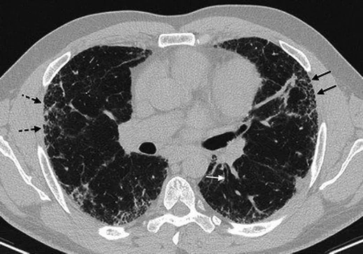
- Bazal-subplevral retikülasyon — alt lob periferinde ağ biçimli interlobüler ve intralobüler çizgiler.
- Traksiyon bronşiolektazisi — fibrotik retikülasyon içinde düzensiz genişlemiş periferik hava yolu; ardışık kesitte bronş ağacına bağlanır.
- Bal peteği — çoğu benzer çaplı, belirgin duvarlı, subplevral kümeli kistik hava boşlukları; tek sıra da uygun bağlamda görülebilir.
- Kraniokaudal gradyan — fibrotik yükün üst zonlardan çok alt loblarda belirginleşmesi.
- Yamalı mimari distorsiyon — bronkovasküler demetlerin çekilmesi, fissürlerin yer değiştirmesi ve lob hacim kaybı.
- Buzlu cam gölgesi retikülasyonla birlikte görülebilir; ancak baskın veya tek başına yaygın buzlu cam varlığı NSIP lehine yorumlanmalı ve UIP dışında değerlendirilmelidir.
- Alternatif tanı ipuçları — belirgin hava hapsi, sentrilobüler nodül, yaygın konsolidasyon veya plevral plak UIP dışı nedeni düşündürür.
Ölçütler ve sınıflamalar
- Bal peteği kistlerinin tipik çapı
- Tipik bal peteği kistleri 3–10 mm çapındadır ve genellikle subplevral yerleşimlidir.
- ATS/ERS/JRS/ALAT HRCT UIP sınıflaması
- UIP: bazal-subplevral baskın fibrozise bal peteği eşlik eder; traksiyon bronşiektazisi/bronşiolektazisi bulunabilir. Olası UIP paterni, karakteristik bazal-subplevral dağılımda retikülasyon ve traksiyon bronşiolektazisi içerir ancak belirgin bal peteği veya UIP dışı özellikler göstermez. UIP için belirsiz bir kategoridir; tanıyı netleştirmek amacıyla klinik veri, biyopsi veya ileri görüntüleme ile multidisipliner tartışma gerekebilir. Alternatif tanı: başka hastalık lehine bulgular (ör. belirgin mozaik atenüasyon, yaygın buzlu cam, sentrilobüler nodüller veya üst/peribronkovasküler baskınlık) vardır.
Normalde
Normal bazal sekonder lobüllerde subplevral retiküler ağ ve kümeli kistik boşluklar bulunmaz; bronşiyoller düzgün kalibrasyonla periferde sonlanır, fissür ve damar-bronş demetleri korunur. Sağ ve sol alt lobların aynı seviyelerinde periferik çizgilenmenin gerçekten intralobüler ağ oluşturup oluşturmadığını, bronşiyolün çevre fibrozisle düzensiz çekilip çekilmediğini ve plevraya bitişik boşlukların duvar/katman özelliklerini kıyaslayın.
Normal BT ile kıyasla
Aynı bölgenin, kaynak veri setinde patoloji içermediği belirtilen bir BT incelemesini kesit kesit kaydırın; organ sınırlarını açarak anatomiyi hasta görüntüsüyle karşılaştırın. Küçük rastlantısal bulgular tümüyle dışlanmamıştır.
Kaynak: TotalSegmentator v2.01 veri seti (Wasserthal ve ark., Radiology: Artificial Intelligence 2023), CC BY 4.0, vaka s1278; organ sınırları veri setinin otomatik segmentasyonundan, birleştirilerek sadeleştirildi. Görüntüler 2,5 mm kesit aralığına yeniden örneklendi.
Ayırıcı tanı
- Fibrotik NSIP
- daha simetrik bazal buzlu cam ve retikülasyon, görece düzgün fibrozis; bal peteği daha az belirgin olabilir.
- Fibrotik hipersensitivite pnömonisi
- fibrozise eşlik eden lobüler hava hapsi, üç yoğunluk paterni veya sentrilobüler nodüller destekler.
- Paraseptal amfizem
- daha geniş subplevral boşluklar genellikle ince duvarlıdır; çevresinde retikülasyon ve traksiyon bozulması yoktur.
- Fibrozise eşlik eden hava boşluğu genişlemesi
- sigaraya bağlı üst lob amfizemi ve düzensiz, düşük yoğunluklu boşluklar bal peteğini taklit edebilir.
- Bağ dokusu hastalığı ilişkili UIP
- görüntüleme UIP paternini gösterebilir; eşlik eden özofagus genişlemesi, eklem bulguları veya sistemik klinik bağlam, IPF tanısı konulmadan önce dikkate alınmalıdır.
Tuzaklar
- UIP paterni IPF ile eş anlamlı değildir; maruziyet, bağ dokusu hastalığı ve ilaç öyküsü dışlanmadan etiyoloji görüntüden tek başına atanamaz.
- Bal peteği sanılan subplevral boşlukları koronal/oblik düzlemde takip edin: devamlı bronşiyol lümeni traksiyon bronşiolektazisini destekler.
- Sırtüstü çekimde posterior bazal buzlu cam veya çizgisel opasite, yüzükoyun pozisyonda alınan görüntüde kayboluyorsa bağımlı atelektazi olabilir; kalıcı mimari distorsiyon ise fibrozisi destekler.
Kendini dene
Alt loblarda subplevral retikülasyon, traksiyon bronşiolektazisi ve kümeli kistler var. En uygun HRCT paterni hangisi?
Cevabı göster
UIP. Subplevral-bazal retikülasyonla birlikte gerçek kümeli bal peteği UIP paternini karşılar. Subplevral-bazal retikülasyon, traksiyon bronşiolektazisi ve kümeli kistik boşluklar UIP paterninin BT bulgularıdır; ancak NSIP, kronik hipersensitivite pnömonisi veya paraseptal amfizem de benzer görünüş verebilir. Bu nedenle diğer patolojiler çok disiplinli tartışmada dışlanmadan UIP paterni tek başına IPF tanısı koymaz. UIP paterninin görülmesi tek başına IPF tanısı koydurmaz.
Plevra altında kistik boşluklar izleniyor; çevresinde retikülasyon yok ve boşluklar geniş. En olası taklitçi nedir?
Cevabı göster
Paraseptal amfizem. Geniş subplevral boşluklar ve eşlik eden fibrotik mimari distorsiyonun bulunmaması paraseptal amfizemi destekler. Bal peteği kümelenmiş kistik boşlukları fibrozis içinde oluşturur; NSIP yaygın interstisyel patern, organize pnömoni ise çoğunlukla hava boşluğu opasiteleri yapar.
Bu konunun yer aldığı turlar
İlgili konular
Kaynaklar
Bu sayfadaki 43 cümle ve 2 quiz sorusu aşağıdaki kaynaklardan alıntılarla tek tek karşılaştırıldı; 7 cümle bu karşılaştırmada düzeltildi (2026-10-08).
- Diagnosis of Idiopathic Pulmonary Fibrosis. An Official ATS/ERS/JRS/ALAT Clinical Practice Guideline.pubmed.ncbi.nlm.nih.gov
- Idiopathic pulmonary fibrosis.pubmed.ncbi.nlm.nih.gov
- Nintedanib in Progressive Fibrosing Interstitial Lung Diseases.pubmed.ncbi.nlm.nih.gov
- Diagnostic criteria for idiopathic pulmonary fibrosis: a Fleischner Society White Paper.pubmed.ncbi.nlm.nih.gov
- Idiopathic Pulmonary Fibrosis.pubmed.ncbi.nlm.nih.gov
- Pictorial Review of Fibrotic Interstitial Lung Disease on High-Resolution CT Scan and Updated Classification.pubmed.ncbi.nlm.nih.gov
- Diagnosis of early idiopathic pulmonary fibrosis: current status and future perspective.pubmed.ncbi.nlm.nih.gov
- Nintedanib in progressive interstitial lung diseases: data from the whole INBUILD trial.pubmed.ncbi.nlm.nih.gov
- Why is UIP peripheral?pubmed.ncbi.nlm.nih.gov
- Advances in Concept and Imaging of Interstitial Lung Disease.pubmed.ncbi.nlm.nih.gov
- A case of an 81-year-old with cough and dyspnea.pubmed.ncbi.nlm.nih.gov
- Connective tissue disease-related interstitial lung disease (CTD-ILD) and interstitial lung abnormality (ILA): Evolving concept of CT findings, pathology and management.pubmed.ncbi.nlm.nih.gov
- Rheumatoid arthritis and lung disease: from mechanisms to a practical approach.pubmed.ncbi.nlm.nih.gov
- Idiopathic pulmonary fibrosis.pubmed.ncbi.nlm.nih.gov
- Utility of Bronchoalveolar Lavage and Transbronchial Biopsy in Patients with Interstitial Lung Disease.pubmed.ncbi.nlm.nih.gov
- Clinical and radiological features of idiopathic interstitial pneumonias (IIPs): a pictorial review.pubmed.ncbi.nlm.nih.gov
- Comparison between the ATS/ERS/JRS/ALAT criteria of 2011 and 2018 for Usual Interstitial Pneumonia on HRCT: a cross-sectional study.pubmed.ncbi.nlm.nih.gov
- Idiopathic pulmonary fibrosis: evolving concepts.pubmed.ncbi.nlm.nih.gov
- Central Role of CT in Management of Pulmonary Fibrosis.pubmed.ncbi.nlm.nih.gov
- High-Resolution Computed Tomography of Fibrotic Interstitial Lung Disease.pubmed.ncbi.nlm.nih.gov
- Acute Exacerbation of Interstitial Lung Disease: Early Diagnosis and Treatment.pubmed.ncbi.nlm.nih.gov
- [Not Available].pubmed.ncbi.nlm.nih.gov
- Acute Exacerbation in Interstitial Lung Disease.pubmed.ncbi.nlm.nih.gov
- Diagnostic accuracy of ultrashort echo times-MRI in patients with and without idiopathic pulmonary fibrosis.pubmed.ncbi.nlm.nih.gov
- Smoking-related lung abnormalities on computed tomography images: comparison with pathological findings.pubmed.ncbi.nlm.nih.gov
- Characteristics, clinical findings, and treatment of idiopathic pulmonary fibrosis in Japanese patients registered with a medical subsidy system for intractable diseases.pubmed.ncbi.nlm.nih.gov
- Comprehensive radiologic-pathologic correlation in systemic sclerosis-associated interstitial lung disease: identification of an early-stage CT findings.pubmed.ncbi.nlm.nih.gov
- Honeycombing on CT; its definition, pathologic correlation, and future direction of its diagnosis.pubmed.ncbi.nlm.nih.gov
- Smoking-related interstitial fibrosis combined with pulmonary emphysema: computed tomography-pathologic correlative study using lobectomy specimens.pubmed.ncbi.nlm.nih.gov
- Radiological Insights into UIP Pattern: A Comparison Between IPF and Non-IPF Patients.pubmed.ncbi.nlm.nih.gov
- Imaging of pulmonary fibrosis in children: A review, with proposed diagnostic criteria.pubmed.ncbi.nlm.nih.gov
- Traction Bronchiectasis/Bronchiolectasis is Associated with Interstitial Lung Abnormality Mortality.pubmed.ncbi.nlm.nih.gov
- Unusual Presentation of Orofacial Granulomatosis Leading to the Incidental Diagnosis of Idiopathic Pulmonary Fibrosis: A Case Report.pubmed.ncbi.nlm.nih.gov
- Advanced Imaging and Occupational History in the Diagnosis of Bird Fancier's Lung: A Case Report.pubmed.ncbi.nlm.nih.gov
- Clustered Cystic Changes in Long-Term Follow-Up Thin-Section Computed Tomographic Findings in Fibrotic Nonspecific Interstitial Pneumonia.pubmed.ncbi.nlm.nih.gov
- Endoscopic resection of upper gastrointestinal lymphangioma: A single-center experience.pubmed.ncbi.nlm.nih.gov
- Equine granulosa cell tumours among other ovarian conditions: Diagnostic challenges.pubmed.ncbi.nlm.nih.gov
- Thin-section CT of the secondary pulmonary lobule: anatomy and the image--the 2004 Fleischner lecture.pubmed.ncbi.nlm.nih.gov
- Imaging of pulmonary emphysema: a pictorial review.pubmed.ncbi.nlm.nih.gov
- Meta-Analysis of Interobserver Agreement in Assessment of Interstitial Lung Disease Using High-Resolution CT.pubmed.ncbi.nlm.nih.gov
- The ATS/ERS/JRS/ALAT statement "IPF by HRCT" could predict acute exacerbation of interstitial lung disease in non-small cell lung cancer.pubmed.ncbi.nlm.nih.gov
- Chronic hypersensitivity pneumonitis.pubmed.ncbi.nlm.nih.gov
- Effects of nintedanib on symptoms in patients with progressive pulmonary fibrosis.pubmed.ncbi.nlm.nih.gov
- Nintedanib in patients with progressive fibrosing interstitial lung diseases-subgroup analyses by interstitial lung disease diagnosis in the INBUILD trial: a randomised, double-blind, placebo-controlled, parallel-group trial.pubmed.ncbi.nlm.nih.gov
- Interstitial Lung Fibrosis Imaging Reporting and Data System: What Radiologist Wants to Know?pubmed.ncbi.nlm.nih.gov
- Effect of Nintedanib in Subgroups of Idiopathic Pulmonary Fibrosis by Diagnostic Criteria.pubmed.ncbi.nlm.nih.gov
- Chest High-Resolution CT Findings of Microscopic Polyangiitis: A Japanese First Nationwide Prospective Cohort Study.pubmed.ncbi.nlm.nih.gov
- Durvalumab with etoposide and carboplatin for patients with extensive-stage small cell lung cancer and interstitial lung disease: A multicenter, open-label prospective trial.pubmed.ncbi.nlm.nih.gov
- [The updated S2k guideline for the diagnosis of idiopathic pulmonary fibrosis : Essential aspects for pathology].pubmed.ncbi.nlm.nih.gov
- Comparison of pulmonary abnormalities on high-resolution computed tomography in patients with early versus longstanding rheumatoid arthritis.pubmed.ncbi.nlm.nih.gov
- Nonspecific interstitial pneumonia versus usual interstitial pneumonia: differences in the density histogram of high-resolution CT.pubmed.ncbi.nlm.nih.gov
- Hypersensitivity pneumonitis: new concepts and classifications.pubmed.ncbi.nlm.nih.gov
- Pathology of Chronic Hypersensitivity Pneumonitis What Is It? What Are the Diagnostic Criteria? Why Do We Care?pubmed.ncbi.nlm.nih.gov
- Bird fancier's lung: a state-of-the-art review.pubmed.ncbi.nlm.nih.gov
- Cystic Lung Diseases: Algorithmic Approach.pubmed.ncbi.nlm.nih.gov
- Interstitial lung abnormalities and interstitial lung diseases associated with cigarette smoking in a rural cohort undergoing surgical resection.pubmed.ncbi.nlm.nih.gov
- CT features of pathologically proven smoking-related interstitial fibrosis: compared with emphysema and usual interstitial pneumonia.pubmed.ncbi.nlm.nih.gov
- Overview of Rheumatoid Arthritis-Associated Interstitial Lung Disease and Its Treatment.pubmed.ncbi.nlm.nih.gov
- COVID-19 pneumonia: pathophysiology and management.pubmed.ncbi.nlm.nih.gov
- CT findings in biomass smoke-associated interstitial lung disease: A retrospective analysis.pubmed.ncbi.nlm.nih.gov
- Neurofibromatosis-Associated Diffuse Lung Disease: A Case Report and Review of the Literature.pubmed.ncbi.nlm.nih.gov
- Dual-Energy Computed Tomography Lung in patients of Pulmonary Tuberculosis.pubmed.ncbi.nlm.nih.gov
- Predictors of pulmonary sequelae after COVID-19 pneumonia: A 12-month follow-up study.pubmed.ncbi.nlm.nih.gov
- Progression of probable UIP and UIP on HRCT.pubmed.ncbi.nlm.nih.gov
- Tissue Continues to Be the Issue: Role of Histopathology in the Context of Recent Updates in the Radiologic Classification of Interstitial Lung Diseases.pubmed.ncbi.nlm.nih.gov
- [Pulmonary manifestations of connective tissue diseases].pubmed.ncbi.nlm.nih.gov
- CT Features of the Usual Interstitial Pneumonia Pattern: Differentiating Connective Tissue Disease-Associated Interstitial Lung Disease From Idiopathic Pulmonary Fibrosis.pubmed.ncbi.nlm.nih.gov
- Pulmonary fibrosis.pubmed.ncbi.nlm.nih.gov
- Mycophenolate and azathioprine in fibrotic interstitial lung disease.pubmed.ncbi.nlm.nih.gov
- The 2025 ATS/ERS update of the international multidisciplinary classification of the interstitial pneumonias: implications for the pathologist.pubmed.ncbi.nlm.nih.gov
- A multidimensional index with SpO<sub>2</sub> at rest for predicting progressive pulmonary fibrosis.pubmed.ncbi.nlm.nih.gov
- High-resolution computed tomography assessment of connective tissue disease-associated interstitial lung disease: Advances in diagnosis and prognostic evaluation.pubmed.ncbi.nlm.nih.gov
- Contemporary Concise Review 2025: Interstitial Lung Disease.pubmed.ncbi.nlm.nih.gov
- Diagnostic, prognostic, and therapeutic challenges in patients with hypersensitivity pneumonitis.pubmed.ncbi.nlm.nih.gov
- Understanding cystic lung lesions in smokers with interstitial lung disease: radiologic-pathological correlation.pubmed.ncbi.nlm.nih.gov
- Smoker's Lungs: Quo Vadis? Exploring the Shared and Individual Smoking-Related Molecular Pathways of Emphysema, Carcinoma, and Fibrosis - An Illustrative Review.pubmed.ncbi.nlm.nih.gov
- Diffuse cystic lung diseases-a primer for radiologists.pubmed.ncbi.nlm.nih.gov
- Acute exacerbation in fibrotic interstitial lung disease: an international working group report.pubmed.ncbi.nlm.nih.gov
- A biomarker-guided framework for corticosteroid treatment stratification in acute exacerbation of idiopathic pulmonary fibrosis.pubmed.ncbi.nlm.nih.gov
- Comparative study on mouse models of acute exacerbation of pulmonary fibrosis.pubmed.ncbi.nlm.nih.gov
- High-Resolution CT Findings in Interstitial Lung Disease Associated with Connective Tissue Diseases: Differentiating Patterns for Clinical Practice-A Systematic Review with Meta-Analysis.pubmed.ncbi.nlm.nih.gov
- Hypersensitivity Pneumonitis on Thin-Section Chest CT Scans: Diagnostic Performance of the ATS/JRS/ALAT versus ACCP Imaging Guidelines.pubmed.ncbi.nlm.nih.gov
- Deep learning-based quantitative CT assessment of interstitial lung abnormalities: prognostic risk thresholds in a health screening population.pubmed.ncbi.nlm.nih.gov
- Pulmonary AL Amyloidosis as a Cause of Interstitial Lung Disease Diagnosed With Transbronchial Lung Biopsy.pubmed.ncbi.nlm.nih.gov
- Nintedanib-Associated Generalized Body Swelling in a Patient With Idiopathic Pulmonary Fibrosis: A Case Report.pubmed.ncbi.nlm.nih.gov
- Impact of Severe COVID-19 on Interstitial Lung Disease in Autoimmune Systemic Diseases.pubmed.ncbi.nlm.nih.gov
- Successful immunosuppressive treatment of hypersensitivity pneumonitis secondary to lycoperdonosis in a dog.pubmed.ncbi.nlm.nih.gov
- A Rare Case of COPA Syndrome: Multisystem Relapse and Fatal Septic Complication.pubmed.ncbi.nlm.nih.gov
- Honeycomb wall thickening during acute deterioration of idiopathic pulmonary fibrosis: clinical, imaging and prognostic characteristics.pubmed.ncbi.nlm.nih.gov
- Pleuroparenchymal Fibroelastosis Lesion in Explanted Lungs of a Patient with Fibrotic Pulmonary Sarcoidosis.pubmed.ncbi.nlm.nih.gov
- Monocyte-Derived LGMN<sup>+</sup> Macrophages Divert Lung Injury Outcomes toward Fibrosis through Matrix Remodeling.pubmed.ncbi.nlm.nih.gov
- CT Features of Granulomatous-Lymphocytic Interstitial Lung Disease (GLILD): The "Kebab Sign" as a Marker to Support Differential Diagnosis.pubmed.ncbi.nlm.nih.gov
- AWMF S2k Guideline: Diagnosis and Assessment of Lung Disease Caused by Occupational Silica Dust Exposure (Silicosis) - Disease No. 4101 as per Occupational Diseases Ordinance (BKV) Annex 1.pubmed.ncbi.nlm.nih.gov
- Fleischner Society: Glossary of Terms for Thoracic Imaging.pubmed.ncbi.nlm.nih.gov
- Consensus statement on thoracic radiology terminology in Portuguese used in Brazil and in Portugal.pubmed.ncbi.nlm.nih.gov
- Lung Structure and the Intrinsic Challenges of Gas Exchange.pubmed.ncbi.nlm.nih.gov
- Diagnostic significance of HRCT imaging features in idiopathic inflammatory myopathy-associated interstitial lung disease.pubmed.ncbi.nlm.nih.gov
- Quantitative CT analysis in interstitial pneumonia with autoimmune features: diagnostic and prognostic insights from a retrospective cohort study.pubmed.ncbi.nlm.nih.gov
- Lung imaging methods: indications, strengths and limitations.pubmed.ncbi.nlm.nih.gov
- Tips and Tricks in Thoracic Radiology for Beginners: A Findings-Based Approach.pubmed.ncbi.nlm.nih.gov
- ERS/ESTS/ESTRO/ESR/ESTI/EFOMP statement on management of incidental findings from low dose CT screening for lung cancer.pubmed.ncbi.nlm.nih.gov
- Assessment of Comorbidities in Chronic Obstructive Pulmonary Disease Patients by Chest High Resolution Computed Tomography (HRCT).pubmed.ncbi.nlm.nih.gov
- Seeing is believing-on the utility of CT in phenotyping chronic obstructive pulmonary disease.pubmed.ncbi.nlm.nih.gov
- The role of vascularity and the fibrovascular interface in interstitial lung diseases.pubmed.ncbi.nlm.nih.gov
- Uncommon Causes of Interlobular Septal Thickening on CT Images and Their Distinguishing Features.pubmed.ncbi.nlm.nih.gov
- Practical Imaging Interpretation in Patients Suspected of Having Idiopathic Pulmonary Fibrosis: Official Recommendations from the Radiology Working Group of the Pulmonary Fibrosis Foundation.pubmed.ncbi.nlm.nih.gov
- Drug-Induced Interstitial Lung Disease in Lung Cancer Patients.pubmed.ncbi.nlm.nih.gov
- Differential diagnosis of pulmonary sarcoidosis: a review.pubmed.ncbi.nlm.nih.gov
- Advances in diagnosis of lung fibrosis: focus on present and future approaches.pubmed.ncbi.nlm.nih.gov
- Characteristics and outcomes of anti-Ku antibody-associated interstitial lung disease: a retrospective study of 60 patients.pubmed.ncbi.nlm.nih.gov
Eğitim amaçlıdır; hasta başında tanı ve tedavi kararının yerine geçmez. İçerik yapay zekâ destekli bir süreçle güncel literatüre göre cümle cümle kaynaklarla karşılaştırıldı, ancak uzman radyolog incelemesinden geçmedi. Klinik kararlarda güncel kılavuzları, kurum protokollerini ve radyoloji raporunu esas alın.