
Özet
Özofagus kanserinde disfaji en sık görülen belirti, kilo kaybı ise diğer yaygın belirtilerden biridir; tanı endoskopik değerlendirmeye dayanır. Kontrastlı BT, tümörün yayılımını, komşu mediastinal yapılarla ilişkisini, bölgesel lenf nodlarını ve uzak metastazları değerlendirmeye katkı verir. BT, erken T evresini güvenilir biçimde belirleyemez; boyut ölçütleri ve mikrometastazlar nedeniyle lenf nodu metastazını gözden kaçırabilir. Kollabe özofagusta yalancı duvar kalınlaşması aşırı evrelemeye; lenf nodlarında boyut ölçütleri ve mikrometastazlar ise nodal hastalığın gözden kaçmasına yol açabilir. Lokal evrelemede endoskopik ultrasonografi T ve bölgesel N değerlendirmesine katkı sağlar; uzak yayılım taramasında kontrastlı BT ve/veya FDG PET/BT kullanılır.
Faz ve pencere
- Portal tanısal
- Evrelemede boyun, toraks ve abdomeni kapsayan tek fazlı kontrastlı BT kullanılır; PET/BT de evrelemede kullanılan görüntüleme yöntemlerindendir. Tek fazlı kontrastlı BT protokolünde yeterli özofagus distansiyonu için oral kontrast verilebilir; aspirasyon riski yüksekse oral su kullanılabilir.
Önerilen pencereler G genişlik, M merkez; değerler Hounsfield birimi (HU)
- MediastenG 350 / M +50
- AkciğerG 1500 / M −600
- BatınG 400 / M +50
BT bulguları
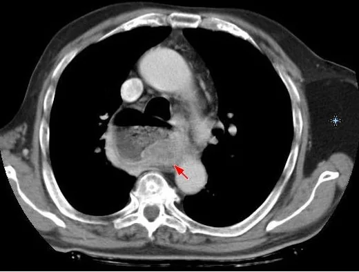
- Özofagus boyunca fokal, asimetrik veya düzensiz çevresel yumuşak doku kalınlaşması; lezyonun üst ve alt sınırını multiplanar görüntülerde belirleyin.
- Tümör segmentinde lümen daralması, geçişin proksimalinde sıvı/hava birikimi ve özofagus genişlemesi görülebilir.
- Özofagus kanseri BT'de infiltratif, polipoid, ülseratif veya yüzeyel yayılan görünümde olabilir.
- BT'de periezofageal yağ infiltrasyonu T3 hastalıkla, tümör ile komşu mediastinal yapı arasındaki yağ planı kaybı ise T4 hastalık şüphesiyle ilişkilidir. T4 kategorisi, tümörün komşu yapılara doğrudan invazyonunu ifade eder; BT'de komşu yapı ile yağ planı kaybı T4 hastalığı düşündürür.
- Özofagus tümörünün trakea, ana bronş, aorta ve diğer komşu mediastinal yapılarla ilişkisini inceleyin; tümör ile komşu mediastinal yapı arasındaki yağ planı kaybı T4 uzanımını düşündürür.
- Paratrakeal, subkarinal, paraözofageal, gastrohepatik ve çölyak nodlar ile akciğer, karaciğer, adrenal ve kemik metastazları gözden geçirilir.
- Özofagus kanserine bağlı trakeoözofageal veya özofagobronşiyal fistülde BT'de kontrastın trakea ya da ana bronşa ekstravazasyonu görülebilir.
- T4a plevra, perikard, azigos ven veya diyafragma gibi yapılara; T4b aorta, vertebra ya da trakea gibi yapılara doğrudan invazyonu ifade eder. Yağ planı kaybı tek başına T4 tanısı koydurmaz.
Ölçütler ve sınıflamalar
- Distandü normal özofagusta beklenen duvar görünümü; kollabe segmentte tek başına eşik olarak kullanılmaz
- 2 mm'den az
- AJCC 8. baskı klinik T kategorisi
- T1 mukoza/submukozaya (T1a lamina propria veya muskularis mukoza, T1b submukoza), T2 muskularis propriaya, T3 adventisyaya uzanımı belirtir. T4 komşu yapılara invazyondur; T4a plevra, perikard, azigos ven, diyafragma/periton gibi yapılara, T4b aorta, vertebra veya trakea gibi yapılara invazyonu tanımlar. BT duvar katlarını güvenilir ayıramadığından T1-T3 ayrımını tek başına yapamaz.
- AJCC 8. baskı bölgesel nod kategorileri
- N0 bölgesel nod metastazı yok; N1 bir-iki, N2 üç-altı, N3 yedi veya daha fazla bölgesel lenf nodu metastazını ifade eder. BT nod boyutuna dayandığı için nodal evreyi kesinleştiremez; normal boyutlu nodda metastaz olabilir.
Normalde
Oral kontrastla distandü normal özofagus oval tübüler görünür, duvarı 2 mm'den incedir ve çevresindeki yağ planları belirgindir. Kollabe özofagusta yalancı duvar kalınlaşmasını azaltmak için oral kontrastla yeterli distansiyon sağlayın.
Normal BT ile kıyasla
Aynı bölgenin, kaynak veri setinde patoloji içermediği belirtilen bir BT incelemesini kesit kesit kaydırın; organ sınırlarını açarak anatomiyi hasta görüntüsüyle karşılaştırın. Küçük rastlantısal bulgular tümüyle dışlanmamıştır.
Kaynak: TotalSegmentator v2.01 veri seti (Wasserthal ve ark., Radiology: Artificial Intelligence 2023), CC BY 4.0, vaka s1278; organ sınırları veri setinin otomatik segmentasyonundan, birleştirilerek sadeleştirildi. Görüntüler 2,5 mm kesit aralığına yeniden örneklendi.
Ayırıcı tanı
- Özofagus duvarında kalınlaşma veya kitle yapan enfeksiyöz/inflamatuar süreçler benzer görünüm verebilir; BT yayılımı haritalarken kesin tanı için endoskopi ve biyopsi gereklidir.
- Özofagus kanserinin değerlendirilmesinde BT, mediastinal tutulumu, komşu lenfadenopatiyi ve uzak yayılımı gösterir; tanı endoskopik değerlendirmeye dayanır.
- Leiomyom/GIST
- sıklıkla düzgün sınırlı intramural veya lümene doğru büyüyen submukozal kitle görünümündedir; mukozal düzensizlik ve çevre invazyonu malign epitelyal tümörden daha az beklenir.
- Normal özofagus BT'de kollabe görünebilir; oral kontrastla distandü edildiğinde duvar kalınlığı 2 mm'den azdır ve çevresindeki yağ planları belirgindir.
- Mediastinal kitle özofagusa dıştan bası yaparak lümeni daraltabilir; endoskopide içsel bir duvar lezyonu yerine dış bası izlenir ve özofagus duvarı kaynaklı kalınlaşma yoktur.
Tuzaklar
- Kollabe özofagus duvarı kalın görünür; oral suyla distansiyon veya komşu kesitlerle kıyas yapmadan tek bir aksiyel ölçümden malignite çıkarmayın.
- Periezofageal yağ planı kaybı zayıf yağ dokusu, inflamasyon veya tedavi sonrası fibrozise bağlı olabilir; tek başına T4 invazyonu kanıtlamaz.
- Boyutu normal nod metastaz içerebilir, büyümüş nod reaktif olabilir; BT nodal boyutunu histolojik doğrulama yerine koymayın.
- Özofagusun doğal daralma seviyelerinde ve gastroözofageal bileşkede kasılma yalancı odaksal kalınlaşma yapabilir; lezyonun komşu kesitlerde devam edip etmediğine ve dış kontura bakın.
Kendini dene
Disfajili hastada orta özofagusta asimetrik kitle, lümen daralması ve subkarinal nod var. BT'nin en doğru rol yorumu nedir?
Cevabı göster
Yayılımı haritalar, biyopsi gerekir. BT primer kitlenin yayılımını, komşu organ ilişkisini, nodları ve uzak metastazları haritalar; histoloji endoskopik biyopsiyle konur. BT duvar katmanlarını ayırıp T1 derinliğini gösteremez ve normal boyutlu nod metastazını dışlayamaz.
Distandü özofagus çevresindeki yağ planı tümörle silinmiş, ancak aorta konturu düzgün ve doğrudan invazyon bulgusu yok. En uygun çıkarım nedir?
Cevabı göster
Yağ planı kaybı tek başına kanıt değil. Yağ planı kaybı olası uzanım için şüphe yaratır fakat tek başına komşu organ invazyonunu kanıtlamaz; kontur deformasyonu gibi bulgular aranmalıdır. T4b doğrudan büyük komşu yapıya invazyondur; uzak metastaz ve perforasyon bu görünümle tanımlanamaz.
İlgili konular
Kaynaklar
Bu sayfadaki 49 cümle ve 2 quiz sorusu aşağıdaki kaynaklardan alıntılarla otomatik olarak karşılaştırıldı; 7 cümle bu karşılaştırmada düzeltildi (2026-10-09).
- Imaging in Esophageal Cancer: A Comprehensive Reviewpmc.ncbi.nlm.nih.gov
- Expert Consensus Contouring Guidelines for Intensity Modulated Radiation Therapy in Esophageal and Gastroesophageal Junction Cancer.pubmed.ncbi.nlm.nih.gov
- Dyspepsia: organic versus functional.pubmed.ncbi.nlm.nih.gov
- Radiation-Induced Heart Disease.pubmed.ncbi.nlm.nih.gov
- Esophageal Cancer: Tumor-Node-Metastasis Staging.pubmed.ncbi.nlm.nih.gov
- Carcinoma of Esophagus.pubmed.ncbi.nlm.nih.gov
- Chemoradiation for esophageal cancer.pubmed.ncbi.nlm.nih.gov
- Advanced endoscopic imaging for detecting and guiding therapy of early neoplasias of the esophagus.pubmed.ncbi.nlm.nih.gov
- Staging in Esophageal and Gastric Cancers.pubmed.ncbi.nlm.nih.gov
- Evaluation of NinePoint Medical's Nvision VLE device for gastrointestinal applications.pubmed.ncbi.nlm.nih.gov
- Minimally invasive staging of esophageal cancer.pubmed.ncbi.nlm.nih.gov
- New frontiers in esophageal radiology.pubmed.ncbi.nlm.nih.gov
- Incidence and treatment of brain metastasis in patients with esophageal carcinoma.pubmed.ncbi.nlm.nih.gov
- PET-CT in Clinical Adult Oncology: III. Gastrointestinal Malignancies.pubmed.ncbi.nlm.nih.gov
- Esophagectomy and Gastric Pull-through Procedures: Surgical Techniques, Imaging Features, and Potential Complications.pubmed.ncbi.nlm.nih.gov
- Spindle cell (sarcomatoid) squamous cell carcinoma of the esophagus: A case report and a review of the literature.pubmed.ncbi.nlm.nih.gov
- Intestinal Metaplasia of the Distal Esophagus in a Patient With Untreated Achalasia-An Unusual Case and Review of Literature.pubmed.ncbi.nlm.nih.gov
- Advanced esophageal adenocarcinoma in a pediatric patient with Down syndrome: a rare case report and literature review.pubmed.ncbi.nlm.nih.gov
- Diagnostic performance of whole-body diffusion-weighted imaging for staging esophageal cancer: a comparative study with PET-CT.pubmed.ncbi.nlm.nih.gov
- CA19-9-producing esophagogastric junction cancer with nodal recurrence outside the planned dissection field: a case report.pubmed.ncbi.nlm.nih.gov
- Esophageal adenosquamous carcinoma with mucinous carcinoma-predominant lymph node metastases: a case report.pubmed.ncbi.nlm.nih.gov
- A Weighted Neural Network Model Based on Laboratory Tests for Identifying Lymph Node Metastases in Esophageal Squamous Cell Carcinomas.pubmed.ncbi.nlm.nih.gov
- A Retrospective Study on Predicting Ki-67 Expression in Esophageal Cancer Patients Based on Delta Radiomics.pubmed.ncbi.nlm.nih.gov
- Surgical management of small bowel metastasis from esophageal squamous cell carcinoma: a case report and literature review.pubmed.ncbi.nlm.nih.gov
- Optimizing Care for Veterans at Risk of Cancer From Camp Lejeune Water Exposure.pubmed.ncbi.nlm.nih.gov
- Association between postoperative nasogastric tube retention duration and total postoperative opioid consumption in patients undergoing minimally invasive esophagectomy: A single-center retrospective cohort study.pubmed.ncbi.nlm.nih.gov
- Lung squamous cell carcinoma complicated with immune checkpoint inhibitor-associated active tuberculosis: a case report and literature review.pubmed.ncbi.nlm.nih.gov
- Clinical and imaging manifestations of metastases from rare sites.pubmed.ncbi.nlm.nih.gov
- Case Report: Refractory desmoplastic small round cell tumor: a report of a molecularly confirmed case with <i>EWSR1-WT1</i> fusion.pubmed.ncbi.nlm.nih.gov
- Clinical TNM Lung Cancer Staging: A Diagnostic Algorithm with a Pictorial Review.pubmed.ncbi.nlm.nih.gov
- Small Intestine Tumors: Diagnostic Role of Multiparametric Ultrasound.pubmed.ncbi.nlm.nih.gov
- Reconstructive flap surgery in head and neck cancer patients: an interdisciplinary view of the challenges encountered by radiation oncologists in postoperative radiotherapy.pubmed.ncbi.nlm.nih.gov
- An Atypical Case of Metastatic Esophageal Adenocarcinoma Causing Biliary Obstruction and Duodenal Infiltration Requiring EUS-Guided Choledochoduodenostomy.pubmed.ncbi.nlm.nih.gov
- Endo-hepatology: Bridging the gap between lumen and liver.pubmed.ncbi.nlm.nih.gov
- Mechanomedicine in digestive surgery: a theranostic framework integrating mechanical diagnostics and therapeutic modulation across the perioperative continuum.pubmed.ncbi.nlm.nih.gov
- Diagnostic Imaging of Extrapulmonary Tuberculosis Across Organ Systems.pubmed.ncbi.nlm.nih.gov
- Primary gastric SMARCA4-deficient carcinosarcoma and sarcomatoid carcinoma: Two case reports.pubmed.ncbi.nlm.nih.gov
- Clinical Features and Diagnostic Challenges of <i>Talaromyces marneffei</i> Infection in Non-Endemic Regions: A 6-Case Study Highlighting NGS and Tailored Antifungal Therapy.pubmed.ncbi.nlm.nih.gov
- MRI outperforms CT for tracheal and vascular invasion staging in esophageal cancer.pubmed.ncbi.nlm.nih.gov
- Imaging in Esophageal Cancer: A Comprehensive Review.pubmed.ncbi.nlm.nih.gov
- Findings of Endoscopic US and CT of Esophageal Disease.pubmed.ncbi.nlm.nih.gov
- State-of-the-Art Mediastinal Staging in Non-Small-Cell Lung Cancer: Integration of Combined Endosonographic Techniques with Updated IASLC TNM 9th Classification.pubmed.ncbi.nlm.nih.gov
- From Migration to Emergency: A Systematic Review of Acute Presentations Following Late Esophageal Stent Migration.pubmed.ncbi.nlm.nih.gov
- Predictive modeling of peripheral blood EGFR positivity in patients with advanced primary non-small cell lung cancer.pubmed.ncbi.nlm.nih.gov
- Extensive-stage small cell esophageal carcinoma: a retrospective analysis of treatment modalities.pubmed.ncbi.nlm.nih.gov
- Organ-specific response to atezolizumab + bevacizumab combination therapy for unresectable hepatocellular carcinoma with extrahepatic metastases: a retrospective cohort study.pubmed.ncbi.nlm.nih.gov
- Respiratory Morbidity After Repair of Type C Esophageal Atresia with Tracheoesophageal Fistula: A Pediatric Case Series.pubmed.ncbi.nlm.nih.gov
- Thin bronchoscope-parallel, endotracheal tube-guided stent deployment for cervical tracheoesophageal fistula after anti-cancer therapy: a case report and literature review.pubmed.ncbi.nlm.nih.gov
- Congenital aerodigestive fistula with extraordinarily delayed adult presentation: a case report and review of endoscopic closure for bronchoesophageal fistula.pubmed.ncbi.nlm.nih.gov
- Risk Factors for Mediastinal Lymph-Node Metastasis in Siewert Type I/II Esophagogastric Junction Cancers.pubmed.ncbi.nlm.nih.gov
- Development and validation of a CT radiomics nomogram for preoperative prediction of right recurrent laryngeal nerve lymph node metastasis in thoracic esophageal squamous cell carcinoma.pubmed.ncbi.nlm.nih.gov
- Imaging findings of gastrointestinal tract tumors in children and adolescents.pubmed.ncbi.nlm.nih.gov
- Neoplastic pathology at the crossroads between neck imaging and cardiothoracic imaging.pubmed.ncbi.nlm.nih.gov
- Commentary: an eye on PET quantification.pubmed.ncbi.nlm.nih.gov
- Long-term safety and effectiveness of the "OptEase" vena cava filter.pubmed.ncbi.nlm.nih.gov
- Comparative analysis of diagnostic techniques and treatment modalities for early-stage esophageal carcinoma: a comprehensive review.pubmed.ncbi.nlm.nih.gov
- Endoscopic Ultrasound in Squamous Cell Esophageal Cancer: From Staging to Strategy-A Narrative Review.pubmed.ncbi.nlm.nih.gov
- Role of Endoscopy in the Diagnosis and Management of Esophageal Cancer.pubmed.ncbi.nlm.nih.gov
- The treatments and postoperative complications of esophageal cancer: a review.pubmed.ncbi.nlm.nih.gov
- Endoscopic Ultrasound (EUS)-Based Multimodal Diagnosis of a Rare Intramural Esophageal Squamous Cell Carcinoma: Case Report and Literature Review.pubmed.ncbi.nlm.nih.gov
- Advancing Esophageal Cancer Staging and Restaging: The Role of MRI in Precision Diagnosis.pubmed.ncbi.nlm.nih.gov
- Prognostic significance and optimal staging of supraclavicular lymph node metastasis in upper esophageal squamous cell carcinoma undergoing chemoradiotherapy.pubmed.ncbi.nlm.nih.gov
- The prognostic value of log odds of positive lymph nodes in patients with resectable locally advanced gastric cancer following neoadjuvant therapy: results from the SEER database.pubmed.ncbi.nlm.nih.gov
- Historical evolution and current research status of lymph node staging in gastric cancer: a review.pubmed.ncbi.nlm.nih.gov
- Assessment of inter-reader agreement and diagnostic performance of Node-RADS in colon cancer.pubmed.ncbi.nlm.nih.gov
- <sup>18</sup>F-FDG PET/CT metabolic parameter to predict pathological complete response and survival after neoadjuvant immunotherapy plus chemoradiotherapy in resectable esophageal squamous cell carcinoma.pubmed.ncbi.nlm.nih.gov
- Intermittent Ileitis and Partial Small-Bowel Obstruction Due to Intestinal Helminth Infection Mimicking Crohn's Disease.pubmed.ncbi.nlm.nih.gov
- Anticoagulation-Amplified Recurrent Esophageal Hemorrhage in Triple-Positive Antiphospholipid Syndrome: The Inescapable Anticoagulation Dilemma.pubmed.ncbi.nlm.nih.gov
- Eosinophilic Cardiomyopathy in Eosinophilic Granulomatosis With Polyangiitis After Dupilumab Therapy.pubmed.ncbi.nlm.nih.gov
- Cystic lesions and their mimics involving the intrahepatic bile ducts and peribiliary space: diagnosis, complications, and management.pubmed.ncbi.nlm.nih.gov
- The Role of Imaging in Portal Vein Thrombosis: From the Diagnosis to the Interventional Radiological Management.pubmed.ncbi.nlm.nih.gov
- Development and validation of the OSASH score to predict overall survival of hepatocellular carcinoma after surgical resection: a dual-institutional study.pubmed.ncbi.nlm.nih.gov
- Giant Mid-Esophageal Leiomyoma in a Young Adult Managed by Video-Assisted Thoracoscopic Enucleation: A Case Report.pubmed.ncbi.nlm.nih.gov
- The clinical value of endoscopic ultrasonography combined with endoscopic treatment for upper gastrointestinal lifting lesions.pubmed.ncbi.nlm.nih.gov
- High-Frequency Miniprobe Endoscopic Ultrasonography Across the Gastrointestinal Tract.pubmed.ncbi.nlm.nih.gov
- Case Report: Spectral CT characterization of giant esophageal schwannoma with reactive lymphoid hyperplasia mimicking lymphoproliferative disease.pubmed.ncbi.nlm.nih.gov
- Dysphagia Secondary to Extrinsic Esophageal Compression From an Anterior Mediastinal Squamous Cell Carcinoma.pubmed.ncbi.nlm.nih.gov
- Comprehensive Insights into Tracheoesophageal Fistula Pathophysiology, Diagnosis, Treatment, and Future Directions.pubmed.ncbi.nlm.nih.gov
- Home Ultrasound: A Contemporary and Valuable Tool for Palliative Medicine.pubmed.ncbi.nlm.nih.gov
- Evaluation of Dynamic CT Scans and Analyzing Its Efficacy Accuracy in Staging of Laryngeal CA: A Prospective Analysis of 30 Patients and Review of Literature.pubmed.ncbi.nlm.nih.gov
- Interstitial Lung Abnormalities in Patients With Locally Advanced Esophageal Cancer: Prevalence, Risk Factors, and Clinical Implications.pubmed.ncbi.nlm.nih.gov
- Imaging of the chemotherapy-induced hepatic damage: Yellow liver, blue liver, and pseudocirrhosis.pubmed.ncbi.nlm.nih.gov
- Patterns of injury and violence in Yaoundé Cameroon: an analysis of hospital data.pubmed.ncbi.nlm.nih.gov
- Oesophagogastrostomy Using Double-Flap Technique Following Resection of an Extensive Leiomyosarcoma in a Cat.pubmed.ncbi.nlm.nih.gov
- Portal vein recanalization and transjugular intrahepatic portosystemic shunt in chronic portal vein thrombosis: a narrative review.pubmed.ncbi.nlm.nih.gov
- Selection of dilution material for non-iodinated iodine as an oral contrast agent for esophageal cancer: a preliminary clinical trial.pubmed.ncbi.nlm.nih.gov
- Endoscopic Ultrasound-Guided Hepaticogastrostomy via a Retrosternal Gastric Tube Conduit for Obstructive Cholangitis Caused by Ampullary Carcinoma: A Case Report.pubmed.ncbi.nlm.nih.gov
- Role of Endoscopic Ultrasonography in Management of Pancreaticobiliary Cancers: Recent Trends and Advances.pubmed.ncbi.nlm.nih.gov
- Endoscopic Management of Post-Bariatric Surgery Complications: Diagnostic Work-Up and Innovative Approaches for Leak, Fistula, and Stricture Management.pubmed.ncbi.nlm.nih.gov
- International Harmonization of Nomenclature and Diagnostic Criteria (INHAND): Non-proliferative and Proliferative Lesions of the Non-human Primate ( M. fascicularis ).pubmed.ncbi.nlm.nih.gov
- Biological Characteristics and Therapeutic Strategies for Resectable Locally Advanced Esophagogastric Junction Cancer: A Comprehensive Update.pubmed.ncbi.nlm.nih.gov
- 2025 American Thyroid Association Management Guidelines for Adult Patients with Differentiated Thyroid Cancer.pubmed.ncbi.nlm.nih.gov
- A core curriculum for basic EUS skills: An international consensus using the Delphi methodology.pubmed.ncbi.nlm.nih.gov
- PET Imaging in Vulvar Cancer: A Literature Review of the Current Evidence and Clinical Applications.pubmed.ncbi.nlm.nih.gov
- Clinicopathological Features of Esophageal Carcinosarcoma Treated with Surgery: A Series of Nine Cases.pubmed.ncbi.nlm.nih.gov
- AI in esophageal cancer: advances, barriers to clinical translation, and perspectives for digital health.pubmed.ncbi.nlm.nih.gov
- Uncommon Causes of Interlobular Septal Thickening on CT Images and Their Distinguishing Features.pubmed.ncbi.nlm.nih.gov
- Usefulness of 18F-fluorodeoxyglucose positron emission tomography for T1 esophageal squamous cell carcinoma.pubmed.ncbi.nlm.nih.gov
- Trans-hiatal small-bowel herniation after laparoscopic radical total gastrectomy for gastric cancer: a case report and literature review.pubmed.ncbi.nlm.nih.gov
- Long-term nutritional outcomes after esophagectomy following induction chemotherapy versus chemoradiotherapy for T4b esophageal cancer.pubmed.ncbi.nlm.nih.gov
- Stricturing Diseases of the Gastrointestinal Tract-Current Clinical Practice.pubmed.ncbi.nlm.nih.gov
- Clinical evaluation of right-sided overlap with single flap valvuloplasty and double tract reconstruction after proximal gastrectomy.pubmed.ncbi.nlm.nih.gov
- Salvage involved-field versus extended-field chemoradiotherapy for postoperative lymph node metastasis in esophageal squamous cell carcinoma: a retrospective clinical study.pubmed.ncbi.nlm.nih.gov
- Comparison of Outcomes From Sequential Endoscopic Therapy in 60 Patients With Cirrhosis and Esophagogastric Varices Treated at a Provincial and a Tertiary District Hospital.pubmed.ncbi.nlm.nih.gov
- Splenic infarction after endoscopic glue injection for gastric varices: proportion, exploratory correlates, and clinical outcomes from a propensity score-matched analysis.pubmed.ncbi.nlm.nih.gov
- The Predilection of Specific Diseases to Affect Different Sections of the Esophagus.pubmed.ncbi.nlm.nih.gov
- Performance of Large Language Models (ChatGPT and Gemini Advanced) in Gastrointestinal Pathology and Clinical Review of Applications in Gastroenterology.pubmed.ncbi.nlm.nih.gov
- How to update esophageal masses imaging using literature review (MRI and CT features).pubmed.ncbi.nlm.nih.gov
- Bronchoscopy and the Pulmonologist in Contemporary Lung Cancer Care.pubmed.ncbi.nlm.nih.gov
- International Society of Paediatric Surgical Oncology (IPSO) Minimally Invasive Surgery (MIS) Guidelines.pubmed.ncbi.nlm.nih.gov
- Airway stent intervention in a high-volume center: safe procedures and educational perspectives.pubmed.ncbi.nlm.nih.gov
Eğitim amaçlıdır; hasta başında tanı ve tedavi kararının yerine geçmez, tıbbi tavsiye değildir. Metinler yapay zekâ desteğiyle yazıldı ve otomatik bir adımda kaynak alıntılarıyla karşılaştırıldı; bu bir tıbbi doğrulama değildir ve içerik uzman radyolog incelemesinden geçmedi. Klinik kararlarda güncel kılavuzları, kurum protokollerini ve radyoloji raporunu esas alın.