Özet
Özofagus, farinksi mideye bağlayan ve servikal, torasik, kısa abdominal bölümleri olan kaslı bir kanaldır. Özofagus toraks BT'sinde kollabe olabilir; oral kontrastla yeterli distansiyon sağlanması duvar kalınlığını değerlendirmeye yardımcı olur ve kollabe durumdaki yalancı kalınlaşmayı azaltır. Toraks BT'sinde özofagusun dış konturu, çevresindeki mediastinal yağ planları ve lümenin kollabe ya da distandü görünümü değerlendirilir; oral kontrastla yeterli distansiyon, kollabe özofagusta yalancı duvar kalınlaşmasını azaltır. Özofagus kollapsı nedeniyle oluşan yalancı duvar kalınlaşması, uygun distansiyon sağlanmadığında patolojik kitle veya tutulum olarak yanlış yorumlanabilir.
Okuma sırası
- karina arkasındaki özofagus. Özofagusun torasik bölümünü posterior mediastinumda, desendan aortanın önünde ve sağında izleyin; T4-T5 düzeyinde aort kavsi ve sol ana bronşla çaprazlaşmasını ardışık kesitlerde doğrulayın.
- karina ve sol ana bronş. T4-T5 seviyesinde özofagus, sol ana bronş ile çakışan daralma bölgesindedir ve komşu yapının ayrımında bu anatomik ilişki referans alınır.
- özofagus-aorta aralığı. Desendan aortanın ön-sağ komşuluğundaki özofagus dış konturunu izleyin; aradaki yağ planının kesintisiz olup olmadığını karşı taraf ve komşu seviyelerle kıyaslayın.
- özofagusun tüm seyri ve hiatus. Gastroözofageal bileşke ve alt torasik özofagus, yeterli oral distansiyon sağlanmış aksiyel ve sagittal çapraz kesitlerde komşu yapılarla ilişkisiyle birlikte değerlendirilmeli; kollaps nedeniyle yalancı duvar kalınlaşması riskine dikkat edilmelidir.
Faz ve pencere
- Portal tanısal
- Rutin kontrastlı toraks BT'de özofagus lümeni sıklıkla hava ya da az miktarda sıvı içerir; duvar kalınlığı distansiyon ve kasılmaya göre değişir. İntravenöz kontrast, duvar dışı yağ planları ile mediastinal damar ve organ komşuluğunu seçmeyi kolaylaştırır; normal özofagus için ayrı bir arteriyel faz gerekmez.
Önerilen pencereler G genişlik, M merkez; değerler Hounsfield birimi (HU)
- MediastenG 350 / M +50
BT bulguları
- Torasik özofagus posterior mediastinumda, desendan torasik aortanın önünde ve sağında seyreder.
- Özofagusun ikinci fizyolojik daralması, aort kavsi ile sol ana bronşun çaprazlaştığı T4-T5 düzeyindedir.
- Üçüncü fizyolojik daralma, alt özofageal bileşke düzeyinde T11 seviyesinde tanımlanır.
- Lümen değişken biçimde hava, sıvı veya kollabe görünür; tek kesitteki duvar kalınlığı distansiyon düzeyinden bağımsız yorumlanmamalıdır.
- Özofagus dış çevresindeki mediastinal yağ planı belirgin olmalı; fokal yumuşak doku, çevre yağda çizgilenme veya komşu yapı konturunda bozulma normal varyasyon sayılmaz.
- Hiatal hernide abdominal içerikler özofageal hiatus üzerinden mediastinuma uzanabilir.
Ölçütler ve sınıflamalar
- Oral kontrastla yeterince distandü normal özofagusta duvar kalınlığı; kollabe segmentte eşik olarak kullanılmaz
- <2 mm
Normalde
Normal karşılaştırmada torasik özofagusun posterior mediastinumda desendan aortanın önünde ve sağında seyrettiğini, ikinci fizyolojik daralmanın ise T4-T5 düzeyinde aort kavsi ile sol ana bronşun çaprazlaşmasında olduğunu izleyin. Desendan aorta ile özofagus arasındaki yağ planı seçilir; lümen kollabe olsa bile odaksal dış kontur çıkıntısı, asimetrik duvar kalınlaşması veya çevre yağda silinme olmamalıdır. Özofagus duvar kalınlığı distansiyona bağlıdır; oral kontrastla yeterince distandü segmentte 2 mm’den azdır ve kollabe segmentte yalancı kalınlaşma görülebilir.
Normal BT ile kıyasla
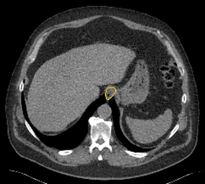
Aynı bölgenin, kaynak veri setinde patoloji içermediği belirtilen bir BT incelemesini kesit kesit kaydırın; organ sınırlarını açarak anatomiyi hasta görüntüsüyle karşılaştırın. Küçük rastlantısal bulgular tümüyle dışlanmamıştır.
Kaynak: TotalSegmentator v2.01 veri seti (Wasserthal ve ark., Radiology: Artificial Intelligence 2023), CC BY 4.0, vaka s1278; organ sınırları veri setinin otomatik segmentasyonundan, birleştirilerek sadeleştirildi. Görüntüler 2,5 mm kesit aralığına yeniden örneklendi.
Ayırıcı tanı
- Kollabe özofagus
- uzun bir segmentte simetrik duvar kıvrımları ve lümen daralması izlenir; sıvı ya da oral kontrastla açılan segmentte kalınlık azalır.
- Hiatal herni
- hiatus üstündeki mide kardiyası/fundus devamlılığı ve gastroözofageal bileşkenin yukarı yer değiştirmesi görülür.
- Özofagus divertikülü
- lümenle boyun aracılığıyla devam eden, çoğunlukla hava veya oral kontrast içeren kese biçimli çıkıntıdır.
Tuzaklar
- Kollabe lümen duvarı yalancı kalın gösterir; fokal asimetriyi başka kesitlerde ve koronal/sagittal reformasyonlarda izleyin, yalnız bir aksiyel ölçüme dayanmayın.
- Sol ana bronş veya sol atriuma ait yumuşak doku dansitesi özofagus duvarı sanılabilir; komşu kesitlerde tübüler lümen devamlılığını takip edin.
Kendini dene
Desendan torasik aortanın ön-sağ tarafında, T4-T5 seviyesinde kollabe özofagus lümeni izlenir; çevresindeki yağ planları belirgindir. En olası görünüm nedir?
Cevabı göster
Normal kollabe özofagus. Çevresinde belirgin yağ planı bulunan, T4-T5 seviyesinde desendan aortanın önünde yer alan kollabe özofagus yapısı normal anatomidir. Özofagus kanseri BT'de infiltratif veya polipoidal yumuşak doku kütlesi olarak görünür ve komşu yapılarda yağ planı kaybına neden olabilir.
Hiatal herniyi tanımlayan bulgu hangisidir?
Cevabı göster
Abdominal içeriklerin özofageal hiatus üzerinden mediastinuma protrüzyonu. Hiatal herni, abdominal içeriklerin diyaframın özofageal hiatusundan mediastinuma protrüde olmasıdır.
İlgili konular
Kaynaklar
Bu sayfadaki 32 cümle ve 2 quiz sorusu aşağıdaki kaynaklardan alıntılarla otomatik olarak karşılaştırıldı; 9 cümle bu karşılaştırmada düzeltildi (2026-10-09).
- Imaging in Esophageal Cancer: A Comprehensive Reviewpmc.ncbi.nlm.nih.gov
- Barrett's oesophagus and oesophageal adenocarcinoma.pubmed.ncbi.nlm.nih.gov
- Variants and Pitfalls in PET/CT Imaging of Gastrointestinal Cancers.pubmed.ncbi.nlm.nih.gov
- Accuracy of 18 F-FDG PET/CT in Predicting Residual Disease After Neoadjuvant Chemoradiotherapy for Esophageal Cancer.pubmed.ncbi.nlm.nih.gov
- Esophageal insufflation computed tomography in clinically normal dogs.pubmed.ncbi.nlm.nih.gov
- Automatic tracking of arbitrarily shaped implanted markers in kilovoltage projection images: a feasibility study.pubmed.ncbi.nlm.nih.gov
- Central adiposity is associated with increased risk of esophageal inflammation, metaplasia, and adenocarcinoma: a systematic review and meta-analysis.pubmed.ncbi.nlm.nih.gov
- Surveillance of Clinically Complete Responders Using Serial 18 F-FDG PET/CT Scans in Patients with Esophageal Cancer After Neoadjuvant Chemoradiotherapy.pubmed.ncbi.nlm.nih.gov
- Characterization of RNA isolated from eighteen different human tissues: results from a rapid human autopsy program.pubmed.ncbi.nlm.nih.gov
- Measuring tumor hypoxia with ¹⁸F-FETNIM PET in esophageal squamous cell carcinoma: a pilot clinical study.pubmed.ncbi.nlm.nih.gov
- Normal radiological lymph node appearance in the thorax.pubmed.ncbi.nlm.nih.gov
- [Dysphagia or dysphagias during neuroleptic medication?].pubmed.ncbi.nlm.nih.gov
- Esophageal cancer characterization with pneumo-64-MDCT.pubmed.ncbi.nlm.nih.gov
- The correlation between the metabolic tumor volume and hematological parameters in patients with esophageal cancer.pubmed.ncbi.nlm.nih.gov
- Intensity-modulated proton therapy (IMPT) interplay effect evaluation of asymmetric breathing with simultaneous uncertainty considerations in patients with non-small cell lung cancer.pubmed.ncbi.nlm.nih.gov
- Cervical esophagogastric anastomosis using a retrosternal gastric conduit in an adult patient with congenital type A long-gap esophageal atresia after 18 years of gastrostomy: a case report.pubmed.ncbi.nlm.nih.gov
- Emergency Minimally Invasive Esophagogastrectomy and Delayed Esophagocoloplasty for Caustic Ingestion: A Stepwise Video-Based Approach.pubmed.ncbi.nlm.nih.gov
- Cross-sectional atlas of the tree shrew (<i>Tupaia belangeri</i>) head, neck, and trunk: a comparison of micro-CT imaging and cadaveric sections.pubmed.ncbi.nlm.nih.gov
- Gastrointestinal Ultrasound: Measurements and Normal Findings - What Do You Need to Know?pubmed.ncbi.nlm.nih.gov
- The Positioning Method of Pulmonary Nodules in Thoracoscopic Surgery Based on CT Simulation Positioning System for Radiotherapy.pubmed.ncbi.nlm.nih.gov
- Clinical Relevance of Distinguishing Between Three Endoscopy-Based Conditions, Bronchiectasis, Bronchomalacia, and Their Combination in Dogs: A Retrospective Study.pubmed.ncbi.nlm.nih.gov
- Swallow contrast-enhanced ultrasonography detects a tiny cervical esophageal diverticulum missed on barium esophagography: a case report.pubmed.ncbi.nlm.nih.gov
- Thoracoscopic-guided paraesophageal abscess drainage in foreign body-induced esophageal perforation: a case report.pubmed.ncbi.nlm.nih.gov
- Mediastinoscopic Esophagectomy for Esophageal Cancer with Right Aortic Arch: A Case Report.pubmed.ncbi.nlm.nih.gov
- Early-Stage Esophago-Mediastinal Fistula After Atrial Fibrillation Radiofrequency Ablation Successfully Treated With Endoscopic Vacuum Therapy.pubmed.ncbi.nlm.nih.gov
- Computed Tomographic Findings and Clinical Manifestations of Acute Severe Hypersensitivity to a Non-Ionic Iodinated Contrast Agent in a Castrated Male Dog: A Case Report.pubmed.ncbi.nlm.nih.gov
- Case Report: Resectability-guided management of an indeterminate periduodenal spindle cell tumor initially suspected to be a gastrointestinal stromal tumor.pubmed.ncbi.nlm.nih.gov
- Pericardial Cyst at the Left Cardiophrenic Angle Mimicking Localized Effusion: A Diagnostic Dilemma.pubmed.ncbi.nlm.nih.gov
- A Rare Case of an Abdominal Pedunculated Bronchogenic Cyst.pubmed.ncbi.nlm.nih.gov
- A giant retropharyngeal space lipoma: a case report and review of the literature.pubmed.ncbi.nlm.nih.gov
- Anatomical Variations in Critical Structures in Esophageal Surgery: Implications for Personalized Surgery.pubmed.ncbi.nlm.nih.gov
- Esophageal Perforation During Dilation in a Patient With Hypopharyngeal Metastatic Squamous Cell Carcinoma.pubmed.ncbi.nlm.nih.gov
- Mediastinal tracheostomy: techniques, outcomes and quality of life-a systematic review.pubmed.ncbi.nlm.nih.gov
- Left atrial anatomy relevant to catheter ablation.pubmed.ncbi.nlm.nih.gov
- Anatomical Basis for the Cardiac Interventional Electrophysiologist.pubmed.ncbi.nlm.nih.gov
- Giant left atrium: a review.pubmed.ncbi.nlm.nih.gov
- Three-dimensional computed tomography reconstruction of the gastroesophageal junction following hiatal hernia repair with Collis gastroplasty and fundoplication: a novel method to demonstrate postoperative morphology and better understand hiatal hernia recurrence.pubmed.ncbi.nlm.nih.gov
- Boerhaave Syndrome Caused by Pancreatitis-Induced Gastric Outlet Obstruction: A Case Report.pubmed.ncbi.nlm.nih.gov
- Grade IV hiatal hernia in a neonate: A radiologic mimic of congenital diaphragmatic hernia.pubmed.ncbi.nlm.nih.gov
- Mechanisms of aortic dissection: From pathological changes to experimental and <i>in silico</i> models.pubmed.ncbi.nlm.nih.gov
- Evidence of neurofibromatosis type 1 in a multi-morbid Inca child mummy: A paleoradiological investigation using computed tomography.pubmed.ncbi.nlm.nih.gov
- Disease- and Treatment-related Complication on F-18-Fluorodeoxyglucose Positron Emission Tomography/Computed Tomography in Oncology Practice: A Pictorial Review.pubmed.ncbi.nlm.nih.gov
- Nodal dissection in thymic surgery: a narrative review of the evolution of practice and evidence in the era of the 9th edition TNM classification (2000-2025).pubmed.ncbi.nlm.nih.gov
- Case Report: Cervical approach for dual mediastinal ectopic thyroid: clinical utility of 3D imaging in surgical planning.pubmed.ncbi.nlm.nih.gov
- Standardized W-H fundoplication procedure for gastroesophageal reflux disease (2026).pubmed.ncbi.nlm.nih.gov
- Robotic Surgical Repair of a Parahiatal Hernia: A Case Report.pubmed.ncbi.nlm.nih.gov
- A Rare Coexistence of Gastric Fundal False Diverticulum and Sliding Hiatal Hernia: Technical Considerations and Literature Review.pubmed.ncbi.nlm.nih.gov
- Spatial associations between esophageal lesions and surrounding tissues in esophageal fistula: a CAM-guided radiomics study.pubmed.ncbi.nlm.nih.gov
- A Retrospective Study on Predicting Ki-67 Expression in Esophageal Cancer Patients Based on Delta Radiomics.pubmed.ncbi.nlm.nih.gov
- Patterns of injury and violence in Yaoundé Cameroon: an analysis of hospital data.pubmed.ncbi.nlm.nih.gov
- Submucosal laryngeal lesions: A puzzling diagnostic conundrum.pubmed.ncbi.nlm.nih.gov
- Findings of Endoscopic US and CT of Esophageal Disease.pubmed.ncbi.nlm.nih.gov
- Primary esophageal lymphoma in immunocompetent patients: Two case reports and literature review.pubmed.ncbi.nlm.nih.gov
- Prevalence of Central Airway Anatomical Variations and Structural Abnormalities Using Virtual Bronchoscopy: A Retrospective Study From a Tertiary Care Center.pubmed.ncbi.nlm.nih.gov
- Long-term safety and effectiveness of the "OptEase" vena cava filter.pubmed.ncbi.nlm.nih.gov
- Age-stratified outcomes of laparoscopic hiatal hernia repair with Nissen fundoplication in children: a single-center experience.pubmed.ncbi.nlm.nih.gov
- Symmetrical anterior-posterior partial fundoplication: technique and outcomes.pubmed.ncbi.nlm.nih.gov
- Symptomatic Paratracheal Air Cyst Mimicking Esophageal Diverticulum: A Rare Presentation and a Diagnostic Challenge.pubmed.ncbi.nlm.nih.gov
- Oral and Pharyngeal Diverticula: A Rare Case of Dysphagia and Diagnostic Challenges.pubmed.ncbi.nlm.nih.gov
- Tracheal Diverticula in People with Cystic Fibrosis on Elexacaftor/Tezacaftor/Ivacaftor: An Italian Multicenter Retrospective Study.pubmed.ncbi.nlm.nih.gov
- Management and Therapeutic Considerations of Hydrogen Peroxide-Induced Necrotizing Gastroenteritis: A Case Report.pubmed.ncbi.nlm.nih.gov
- Post-Caustic Esophagitis: Updated Clinical Insights and Practical Lessons from Case Illustrations.pubmed.ncbi.nlm.nih.gov
- Severe Rectal Syphilis in the Setting of Profound HIV Immunosuppression: A Case Report Highlighting ERG/CD38 Immunophenotyping and a Review of the Literature.pubmed.ncbi.nlm.nih.gov
- A Thorough Review of the Clinical Applications of Artificial Intelligence in Lung Cancer.pubmed.ncbi.nlm.nih.gov
- International Harmonization of Nomenclature and Diagnostic Criteria (INHAND): Nonproliferative and Proliferative Lesions of the Rabbit.pubmed.ncbi.nlm.nih.gov
- International Harmonization of Nomenclature and Diagnostic Criteria (INHAND): Non-proliferative and Proliferative Lesions of the Non-human Primate ( M. fascicularis ).pubmed.ncbi.nlm.nih.gov
- Laparoscopic duodenojejunostomy for recurrent acute pancreatitis associated with superior mesenteric artery syndrome in a medically complex patient: A case report and literature review.pubmed.ncbi.nlm.nih.gov
- Stricturing Diseases of the Gastrointestinal Tract-Current Clinical Practice.pubmed.ncbi.nlm.nih.gov
- Featureless Trachea Mimicking the Esophagus During Awake Fiberoptic Intubation Secondary to Longstanding Thyroid Compression.pubmed.ncbi.nlm.nih.gov
- Airway Ultrasound: Applications in Pediatric Anesthesiology and Critical Care.pubmed.ncbi.nlm.nih.gov
- Review of Artificial Intelligence for Automated Assessment of Lines, Drains, and Airways on Pediatric and Adult Chest Radiographs.pubmed.ncbi.nlm.nih.gov
- Complete Resection of a Giant Middle Mediastinal Castleman Disease Using Robot-Assisted Thoracic Surgery: A Case Report.pubmed.ncbi.nlm.nih.gov
- Pediatric Congenital Tracheobronchial Variants and Their Clinical Significance for Endotracheal Intubation and Airway Management: A Scoping Review of Fetal, Neonatal, and Pediatric Cases.pubmed.ncbi.nlm.nih.gov
- The role of computed tomography in quantitative assessment of tracheal morphology before and after pulmonary artery sling surgery.pubmed.ncbi.nlm.nih.gov
- An Unexpected Diagnosis After a Viral Infection: A Case of a Giant Mediastinal Ganglioneuroma.pubmed.ncbi.nlm.nih.gov
- Revising Fascial Anatomy With a Focus on the Fusion Fascia in Mesenteric Gastrointestinal Cancer Surgery.pubmed.ncbi.nlm.nih.gov
- The Wandering Pancreas: A Rare Case of Complex Hiatal Hernia Presenting With Thoracic Symptoms.pubmed.ncbi.nlm.nih.gov
- Two-stage surgery of atrioesophageal fistula after radiofrequency catheter ablation: case report and literature review.pubmed.ncbi.nlm.nih.gov
- Esophageal schwannoma mimicking non-functional parathyroid adenoma on <sup>99m</sup>Tc-sestamibi imaging: a case report.pubmed.ncbi.nlm.nih.gov
- Guidelines on the Use of Therapeutic Apheresis in Clinical Practice-Evidence-Based Approach From the Writing Committee of the American Society for Apheresis: The Tenth Special Issue.pubmed.ncbi.nlm.nih.gov
- Imaging side effects and complications of chemotherapy and radiation therapy: a pictorial review from head to toe.pubmed.ncbi.nlm.nih.gov
- Imaging in Esophageal Cancer: A Comprehensive Review.pubmed.ncbi.nlm.nih.gov
- Chronic cough: A review and prospects.pubmed.ncbi.nlm.nih.gov
- Features and Outcomes of Inflammatory Bowel Disease among Filipino Children in a Tertiary Hospital: An Eleven-year Review.pubmed.ncbi.nlm.nih.gov
- Laparoscopic Repair of an Incarcerated Parahiatal Hernia with Gastric Necrosis: A Rare Case Report.pubmed.ncbi.nlm.nih.gov
- Diagnostic value of real-time dynamic gastric contrast ultrasound for hiatal hernia.pubmed.ncbi.nlm.nih.gov
- Esophageal and Oropharyngeal Dysphagia: Clinical Recommendations From the United European Gastroenterology and European Society for Neurogastroenterology and Motility.pubmed.ncbi.nlm.nih.gov
- The Other Side of the Coin: Transesophageal Echocardiography Complications following Cardiac Surgery and Transcatheter Structural Heart Interventions.pubmed.ncbi.nlm.nih.gov
- A One Health review of aerodigestive disease in dogs.pubmed.ncbi.nlm.nih.gov
- Carinal resection.pubmed.ncbi.nlm.nih.gov
- Diagnostic challenges in boerhaave syndrome with bilateral pleural contamination: complementary roles of computed tomography, endoscopy, and video-assisted thoracoscopic surgery.pubmed.ncbi.nlm.nih.gov
- Impact of dual-layer spectral CT-derived stopping power ratio on single-energy CT-based carbon-ion treatment planning for thoracic cancer.pubmed.ncbi.nlm.nih.gov
- Intrapulmonary Bronchogenic Cyst Adjacent to the Inferior Pulmonary Ligament Mimicking a Paraesophageal Mediastinal Cyst: A Case Report.pubmed.ncbi.nlm.nih.gov
- A Hybrid Laparoscopic-Endoscopic Approach Enabling Endoscopic Submucosal Dissection for Early Gastric Cancer in a Patient with a Large Hiatal Hernia: A Case Report.pubmed.ncbi.nlm.nih.gov
- Multifactorial late complications 13 years after conversion to Roux-en-Y gastric bypass: a case report.pubmed.ncbi.nlm.nih.gov
- The "ABCD" framework for assessment: a task-oriented approach to reconcile multimodal diagnostic discordance.pubmed.ncbi.nlm.nih.gov
- THE ROLE OF 3D MODELING IN THE SURGICAL MANAGEMENT OF HIATAL HERNIAS: A LITERATURE REVIEW.pubmed.ncbi.nlm.nih.gov
- Multidisciplinary Treatment for Advanced Pancreatic Adenocarcinoma Arising from Esophageal Heterotopic Pancreas in a Young Adult: A Case Report.pubmed.ncbi.nlm.nih.gov
- The Predilection of Specific Diseases to Affect Different Sections of the Esophagus.pubmed.ncbi.nlm.nih.gov
- "Mega" Cisterna Chyli: A Case Report and Review of the Literature.pubmed.ncbi.nlm.nih.gov
- Principles of Treatment and Clinical-Evolutionary Peculiarities of Deep Cervical Spaces Suppurations-Clinical Study.pubmed.ncbi.nlm.nih.gov
- Isolated thoracic duct injury from blunt force trauma.pubmed.ncbi.nlm.nih.gov
- Thoracic and Abdominal Radiologic Anatomy of the Greater Cane Rat (<i>Thryonomys swinderianus</i>).pubmed.ncbi.nlm.nih.gov
- Gastric outlet obstruction in uncomplicated mesentero-axial gastric volvulus associated to hiatal hernia.pubmed.ncbi.nlm.nih.gov
- MDCT Imaging of Non-Traumatic Thoracic Aortic Emergencies and Its Impact on Diagnosis and Management-A Reappraisal.pubmed.ncbi.nlm.nih.gov
- Position paper of the German Society of Oto-Rhino-Laryngology, Head and Neck Surgery and the German Society of Phoniatrics and Pediatric Audiology - Current state of clinical and endoscopic diagnostics, evaluation, and therapy of swallowing disorders in children.pubmed.ncbi.nlm.nih.gov
- Cervical Esophageal Characteristics in Smokers Versus Non-Smokers: An Ultrasonographic Comparative Analysis.pubmed.ncbi.nlm.nih.gov
- Anatomy-resolved digital twin framework for personalized light dosimetry in lung therapies.pubmed.ncbi.nlm.nih.gov
- The three-tube method under fluoroscopy guidance for post-gastrectomy gastric fistula.pubmed.ncbi.nlm.nih.gov
- Retroesophageal aberrant right subclavian artery associated with a non-recurrent laryngeal nerve and absent right internal thoracic artery: a donor case report.pubmed.ncbi.nlm.nih.gov
- Association of esophageal hiatus size with reflux esophagitis and type I hiatal hernia in patients with obesity.pubmed.ncbi.nlm.nih.gov
Eğitim amaçlıdır; hasta başında tanı ve tedavi kararının yerine geçmez, tıbbi tavsiye değildir. Metinler yapay zekâ desteğiyle yazıldı ve otomatik bir adımda kaynak alıntılarıyla karşılaştırıldı; bu bir tıbbi doğrulama değildir ve içerik uzman radyolog incelemesinden geçmedi. Klinik kararlarda güncel kılavuzları, kurum protokollerini ve radyoloji raporunu esas alın.