Özet
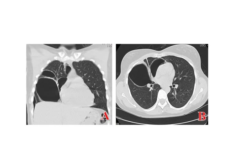
Loküle pnömotoraksta plevral hava, inflame plevranın göğüs duvarına yapışması veya diğer plevral patolojiler nedeniyle serbestçe dağılmak yerine sınırlı bir cepte kalır. BT, lokalize plevral havayı tespit etmek ve yararlı bir yöntem olarak hacim belirlemek için kullanılır; radyografide belirsiz durumlarda havanın tam yerini ve etyolojisini belirlemeye olanak tanır. Bu görünüm, özellikle amfizemli akciğerde dev bülle karışabilir ve yerleşimi yanlış kabul edilirse plevral girişim planlaması yanıltıcı olabilir. Loküle koleksiyonların doğru pozisyonda dren edilmemesi veya kateterin hedef odacığa ulaşamaması, yetersiz drenaj ve komplikasyon riskini artırır.
Faz ve pencere
- Kontrastsız tanısal
- Kontrastsız BT, plevral hava cebini ve eşlik eden hava-sıvı düzeyini gösterebilir. Akciğer penceresinde sınırlı, hilusa uzanmayan ya da plevral yapışıklıkların çevrelediği hava koleksiyonu; koronal ve sagittal planda plevral yüzeyle ilişkisiyle birlikte izlenir.
Önerilen pencereler G genişlik, M merkez; değerler Hounsfield birimi (HU)
- AkciğerG 1500 / M −600
- MediastenG 350 / M +50
BT bulguları
- Kostal, fissüral veya paramediastinal plevra boyunca sınırlı hava cebi; serbest pnömotoraks gibi hemitoraksın bağımlı olmayan tüm alanlarına yayılmaması
- BT’de bül duvarının iki tarafında plevral hava görülmesi çift-duvar işaretini oluşturabilir. Lung point, pnömotoraks sınırında inspiryum sırasında kayan akciğerin aralıklı olarak göğüs duvarına temas ettiği noktadır.
- Komşu plevrada kalınlaşma, kıvrımlı yapışıklık çizgileri veya önceki cerrahiye/plörodeze bağlı plevral kontur değişikliği
- Loküle pnömotoraksta, inflame plevranın göğüs duvarına yapışması veya fibröz bantlar, havanın belirli bir plevral bölgede sınırlı kalmasına neden olur.
- Birbirinden ayrı apikal ve bazal hava odacıkları veya bölmeler arasında sınırlı iletişim
- Drenin lokülün dışından geçmesi, ucunun başka bir plevral bölmede kalması ya da akciğer parankimiyle ilişkisi
- Eşlik eden plevral sıvı, plevral kalınlaşma veya geçirilmiş enfeksiyon/operasyon sekeli
Normalde
Normal plevrada visseral ve parietal yapraklar akciğer ile göğüs duvarı boyunca birbirine yakın seyreder; yapışıklık bandı, fokal hava cebi veya plevral kalınlaşma görülmez. BT, radyografide yeri belirsiz olan torasik hava koleksiyonunu lokalize etmeye ve olası nedenlerini ayırt etmeye yardımcı olur.
Normal BT ile kıyasla
Aynı bölgenin, kaynak veri setinde patoloji içermediği belirtilen bir BT incelemesini kesit kesit kaydırın; organ sınırlarını açarak anatomiyi hasta görüntüsüyle karşılaştırın. Küçük rastlantısal bulgular tümüyle dışlanmamıştır.
Kaynak: TotalSegmentator v2.01 veri seti (Wasserthal ve ark., Radiology: Artificial Intelligence 2023), CC BY 4.0, vaka s1278; organ sınırları veri setinin otomatik segmentasyonundan, birleştirilerek sadeleştirildi. Görüntüler 2,5 mm kesit aralığına yeniden örneklendi.
Ayırıcı tanı
- Periferik dev bül, akciğer parankimi içindeki hava dolu bir boşluktur ve ince duvarlı olabilir; pnömotoraksta hava plevral boşlukta birikir. Çift-duvar işareti destekleyicidir, ancak her kesitte görülmesi beklenmez.
- Paraseptal amfizem, loküle pnömotoraksla birlikte görülebilen bir parankim bulgusudur.
- Pnömomediastinumda Macklin etkisiyle alveollerden kaçan hava perivasküler ve peribronşiyal fasya kılıflarını izleyerek mediastinuma ilerler ve plevraya da uzanabilir.
- İltihaplı plevranın göğüs duvarına yapışması, pnömotoraksı plevral boşluğun bir bölümünde sınırlayabilir.
- Ekstraplevral hava
- gaz parietal plevranın göğüs duvarı tarafındaki dokularda yer alır ve çoğunlukla göğüs duvarı planları boyunca uzanır.
Tuzaklar
- Loküle pnömotoraks büllöz hastalığı taklit edebilir; BT bu ayırıcı tanıda yardımcıdır.
- Büllöz amfizemde çift duvar işareti pnömotoraksı destekleyebilir, ancak tek bir kesitte görünmeyebilir; ardışık ince kesitlerde komşu bül ve plevra kenarını izleyin.
- Plörodez, plevra yapraklarının mekanik appozisyonunu sağlamayı amaçlar.
- Loküle pnömotoraksta BT, hava koleksiyonunu değerlendirmeye ve drenajın görüntüleme eşliğinde yönlendirilmesine yardımcı olabilir.
- Büllöz akciğerde bül ile pnömotoraks birlikte bulunabilir; BT, loküle pnömotoraksı amfizematöz büllerden ayırt etmeye yardımcı olur.
Kendini dene
Plevral yapışıklıkların çevrelediği ve plevral boşluğun bir bölümünde sınırlı kalan bir hava cebi görülüyor. En olası tanı nedir?
Cevabı göster
Loküle pnömotoraks. Plevral boşluğun fokal bir bölümünde sınırlı hava koleksiyonu loküle pnömotoraksla uyumludur. Bül pnömotoraksı taklit edebilir; pnömomediastinal hava perivasküler ve peribronşiyal fasya boyunca mediastinuma ilerleyebilir.
Akciğer parankimindeki büyük bir hava dolu boşluk loküle pnömotoraksı taklit edebilir. En olası taklitçi hangisidir?
Cevabı göster
Periferik bül. Büller ince kıvrımlı duvarlı, avasküler radyolüsent alanlar olarak görülebilir ve loküle pnömotoraksı taklit edebilir; BT ayırıcı tanıda yararlıdır. Loküle pnömotoraks büllöz hastalığı taklit edebilir; BT bu ikisini ayırmada yararlıdır.
İlgili konular
Kaynaklar
Bu sayfadaki 40 cümle ve 2 quiz sorusu aşağıdaki kaynaklardan alıntılarla otomatik olarak karşılaştırıldı; 25 cümle bu karşılaştırmada düzeltildi (2026-10-09).
- Sarcoidosis Presenting as a Giant Pulmonary Bulla With Concurrent COVID-19 Infectionpmc.ncbi.nlm.nih.gov
- Unusual presentations of COVID-19 pneumonia on CT scans with spontaneous pneumomediastinum and loculated pneumothorax: A report of two cases and a review of the literature.pmc.ncbi.nlm.nih.gov
- Multi-loculated pneumothorax post COVID-19 pneumoniapmc.ncbi.nlm.nih.gov
- Development of a Large Pneumatocele in a Patient Recovering from COVID-19 Pneumonitispmc.ncbi.nlm.nih.gov
- Post-traumatic Aspergillus niger empyema in an immunocompetent military patient: a case reportpmc.ncbi.nlm.nih.gov
- Development of a Large Pneumatocele in a Patient Recovering from COVID-19 Pneumonitis.pubmed.ncbi.nlm.nih.gov
- Loculated pneumothorax due to a rare combination resulting in an interesting chest radiograph.pubmed.ncbi.nlm.nih.gov
- A computed tomography-based nomogram to predict pneumothorax caused by preoperative localization of ground glass nodules using hook wire.pubmed.ncbi.nlm.nih.gov
- Unusual presentations of COVID-19 pneumonia on CT scans with spontaneous pneumomediastinum and loculated pneumothorax: A report of two cases and a review of the literature.pubmed.ncbi.nlm.nih.gov
- Sarcoidosis Presenting as a Giant Pulmonary Bulla With Concurrent COVID-19 Infection.pubmed.ncbi.nlm.nih.gov
- Competence in pleural procedures.pubmed.ncbi.nlm.nih.gov
- Can chest ultrasonography replace standard chest radiography for evaluation of acute dyspnea in the ED?pubmed.ncbi.nlm.nih.gov
- Can routine trauma bay chest x-ray be bypassed with an extended focused assessment with sonography for trauma examination?pubmed.ncbi.nlm.nih.gov
- Thoracic ultrasonography for the pulmonary specialist.pubmed.ncbi.nlm.nih.gov
- Nontyphi Salmonella Empyema with Bronchopleural Fistula in a Patient with Human Immunodeficiency Virus.pubmed.ncbi.nlm.nih.gov
- Multi-Loculated Catamenial Pneumothorax: A Rare Complication of Thoracic Endometriosis.pubmed.ncbi.nlm.nih.gov
- Image-guided catheter drainage in loculated pleural space collections, effectiveness, and complications.pubmed.ncbi.nlm.nih.gov
- <i>Paragonimiasis</i> presenting as pyopneumothorax with direct visualization of live worms via video-assisted thoracoscopic surgery: case report.pubmed.ncbi.nlm.nih.gov
- Efficacy and safety of urokinase in the treatment of loculated pleural effusions caused by various etiologies: a systematic review and meta-analysis.pubmed.ncbi.nlm.nih.gov
- Clinical, Echocardiographic, and Procedural Characteristics of Eccentric Versus Circumferential Pericardial Effusions in a Symptomatic Drainage Cohort.pubmed.ncbi.nlm.nih.gov
- Retrospective Review Is the Best View: A Primer for Residents and Radiologists on Missed Findings in Chest Radiography.pubmed.ncbi.nlm.nih.gov
- Descending Necrotizing Mediastinitis, When Mediastinal Drainage By Thoracoscopy Is Not Sufficient: Case Report and Review of the Literature.pubmed.ncbi.nlm.nih.gov
- Unilateral Giant Bullae: Pulmonary Placental Transmogrification Should Be Kept in Mind: Case Reports.pubmed.ncbi.nlm.nih.gov
- A case of lung volume reduction surgery with decortication for a septic patient in respiratory failure.pubmed.ncbi.nlm.nih.gov
- Malignancy Presenting As Recurrent Empyema: A Report of Two Cases and a Systematic Review.pubmed.ncbi.nlm.nih.gov
- Postoperative Pulmonary Complications: Clinical and Imaging Insightspubmed.ncbi.nlm.nih.gov
- Understanding Air in the Body: A Radiologic Perspective on Emphysematous and Traumatic Conditions.pubmed.ncbi.nlm.nih.gov
- Pulmonary Complications of Rheumatoid Arthritis Presenting as Spontaneous Pneumothorax and Empyema: A Case Report.pubmed.ncbi.nlm.nih.gov
- An unusual case of chylothorax.pubmed.ncbi.nlm.nih.gov
- Successful Multidisciplinary Management of Esophageal Perforation Complicated by Septic Shock: A Race Against Time.pubmed.ncbi.nlm.nih.gov
- Chest Wall and Diaphragmatic Injuries: Imaging Evaluation and Clinical Considerations.pubmed.ncbi.nlm.nih.gov
- Spontaneous Pneumothorax: A Review of Underlying Etiologies and Diagnostic Imaging Modalities.pubmed.ncbi.nlm.nih.gov
- Recurrent Spontaneous Pneumothorax in a Nine-Year-Old Patient.pubmed.ncbi.nlm.nih.gov
- Pediatric lung ultrasound: Tips and tricks for better scanning and interpretation.pubmed.ncbi.nlm.nih.gov
- Recurrent Pneumothorax With Bronchopleural Fistula in Primary Multidrug-Resistant Pulmonary Tuberculosis: A Challenging Case.pubmed.ncbi.nlm.nih.gov
- SPOT THE BOT: A comparative quality assessment of AI-generated written instructions for lung ultrasound training.pubmed.ncbi.nlm.nih.gov
- A retrospective analysis of the anatomic characteristics of pulmonary bullae based on chest CT classification and their association with pulmonary function.pubmed.ncbi.nlm.nih.gov
- Ultrasound-Guided Small-Caliber Catheter Drainage for Complex Septated Pleural Effusions: Discrimination of Septal Characteristics for Outcomes.pubmed.ncbi.nlm.nih.gov
- Management of pneumonia by the emergency radiologist.pubmed.ncbi.nlm.nih.gov
- Contemporary Pleural Drainage in Adult Practice: Image-Guided Techniques, Procedural Safety, Training Standards, and Malignant Pleural Effusion Management.pubmed.ncbi.nlm.nih.gov
- Ultrasound-Fluoroscopy Versus Ultrasound Guidance for Catheter Drainage of Loculated Pleural Effusions: A Retrospective Study.pubmed.ncbi.nlm.nih.gov
- Tube Thoracostomy Across Emergency Medicine, Thoracic Surgery, and Intensive Care Residents: A Three-Year Retrospective Analysis of Practice Patterns and Complications at a Tertiary University Hospital.pubmed.ncbi.nlm.nih.gov
- Pleural Effusion Phenotype and Ultrasound Guidance Approach Associated with Unplanned Pigtail Pleural Catheter Removal in an Adult Intensive Care Unit: A Single-Center Retrospective Cohort Study.pubmed.ncbi.nlm.nih.gov
- Ultrasonographic Assessment of the Lungs in Asiatic Elephants (Elephas maximus).pubmed.ncbi.nlm.nih.gov
- Extensive Bilateral Pleural Plaques: A Cadaveric Case Study.pubmed.ncbi.nlm.nih.gov
- Bilateral Lung Transplantation in Patients With Severe Chest Asymmetry: A Case Series From a Single Center.pubmed.ncbi.nlm.nih.gov
- Comparison of surgical approaches for pectus excavatum in children and adolescents: a single-center retrospective study.pubmed.ncbi.nlm.nih.gov
- Fatal multi-organ failure following delayed diagnosis of ruptured pulmonary hydatid cyst in an 11-year-Old boy: The airway management dilemma ('intubate or not intubate').pubmed.ncbi.nlm.nih.gov
- Recurrent pneumonia unmasks congenital lobar emphysema in a 2-month-old infant: Radiological insights from a rare ICU case report.pubmed.ncbi.nlm.nih.gov
- Vanishing Lung Syndrome in a Dog: Giant Pneumatocele or Giant Pulmonary Bulla Mimicking Tension Pneumothorax-First Report.pubmed.ncbi.nlm.nih.gov
- Elevated troponin in a patient with pneumomediastinum and pneumothorax: a diagnostic challenge.pubmed.ncbi.nlm.nih.gov
- A Case of Birt-Hogg-Dubé Syndrome: A Rare but Essential Diagnosis to Consider.pubmed.ncbi.nlm.nih.gov
- Fatal Rapidly Progressive Interstitial Lung Disease in Anti-Melanoma Differentiation-Associated Gene 5 (MDA5) Dermatomyositis in a Brazilian Patient: A Case Report.pubmed.ncbi.nlm.nih.gov
- Secondary Pneumomediastinum Following Bronchoscopic Interventions in Patients With Prolonged Tracheostomy Intubation: A Case Series.pubmed.ncbi.nlm.nih.gov
- Extensive Pneumomediastinum, Pneumopericardium, and Subcutaneous Emphysema Complicating Anti-melanoma Differentiation-Associated Gene 5 (Anti-MDA5)-Positive Dermatomyositis-Associated Fibrosing Interstitial Lung Disease: A CT-Based Case Report.pubmed.ncbi.nlm.nih.gov
- Successful management of fistulous empyema caused by <i>Mycobacterium avium</i> complex in an advanced-age patient: A case report.pubmed.ncbi.nlm.nih.gov
- From tradition to evidence: a narrative review for standardizing chest tube management after anatomic lung resection.pubmed.ncbi.nlm.nih.gov
- Diaphragm-Directed Strategies for Prolonged Air Leak Prevention After Lung Resection: A Narrative Review of the Residual Pleural Space Paradigm.pubmed.ncbi.nlm.nih.gov
- Lung Parenchymal and Airway Injuries: Differential Diagnosis and Severity Grading.pubmed.ncbi.nlm.nih.gov
- Common and Rare Radiological Findings of Pulmonary and Thoracic Hydatid Cysts.pubmed.ncbi.nlm.nih.gov
- When a Giant Bulla Mimics a Tension Pneumothorax: A Case Report and Literature Review.pubmed.ncbi.nlm.nih.gov
- Mapping Bullous Emphysema With Lung Ultrasound: A Prospective Multicentre Study.pubmed.ncbi.nlm.nih.gov
- A Case of Exuberant Type 2 Pneumocyte Hyperplasia in a Young Man with Recurrent Spontaneous Pneumothorax: A Diagnostic Pitfall.pubmed.ncbi.nlm.nih.gov
- Endobronchial epithelioid leiomyoma (leiomyoblastoma) causing complete right-lung atelectasis: A case report.pubmed.ncbi.nlm.nih.gov
- Identification and Management of Intraoperative Pneumothorax During Laparoscopic Surgery: A Rare Complication.pubmed.ncbi.nlm.nih.gov
- The efficacy and safety of medical thoracoscopy compared with tube thoracostomy in the treatment of complicated parapneumonic effusion and empyema: A retrospective single-center study.pubmed.ncbi.nlm.nih.gov
- Point-of-Care Ultrasound and Clinical Findings in 14 Cats With High-Rise Syndrome.pubmed.ncbi.nlm.nih.gov
- ICG combined with medical adhesive in preoperative localization of complex pulmonary nodules: a retrospective study.pubmed.ncbi.nlm.nih.gov
- Extended Focused Assessment with Sonography for Trauma in the Emergency Department: A Comprehensive Review.pubmed.ncbi.nlm.nih.gov
- Fleischner Society: Glossary of Terms for Thoracic Imaging.pubmed.ncbi.nlm.nih.gov
- Clinical phenotypes of secondary spontaneous pneumothorax (SSP) in different underlying lung diseases: a retrospective study and literature review on the clinical heterogeneity in this single entity.pubmed.ncbi.nlm.nih.gov
- Long-term safety and effectiveness of the "OptEase" vena cava filter.pubmed.ncbi.nlm.nih.gov
- Diffuse cystic lung diseases-a primer for radiologists.pubmed.ncbi.nlm.nih.gov
- CT Imaging Features of Pulmonary Sarcoidosis: Typical and Atypical Radiological Features and Their Differential Diagnosis.pubmed.ncbi.nlm.nih.gov
- Thoracic abnormal air collections in patients in the intensive care unit: radiograph findings correlated with CT.pubmed.ncbi.nlm.nih.gov
- Post-traumatic Aspergillus niger empyema in an immunocompetent military patient: a case report.pubmed.ncbi.nlm.nih.gov
- Chinese expert consensus on the diagnosis and management of bronchopleural fistula after pulmonary resection (2026 edition).pubmed.ncbi.nlm.nih.gov
- Robotic-assisted transbronchial biopsy versus computed tomography-guided transthoracic needle biopsy for peripheral pulmonary lesions: a systematic review and meta-analysis of direct comparative studies.pubmed.ncbi.nlm.nih.gov
- Lung Ultrasound in Pediatrics: A Review with Core Principles That Every User Should Know.pubmed.ncbi.nlm.nih.gov
- Radiological insights into a voluminous left-sided Morgagni-Larrey hernia: A case report.pubmed.ncbi.nlm.nih.gov
- Low Diagnostic Accuracy of Transthoracic Ultrasound for the Assessment of Spontaneous Pneumothorax in the Emergency Setting: A Multicentric Study.pubmed.ncbi.nlm.nih.gov
- Infected Emphysematous Bullae of the Lung: A Diagnostic Challenge.pubmed.ncbi.nlm.nih.gov
- Watch Out for the Early Killers: Imaging Diagnosis of Thoracic Trauma.pubmed.ncbi.nlm.nih.gov
- The Evolving Role of Medical Thoracoscopy for the Management of Malignant Pleural Effusion.pubmed.ncbi.nlm.nih.gov
- Imaging of pleural disease.pubmed.ncbi.nlm.nih.gov
- Determinants of Chemical Pleurodesis Recurrence in Malignant Pleural Effusion: Mechanical Apposition or Systemic Inflammation? A Retrospective Cohort Study.pubmed.ncbi.nlm.nih.gov
- Double-Pigtail Drainage Catheter: A New Design for Efficient Pleural Drainage.pubmed.ncbi.nlm.nih.gov
- Complications of Pulmonary Embolism in a Pediatric Patient.pubmed.ncbi.nlm.nih.gov
- Paraseptal Lucencies Mimicking Emphysema in a Non-smoker With Acute Lung Injury in COVID-19.pubmed.ncbi.nlm.nih.gov
- Hybrid deep learning approach for early emphysema diagnosis combining fuzzy C-means, TransUNet, and faster mask R-CNN.pubmed.ncbi.nlm.nih.gov
- Surgical stabilization of rib fractures (SSRF): the WSES and CWIS position paper.pubmed.ncbi.nlm.nih.gov
- Radiologist's Guide to the Ninth Edition TNM Staging System for Quantitative and Qualitative Assessment of Pleural Mesothelioma.pubmed.ncbi.nlm.nih.gov
- Correlation between CT findings and thoracoscopic diagnosis in diffuse pleural disease.pubmed.ncbi.nlm.nih.gov
- Pleural effusion guidelines from ICS and NCCP Section 1: Basic principles, laboratory tests and pleural procedures.pubmed.ncbi.nlm.nih.gov
- Rare presentation of a rare disease: Bilateral congenital lobar overinflation.pubmed.ncbi.nlm.nih.gov
- Chest CT findings of early and progressive phase COVID-19 infection from a US patient.pubmed.ncbi.nlm.nih.gov
- Reporting and management of incidental lung findings on computed tomography: beyond lung nodules.pubmed.ncbi.nlm.nih.gov
- A middle-aged man with dyspnea and hoarseness: an unusual case of vocal cord paralysis.pubmed.ncbi.nlm.nih.gov
- A single-institution outcome of bronchial valve placement for persistent air leak.pubmed.ncbi.nlm.nih.gov
- Diagnostic utility of ultrasonography for thoracic and abdominal bacterial and parasitic diseases in ruminants: a comprehensive overview.pubmed.ncbi.nlm.nih.gov
- Modified enhanced pleural fixation: combined intra- and postoperative hypertonic glucose in elderly and high-risk pulmonary bullae patients - a retrospective cohort study.pubmed.ncbi.nlm.nih.gov
- Secondary spontaneous pneumothorax as an initial manifestation of lung cancer: a case report.pubmed.ncbi.nlm.nih.gov
- Diagnosis and management of pleural infection.pubmed.ncbi.nlm.nih.gov
- A Case of Serotonin Syndrome Associated With Acute Quetiapine Poisoning.pubmed.ncbi.nlm.nih.gov
- Thermal Ablation of Hypervascular Liver Tumors After Selective Intra-Arterial Lipiodol Injection (SIALI): A Technical Narrative Review of Technique, Institutional Variations, and Published Evidence.pubmed.ncbi.nlm.nih.gov
- AI-Based Algorithm to Detect Heart and Lung Disease From Acute Chest Computed Tomography Scans: Protocol for an Algorithm Development and Validation Study.pubmed.ncbi.nlm.nih.gov
- Association Between Emphysema and Coronary Artery Calcium on Low-Dose CT in Urban Chinese Adults: Does Lifestyle Matter?pubmed.ncbi.nlm.nih.gov
- Endoscope-assisted prone lateral retropleural 360° reconstruction of thoracic pathological fracture in metastatic melanoma: a case report and technical note.pubmed.ncbi.nlm.nih.gov
- Emergency Minimally Invasive Esophagogastrectomy and Delayed Esophagocoloplasty for Caustic Ingestion: A Stepwise Video-Based Approach.pubmed.ncbi.nlm.nih.gov
- Residual apical space (RAS) correlation with recurrence in primary spontaneous pneumothorax after surgery: multicenter analysis stratified by type of surgery.pubmed.ncbi.nlm.nih.gov
- Management of pleural effusion, empyema, and lung abscess.pubmed.ncbi.nlm.nih.gov
- Pediatric pleural empyema - decision making beyond imaging - a retrospective cohort study.pubmed.ncbi.nlm.nih.gov
- Imaging of Acute Complications of Community-Acquired Pneumonia in the Paediatric Population-From Chest Radiography to MRI.pubmed.ncbi.nlm.nih.gov
- Breathe easy, baby, breathe. Lung ultrasound in neonatal critical care.pubmed.ncbi.nlm.nih.gov
- International Pain and Spine Intervention Society Emergency Protocols: Pneumothorax.pubmed.ncbi.nlm.nih.gov
Eğitim amaçlıdır; hasta başında tanı ve tedavi kararının yerine geçmez, tıbbi tavsiye değildir. Metinler yapay zekâ desteğiyle yazıldı ve otomatik bir adımda kaynak alıntılarıyla karşılaştırıldı; bu bir tıbbi doğrulama değildir ve içerik uzman radyolog incelemesinden geçmedi. Klinik kararlarda güncel kılavuzları, kurum protokollerini ve radyoloji raporunu esas alın.