Özet
Kronik ve granülomatöz prostatit, benign inflamasyon olmasına karşın prostat kanserini klinik ve özellikle MR'da taklit edebilir; önceki BCG tedavisi, enfeksiyon veya prostat girişimi öyküsü olasılığı etkiler. Granülomatöz prostatit ile adenokarsinomun görüntüleme bulguları örtüşebilir; mpMR tanıyı düşündürse de kesin tanı için biyopsi gerekebilir. Fokal prostat lezyonlarında granülomatöz prostatit ile adenokarsinomun görüntüleme bulguları örtüşebilir; kesin tanı için biyopsi gerekebilir. Granülomatöz prostatit MR'da adenokarsinomu taklit edebilir ve nadiren adenokarsinomla birlikte bulunabilir; görüntüleme bulguları kesin ayrım sağlamayabilir.
Faz ve pencere
- Kontrastsız
- Kontrastsız BT'de lezyonların kontrastlanma özellikleri değerlendirilemez; granülomatöz prostatit ile adenokarsinomun görüntüleme bulguları örtüşebildiğinden tanı gerektiğinde biyopsiyle doğrulanır. Granülomatöz prostatit ile adenokarsinom MR'da örtüşen bulgular gösterebilir; görüntüleme tek başına kesin ayrım sağlayamaz.
Önerilen pencereler G genişlik, M merkez; değerler Hounsfield birimi (HU)
- Yumuşak dokuG 400 / M +40
- KemikG 1800 / M +400
BT bulguları
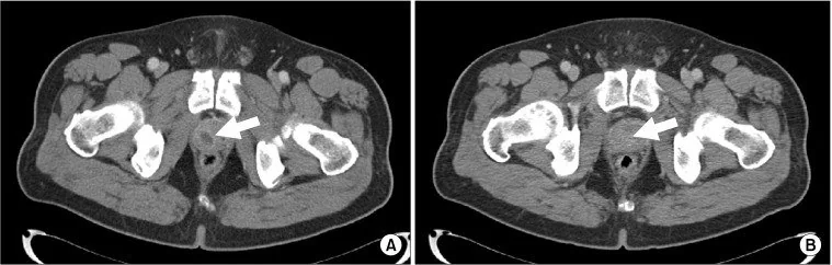
- Prostat parankiminde noktasal veya kaba kalsifikasyonlar izlenebilir; kalsifikasyon, kanser taklidi yapan benign durumlar arasında prostatitten ayrı bir varlık olarak tanımlanır ve tek başına granülomatöz prostatite özgü değildir.
- Granülomatöz prostatit MR'da difüz veya fokal nodüler patern gösterebilir; MR bulguları adenokarsinomla örtüşebildiğinden kesin tanı için biyopsi gerekebilir.
- Prostat girişimleri, biyopsi dahil, granülomatöz prostatitle ilişkili olabilir; işlem öyküsü bu tanı için klinik bağlam sağlar.
- Önceki intravezikal BCG, tekrarlayan üriner enfeksiyonlar veya prostat girişimleri granülomatöz prostatit için klinik bağlam sağlayabilir.
- Prostat apsesi varlığında MR'da lezyon kontrastlanması veya apse çevresinde halka tarzı kontrastlanma görülebilir. Granülomatöz prostatit ile adenokarsinomun MR bulguları örtüşebilir; görüntüleme tek başına kesin ayrım sağlamaz.
- Prostat dışına uzanan düzensiz yumuşak doku ya da patolojik lenf nodu kanseri dışlamaz; granülomatöz inflamasyon ve malignite görüntüde örtüşebilir.
- mpMR klinik olarak anlamlı prostat kanserinin değerlendirilmesinde kullanılır; PI-RADS 3, klinik olarak anlamlı kanser varlığının belirsiz olduğu kategoridir ve biyopsi için ileri değerlendirmeye yol açabilir. Prostat adenokarsinomunun değerlendirilmesinde mpMR kullanıldığından, BT'de belirgin bulgu olmaması mpMR değerlendirmesinin yerini tutmaz.
- Granülomatöz prostatit idiyopatik, enfeksiyöz, iyatrojenik (örneğin intravezikal BCG sonrası) ve sistemik granülomatöz hastalıklarla ilişkili alt gruplara ayrılır.
Normalde
Prostatın zonal anatomisinin bilinmesi, mpMR değerlendirmesinde anatomik yapıları prostat kanserinden ayırmaya yardımcı olur. Kıyaslamada kalsifikasyonların konumunu, bez simetrisini, işlem yatağını ve periprostatik yağ planlarını inceleyin; ancak granülomatöz prostatit ile adenokarsinom görüntüde kesin ayrışamadığından mpMR değerlendirmesi ve gerektiğinde histolojik doğrulama gerekir.
Normal BT ile kıyasla
Aynı bölgenin, kaynak veri setinde patoloji içermediği belirtilen bir BT incelemesini kesit kesit kaydırın; organ sınırlarını açarak anatomiyi hasta görüntüsüyle karşılaştırın. Küçük rastlantısal bulgular tümüyle dışlanmamıştır.
Kaynak: TotalSegmentator v2.01 veri seti (Wasserthal ve ark., Radiology: Artificial Intelligence 2023), CC BY 4.0, vaka s0119; organ sınırları veri setinin otomatik segmentasyonundan, birleştirilerek sadeleştirildi. Görüntüler 2,5 mm kesit aralığına yeniden örneklendi.
Ayırıcı tanı
- Prostat adenokarsinomu
- fokal lezyon, ekstrakapsüler uzanım veya nodal/kemik hastalığı kuşkusu yaratabilir; BT intraprostatik ayrımda yetersizdir ve mpMR/klinik-histolojik değerlendirme gerekir.
- İyi huylu prostat hiperplazisi nodülleri granülomatöz prostatiti taklit edebilir; bu lezyonların özellikleri mpMR'da değerlendirilebilir.
- Prostat apsesi
- sıvı merkezli fokal kavite ve çevresel kontrastlanan duvar, yalnız kalsifikasyon/fibrozisten ayrılır.
- Prostat taşı/kalsifikasyonu
- kalsifikasyon, kanser taklidi yapan benign durumlar arasında ayrı bir varlıktır; taş veya kalsifikasyon varlığı granülomatöz inflamasyon kanıtı değildir.
- Prostat biyopsisi gibi girişimler granülomatöz prostatitle ilişkili olabilir; bu öykü klinik değerlendirmede dikkate alınmalıdır.
Tuzaklar
- Prostat içi kalsifikasyonu granülomatöz hastalığın işareti olarak sunmayın; kalsifikasyon, kanser taklidi yapan benign durumlar arasında prostatitten ayrı olarak tanımlanan bir varlıktır.
- PI-RADS, şüpheli prostat kanserinin mpMR değerlendirmesi için geliştirilmiş yapılandırılmış bir raporlama sistemidir; granülomatöz prostatit mpMR'da kanseri taklit edebilir.
- Önceki intravezikal BCG veya prostat girişimi öyküsü granülomatöz prostatit olasılığını ayırıcı tanıda düşündürmelidir.
- Granülomatöz prostatit bazı olgularda ekstraprostatik uzanım ve lenfadenopati gösterebilir; bu bulgular lokal ileri prostat kanserini taklit edebilir.
Kendini dene
İntravezikal BCG öyküsü ve prostat kalsifikasyonu granülomatöz prostatit tanısını tek başına doğrular mı?
Cevabı göster
Hayır; BCG öyküsü ayırıcı tanıda önemlidir, kesin tanı biyopsiyle doğrulanır. BCG öyküsü granülomatöz prostatiti ayırıcı tanıda düşündürmelidir; görüntüleme bulguları adenokarsinomla örtüşebilir. Kesin tanı için biyopsiyle doğrulama gerekir.
Granülomatöz prostatit öyküsü olan hastada mpMR'da şüpheli fokal lezyon görülürse hangi ifade doğrudur?
Cevabı göster
mpMR bulguları adenokarsinomla örtüşebilir; doğrulayıcı tanı için biyopsi gerekebilir. Granülomatöz prostatit ile adenokarsinomun görüntüleme bulguları örtüşebilir; kesin tanı için biyopsi gerekebilir.
İlgili konular
Kaynaklar
Bu sayfadaki 37 cümle ve 2 quiz sorusu aşağıdaki kaynaklardan alıntılarla otomatik olarak karşılaştırıldı; 30 cümle bu karşılaştırmada düzeltildi (2026-10-09).
- Abscess Adjacent to the Vas Deferens Mimicking Recurrent Inguinal Hernia: A Case Report and Review of the Literaturepmc.ncbi.nlm.nih.gov
- Granulomatous prostatitis following Bacillus Calmette–Guérin therapypmc.ncbi.nlm.nih.gov
- Rectal tuberculosis in a 48-years-old immuno-competent man: A rare case reportpmc.ncbi.nlm.nih.gov
- Atypical Presentations in Melioidosis: A Case-Based Review from Endemic Regionspmc.ncbi.nlm.nih.gov
- Imaging findings in inflammatory disease of the genital organspmc.ncbi.nlm.nih.gov
- Rectal malakoplakia mimicking advanced rectal cancer: A case report.pubmed.ncbi.nlm.nih.gov
- The Curious Case of the Black Buboes.pubmed.ncbi.nlm.nih.gov
- Benign Conditions That Mimic Prostate Carcinoma: MR Imaging Features with Histopathologic Correlation.pubmed.ncbi.nlm.nih.gov
- Granulomatous Prostatitis Mimicking Prostate Cancer.pubmed.ncbi.nlm.nih.gov
- Best practices recommendations in the application of immunohistochemistry in the prostate: report from the International Society of Urologic Pathology consensus conference.pubmed.ncbi.nlm.nih.gov
- Metastatic prostate cancer on the thyroid cartilage: unusual symptoms of prostatic adenocarcinoma. Case report.pubmed.ncbi.nlm.nih.gov
- Metachronous Schnitzler's Metastasis of Gastric Adenocarcinoma 13 Years After Curative Resection: A Case Report.pubmed.ncbi.nlm.nih.gov
- More advantages in detecting bone and soft tissue metastases from prostate cancer using 18 F-PSMA PET/CT.pubmed.ncbi.nlm.nih.gov
- An Update on the Role of mpMRI and 68 Ga-PSMA PET Imaging in Primary and Recurrent Prostate Cancer.pubmed.ncbi.nlm.nih.gov
- Final analysis of a prospective trial on functional imaging for nodal staging in patients with prostate cancer at high risk for lymph node involvement.pubmed.ncbi.nlm.nih.gov
- Comparison of Prostate-Specific Membrane Antigen PET/CT and Contrast-Enhanced Magnetic Resonance Imaging in Follow-up Assessment of Juvenile Nasal Angiofibroma-A Novel Pilot Study.pubmed.ncbi.nlm.nih.gov
- Interpreting Prostate Multiparametric MRI: Beyond Adenocarcinoma - Anatomical Variations, Mimickers, and Post-Intervention Changes.pubmed.ncbi.nlm.nih.gov
- Imaging findings in inflammatory disease of the genital organs.pubmed.ncbi.nlm.nih.gov
- CT and MRI in Urinary Tract Infections: A Spectrum of Different Imaging Findings.pubmed.ncbi.nlm.nih.gov
- Aggressive chronic prostatitis mimicking locally advanced prostate cancer with apparent extraprostatic extension: a case report.pubmed.ncbi.nlm.nih.gov
- Challenges of Prostatic Tuberculosis: When Infection and Neoplasia Coexist.pubmed.ncbi.nlm.nih.gov
- Hidden in plain sight: Extrapulmonary manifestations of tuberculosis in Asia.pubmed.ncbi.nlm.nih.gov
- Association of PI-RADS Score with Clinically Significant Prostate Cancer and Histopathological Upgrading in Patients Undergoing Radical Prostatectomy: A Retrospective Single-Centre Study.pubmed.ncbi.nlm.nih.gov
- An Assessment of PI-RADS v2.1: Inter-Reader Agreement, Diagnostic Accuracy, and the Added Value of PSA Density in Prostate Cancer Prediction.pubmed.ncbi.nlm.nih.gov
- Clinical efficacy of AI-navigated transperineal MRI-TRUS fusion biopsy for prostate cancer diagnosis: A retrospective cohort study.pubmed.ncbi.nlm.nih.gov
- <sup>18</sup>F-labeled prostate-specific membrane antigen positron emission tomography/computed tomography vs. magnetic resonance imaging in diagnosis of prostate cancer A meta-analysis.pubmed.ncbi.nlm.nih.gov
- The Role of Artificial Intelligence in Optimizing Diagnosis in Prostate Cancer-A Narrative Review.pubmed.ncbi.nlm.nih.gov
- Head-to-head Comparison of Multimodal Imaging for Prostate Cancer Detection and Local Staging with Whole-mount Histopathology Reference.pubmed.ncbi.nlm.nih.gov
- Multiparametric magnetic resonance imaging for the differential diagnosis between granulomatous prostatitis and prostate cancer: a literature review to an intriguing diagnostic challenge.pubmed.ncbi.nlm.nih.gov
- Granulomatous Prostatitis, the Great Mimicker of Prostate Cancer: Can Multiparametric MRI Features Help in This Challenging Differential Diagnosis?pubmed.ncbi.nlm.nih.gov
- Differential diagnosis of uncommon prostate diseases: combining mpMRI and clinical information.pubmed.ncbi.nlm.nih.gov
- Abscess Adjacent to the Vas Deferens Mimicking Recurrent Inguinal Hernia: A Case Report and Review of the Literature.pubmed.ncbi.nlm.nih.gov
- Rectal tuberculosis in a 48-years-old immuno-competent man: A rare case report.pubmed.ncbi.nlm.nih.gov
- Granulomatous prostatitis following Bacillus Calmette-Guérin therapy.pubmed.ncbi.nlm.nih.gov
- Long-term safety and effectiveness of the "OptEase" vena cava filter.pubmed.ncbi.nlm.nih.gov
- Clinical Impact of Prostate-Specific Membrane Antigen Positron Emission Tomography (PSMA-PET) on Staging, Biochemical Recurrence Detection, and Treatment Decision-Making in Prostate Cancer.pubmed.ncbi.nlm.nih.gov
- Present status of prostate cancer diagnosis, limitations, challenges, and future endeavors.pubmed.ncbi.nlm.nih.gov
- Biomarkers for Precision Prognosis in Prostate Cancer: Imaging, Molecular, and Integrated Approaches.pubmed.ncbi.nlm.nih.gov
- Etiopathogenesis and Antibacterial Therapy Approach in Patients with Acute Obstructive Pyelonephritis-A Retrospective Study.pubmed.ncbi.nlm.nih.gov
- Imaging and Clinical Findings of Xanthogranulomatous Inflammatory Disease of Various Abdominal and Pelvic Organs: A Pictorial Essay.pubmed.ncbi.nlm.nih.gov
- MR Virtual Cytometry for Zone-specific Prostate Cancer Characterization with Histologic and Molecular Validation.pubmed.ncbi.nlm.nih.gov
- Xanthogranulomatous prostatitis mimicking prostate carcinoma: a case report and comprehensive literature review.pubmed.ncbi.nlm.nih.gov
- Diagnosis of metastatic prostate cancer by supraclavicular lymph node excision in a patient with persistent macroscopic hematuria: A case report.pubmed.ncbi.nlm.nih.gov
- Melioidosis: diverse clinical manifestations and patient outcomes in a case series.pubmed.ncbi.nlm.nih.gov
- A Case of Necrotic Metastases Imitating Hepatic Abscesses in a Patient With Multiple Primary Cancers.pubmed.ncbi.nlm.nih.gov
- Zinner Syndrome: A Narrative Review of Imaging Findings with an Illustrative Case Report.pubmed.ncbi.nlm.nih.gov
- Precision Diagnostics in Prostate Cancer: Integrating Biomarkers, Imaging, Genomics, and Artificial Intelligence in Contemporary United States Practice.pubmed.ncbi.nlm.nih.gov
- Prostate-specific membrane antigen in malignant brain tumors: vascular heterogeneity, imaging promise, and therapeutic challenges.pubmed.ncbi.nlm.nih.gov
- Impact of 18F-DCFPyL-PSMA PET/CT on Radiotherapy Planning and Treatment Intensification in Occult Biochemical Recurrence of Prostate Cancer After Radical Prostatectomy.pubmed.ncbi.nlm.nih.gov
- Serum PSA levels fluctuations in patients with non-muscle-invasive bladder cancer (NMIBC) undergoing BCG therapy: a prospective study.pubmed.ncbi.nlm.nih.gov
- A Case Report and Literature Review of Prostatic Tuberculosis Masquerading as Prostate Cancer: A Diagnostic Challenge in a Tuberculosis-Endemic Region.pubmed.ncbi.nlm.nih.gov
- [ 68 Ga]Ga-PSMA-11 in prostate cancer: a comprehensive review.pubmed.ncbi.nlm.nih.gov
- Imaging in Prostate Cancer: Magnetic Resonance Imaging and Beyond.pubmed.ncbi.nlm.nih.gov
- Prostatic Tuberculosis Mimicking Clinically Significant Prostate Cancer in a Patient Presenting With Low Ejaculate Volume: A Case Report.pubmed.ncbi.nlm.nih.gov
- Xanthogranulomatous Prostatitis Mimicking Prostate Carcinoma in A 78-Year-Old Male.pubmed.ncbi.nlm.nih.gov
- Mucinous adenocarcinoma of the prostate: a rare tumor - case report and literature review.pubmed.ncbi.nlm.nih.gov
- Future opportunities and nuances with the use of PSMA PET in prostate cancer (MD PET 1).pubmed.ncbi.nlm.nih.gov
- Prostatic incidental uptake on PET/CT: an updated systematic review and meta-analysis of prevalence and malignancy risk.pubmed.ncbi.nlm.nih.gov
- Granulomatous and xanthogranulomatous prostatitis: A case report.pubmed.ncbi.nlm.nih.gov
- Identifying the deceiver: the non-neoplastic mimickers of genital system neoplasms.pubmed.ncbi.nlm.nih.gov
- Extravertebral low back pain: a scoping review.pubmed.ncbi.nlm.nih.gov
- Imaging Spectrum of Granulomatous Diseases of the Abdomen and Pelvis.pubmed.ncbi.nlm.nih.gov
- The addition of PSMA PET in selecting candidates for prostate cancer focal therapy.pubmed.ncbi.nlm.nih.gov
- Patient and Technology Selection for Focal Therapy in Prostate Cancer.pubmed.ncbi.nlm.nih.gov
- Integrating MRI-based tumour staging within the TNM classification system in modern prostate cancer management.pubmed.ncbi.nlm.nih.gov
- Emphysematous conditions of the abdomen and pelvis: pearls and pitfalls.pubmed.ncbi.nlm.nih.gov
- Multiparametric MRI before and after Focal Therapy for Prostate Cancer: Pearls and Pitfalls for the Reporting Radiologist.pubmed.ncbi.nlm.nih.gov
- Isolated epididymal tuberculosis presenting as a chronic hydrocele: A rare manifestation of genitourinary TB.pubmed.ncbi.nlm.nih.gov
- Focal IgG4-related periprostatic "PI-RADS 5" pseudotumor mimicking prostatic adenocarcinoma.pubmed.ncbi.nlm.nih.gov
- Primary diffuse large B cell lymphoma (DLBCL) of the prostate presenting with lower urinary tract symptoms (LUTS).pubmed.ncbi.nlm.nih.gov
- Low PSA but high-grade prostate cancer presenting as a bladder-neck mass in a young patient with refractory bladder outlet obstruction.pubmed.ncbi.nlm.nih.gov
- The evolving PI-RADS paradigm.pubmed.ncbi.nlm.nih.gov
- A rare case of successful treatmeat of xanthogranulomatous prostatitis and literature reviews.pubmed.ncbi.nlm.nih.gov
- Comparison of percutaneous and transarterial fiducial marker placement before proton therapy for pancreatic cancer.pubmed.ncbi.nlm.nih.gov
- Primary Diffuse Large B-cell Lymphoma of the Urinary Bladder: A Case Report and Narrative Review of Treatment Modalities and Clinical Outcomes.pubmed.ncbi.nlm.nih.gov
- Cryoablation combined with immunotherapy: new strategies and progress in the treatment of tumors.pubmed.ncbi.nlm.nih.gov
- In the Era of mpMRI and PSMA PET/CT: Does Digital Rectal Examination Still Matter?pubmed.ncbi.nlm.nih.gov
- Patterns of injury and violence in Yaoundé Cameroon: an analysis of hospital data.pubmed.ncbi.nlm.nih.gov
- Xanthogranulomatous Prostatitis: Novel Presentation of a Rare Condition.pubmed.ncbi.nlm.nih.gov
- Incidental findings in and around the prostate on prostate MRI: a pictorial review.pubmed.ncbi.nlm.nih.gov
- Renal-Type Clear Cell Carcinoma of the Prostate: A Histopathological Case Report of a Rare and Underrecognized Variant.pubmed.ncbi.nlm.nih.gov
- Evolution of prostate MRI: from multiparametric standard to less-is-better and different-is better strategies.pubmed.ncbi.nlm.nih.gov
- Pitfalls in Interpreting mp-MRI of the Prostate: A Pictorial Review with Pathologic Correlation.pubmed.ncbi.nlm.nih.gov
- Successful non-surgical management of locally advanced gastric adenocarcinoma with squamous differentiation and programmed death-ligand 1 combined positive score 75: a case report.pubmed.ncbi.nlm.nih.gov
- Primary Synovial Sarcoma of the Prostate: A Case Report and Review of the Literature.pubmed.ncbi.nlm.nih.gov
- Normal 3T MR Anatomy of the Prostate Gland and Surrounding Structures.pubmed.ncbi.nlm.nih.gov
- Primary prostatic seminoma: a rare case report and structured literature review highlighting tumor marker discordance.pubmed.ncbi.nlm.nih.gov
- IgG4-Related Prostatitis Detected by Whole-Body Diffusion-Weighted Imaging With Background Body Signal Suppression (DWIBS).pubmed.ncbi.nlm.nih.gov
Eğitim amaçlıdır; hasta başında tanı ve tedavi kararının yerine geçmez, tıbbi tavsiye değildir. Metinler yapay zekâ desteğiyle yazıldı ve otomatik bir adımda kaynak alıntılarıyla karşılaştırıldı; bu bir tıbbi doğrulama değildir ve içerik uzman radyolog incelemesinden geçmedi. Klinik kararlarda güncel kılavuzları, kurum protokollerini ve radyoloji raporunu esas alın.