Özet
Pelvik venöz bozukluklar over veya internal iliak venlerde genişleme ve işlev bozukluğuyla ilişkili olabilir; pelvik ven genişlemeleri asemptomatik de görülebildiğinden görüntüleme bulguları hastanın yakınmalarıyla ilişkilendirilerek yorumlanır. Kontrastlı BT, uterus ve çevresindeki genişlemiş, kıvrımlı venöz yapıları ve renal ya da iliak ven düzeyindeki olası darlıkları göstermeye yardımcı olur; venöz akımın dinamik değerlendirilmesinde BT sınırlıdır, Doppler US ve venografi reflüyü değerlendirebilir. BT bulguları klinik yakınmalarla uyumlu bir venöz anatomi örüntüsünü tanımlamaya yarar, tek başına sendrom tanısı koymaz. Belirgin venöz kollaterallerin altında yatan renal veya iliak ven obstrüksiyonu atlanırsa konjesyonun nedeni açıklanamayabilir.
Faz ve pencere
- Portal tanısal
- Kontrastlı venöz opasifikasyonda parauterin pleksusta serpijinöz, değişken kalibrasyonlu damarlar ve genişlemiş over venleri izlenir; damarların devamlılığı renal ven, iliak ven ve vena kava çıkışlarına kadar izlenerek olası kompresyon ya da kollateral yol araştırılır. Tek fazlı BT venöz akımın dinamik değerlendirmesini sağlamaz; reflünün gösterilmesi ve derecelendirilmesinde Doppler US, zaman çözünürlüklü MR anjiyografi veya venografi daha elverişlidir. Tek fazlı BT reflü yönünü doğrudan göstermez.
Önerilen pencereler: Batın (G 400 / M 50), Yumuşak doku (G 400 / M 40).
BT bulguları
- BT'de uterus, adneksler ve geniş bağ çevresinde kıvrımlı, genişlemiş ve kontrastlanan tübüler venöz yapılar görülebilir.
- Over veninin olağandan geniş kalibrasyonu; çap ölçümü damar eksenine dik, kontrastla dolu lümenden yapılır.
- Pelvik yan duvar ve adneks çevresinde serpijinöz venöz kollaterallerin birden çok kesitte sürmesi.
- Uterus arkasında ve vajen çevresinde pleksusların kümelenmesi; dağılımı tek kistik adneks kitlesinden ayır.
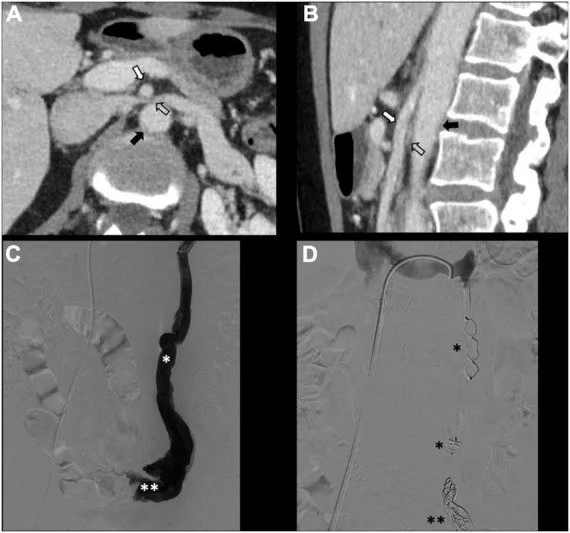
- Sol renal venin aortomezenterik aralıkta daralması veya iliak venlerin kompresyonu gibi olası çıkış engelleri.
- Over veni boyunca trombüs veya kontrastlı BT'de lüminal dolum defekti görülmesi, over ven trombozu olasılığını düşündürür ve ayrıca değerlendirilmelidir.
- Pelvik varisler belirgin olsa da BT'de görünür genişleme kronik pelvik ağrı sendromunu tek başına doğrulamaz.
Ölçütler ve sınıflamalar
- Önerilmiş kesitsel görüntüleme örüntüsü
- Aynı tarafta en az 4 parauterin ven; bunlardan en az 1'i >4 mm veya over veni >8 mm
Normalde
Pelvik venöz drenaj kişiden kişiye değişebilir; uterusun alt bölümü başlıca internal iliak venlere, üst bölümü ise uterin veya over pleksusu üzerinden over venlerine drene olur. Pelvik venöz genişlemeyi değerlendirirken ipsilateral parauterin venlerin sayısı ve çapı ile over veni çapını kaydet; önerilmiş BT ölçütleri en az dört ipsilateral pelvik venden en az birinin 4 mm'den büyük olmasını veya over veninin 8 mm'den büyük olmasını içerir.
Normal BT ile kıyasla
Aynı bölgenin, kaynak veri setinde patoloji içermediği belirtilen bir BT incelemesini kesit kesit kaydırın; organ sınırlarını açarak anatomiyi hasta görüntüsüyle karşılaştırın. Küçük rastlantısal bulgular tümüyle dışlanmamıştır.
Kaynak: TotalSegmentator v2.01 veri seti (Wasserthal ve ark., Radiology: Artificial Intelligence 2023), CC BY 4.0, vaka s0119; organ sınırları veri setinin otomatik segmentasyonundan, birleştirilerek sadeleştirildi. Görüntüler 2,5 mm kesit aralığına yeniden örneklendi.
Ayırıcı tanı
- Over ven trombozu
- geniş ven içinde kontrastlanmayan santral dolum defekti ve bazen çevresel inflamasyon bulunur; basit varikste lümen opaklaşır.
- Adneksiyel kistik kitle
- tek ya da az sayıda yuvarlak lezyon yapar; venöz fazda serpijinöz damar ağı gibi kontrastlanmaz.
- Uterin miyomlar
- miyometriuma bağlı solid kitle ve kontur deformasyonu görülür; parametriumdaki damar kanallarıyla karıştırılmamalıdır.
- Pelvik arteriyovenöz malformasyonda şantın venöz çıkış damarı ve akımı anjiyografide değerlendirilebilir; verilen kaynaklar BT'de erken arteriyel doluş için tanısal ölçüt sunmuyor.
- İliak ya da renal ven obstrüksiyonuna bağlı kollateraller
- venöz ağ benzer görünebilir; merkezi darlık veya bası seviyesini ve kollateral yönlenmesini araştır.
Tuzaklar
- Geniş over veni veya parauterin varisler asemptomatik hastada da bulunabilir; BT morfolojisini klinik ağrı örüntüsü olmadan sendrom tanısı sayma.
- Kontrastlı BT'de uterus çevresindeki kıvrımlı, genişlemiş ve kontrastlanan tübüler yapılar pelvik venöz varis görünümünü destekler; damarların seyri ve devamlılığı değerlendirilmelidir.
- Tek fazlı BT, venöz reflünün yönünü ve sürekliliğini göstermez; çap ölçümü hemodinamik yetmezlik kanıtı değildir.
- Gonadal ven ölçümü kesit eğikliği nedeniyle fazla çıkabilir; lümeni damar eksenine dik planda ölç ve dallanma seviyesini belirt.
Kendini dene
Kontrastlı BT'de pelvik venöz bozuklukla ilişkili damar görünümünü en iyi tanımlayan seçenek hangisidir?
Cevabı göster
Parauterin bölgede kıvrımlı, kalibrasyonu artmış ve kontrastlanan damar kümeleri. Pelvik varisler BT'de uterus ve çevresindeki yapılarda kıvrımlı, genişlemiş, kontrastlanan tübüler yapılar olarak görülebilir; görüntüleme bulguları tanıyı destekler ancak tek başına tanımlamaz.
Sol renal venin aort ile üst mezenterik arter arasında basıya uğraması hangi anatomik bulguyu tanımlar?
Cevabı göster
Aortomezenterik sol renal ven kompresyonu. Sol renal venin üst mezenterik arter ile aort arasında sıkışması anterior nutcracker anatomisini tanımlar; renal ven çıkışındaki obstrüksiyon pelvik venöz konjesyona ikincil katkıda bulunabilir.
İlgili konular
- Over ven trombozu ve septik pelvik tromboflebit
- Postpartum pelvik komplikasyonlar ve normal postpartum görünüm
Kaynaklar
Bu sayfadaki 35 cümle ve 2 quiz sorusu aşağıdaki kaynaklardan alıntılarla otomatik olarak karşılaştırıldı; 13 cümle bu karşılaştırmada düzeltildi (2026-10-09).
- Comprehensive overview of the venous disorder known as pelvic congestion syndromepmc.ncbi.nlm.nih.gov
- Noninvasive diagnostic imaging of pelvic venous disorderspmc.ncbi.nlm.nih.gov
- Nutcracker syndrome with pelvic congestion syndrome: A case reportpmc.ncbi.nlm.nih.gov
- Initial Clinical Experience with Shape Memory Polymer Plugs for Various Venous Embolizationpmc.ncbi.nlm.nih.gov
- Compression and Superficial Varicosities Outperform Gonadal Vein Diameter in Differentiating Symptomatic from Asymptomatic Pelvic Venous Disorders: A Case–Control Studypmc.ncbi.nlm.nih.gov
- Pelvic Congestion Syndrome.pubmed.ncbi.nlm.nih.gov
- Pelvic venous congestion syndrome: female venous congestive syndromes and endovascular treatment options.pubmed.ncbi.nlm.nih.gov
- Pelvic Congestion Syndrome: A Missed Opportunity.pubmed.ncbi.nlm.nih.gov
- Ovarian Vein Embolization: How and When Should It Be Done?pubmed.ncbi.nlm.nih.gov
- Imaging of Pelvic Venous Disorders (PeVD); Should Every Patient Get an MRI?pubmed.ncbi.nlm.nih.gov
- Hemodynamic and Radiological Classification of Ovarian Veins System Insufficiency.pubmed.ncbi.nlm.nih.gov
- Pelvic congestion syndrome and left renal compression syndrome - clinical features and therapeutic approaches.pubmed.ncbi.nlm.nih.gov
- The role of duplex ultrasound in the workup of pelvic congestion syndrome.pubmed.ncbi.nlm.nih.gov
- Clinical Presentation, Patient Assessment, Anatomy, Pathophysiology, and Imaging of Pelvic Venous Disease.pubmed.ncbi.nlm.nih.gov
- [A CASE OF PELVIC CONGESTION SYNDROME SUCCESSFULLY TREATED WITH ENDOVASCULAR THERAPY].pubmed.ncbi.nlm.nih.gov
- Spontaneous rupture of the ovarian vein in association with nutcracker syndrome: a case report.pubmed.ncbi.nlm.nih.gov
- Secondary pelvic congestion syndrome: description and radiographic diagnosis.pubmed.ncbi.nlm.nih.gov
- Pelvic congestion syndrome: the role of interventional radiology in the treatment of chronic pelvic pain.pubmed.ncbi.nlm.nih.gov
- Venous angioplasty and stenting improve pelvic congestion syndrome caused by venous outflow obstruction.pubmed.ncbi.nlm.nih.gov
- Diagnosis and Management of Pelvic Congestion Syndrome: Comprehensive Review.pubmed.ncbi.nlm.nih.gov
- Symptoms in Dilating Venous Disease.pubmed.ncbi.nlm.nih.gov
- [Pelvic congestion syndrom].pubmed.ncbi.nlm.nih.gov
- [Imaging representation of peripheral veins].pubmed.ncbi.nlm.nih.gov
- Current trends in the diagnosis and management of renal nutcracker syndrome: a review.pubmed.ncbi.nlm.nih.gov
- Endovascular management of pelvic congestion syndrome: An expert consensus statement from the French Society of Cardiovascular Imaging (SFICV), Interventional Radiology Federation (FRI), College of French Radiology Teachers (CERF), and French Society of Women's Imaging (SIFEM).pubmed.ncbi.nlm.nih.gov
- Diagnosis and Management of Pelvic Venous Disorders in Women.pubmed.ncbi.nlm.nih.gov
- Review of the literature supporting international clinical practice guidelines on iliac venous stenting and their applicability to Australia and New Zealand practice.pubmed.ncbi.nlm.nih.gov
- Quantitative 4D-Flow MR Imaging of Abdominopelvic Vasculature in Pelvic Venous Disorders.pubmed.ncbi.nlm.nih.gov
- Efficacy of two micronized purified flavonoid fraction dosing regimens in the pelvic venous pain relief.pubmed.ncbi.nlm.nih.gov
- Transcatheter foam sclerotherapy of symptomatic female varicocele with sodium-tetradecyl-sulfate foam.pubmed.ncbi.nlm.nih.gov
- Medical treatment for pelvic congestion syndrome with flavonoid: A pilot study.pubmed.ncbi.nlm.nih.gov
- Nutcracker syndrome: diagnosis and therapy.pubmed.ncbi.nlm.nih.gov
- Nutcracker syndrome: a case-based review.pubmed.ncbi.nlm.nih.gov
- Nutcracker syndrome (a Delphi consensus).pubmed.ncbi.nlm.nih.gov
- A novel endovascular thermal ablation technique for pelvic venous disorders via basilic vein access: A prospective descriptive study.pubmed.ncbi.nlm.nih.gov
- Comprehensive overview of the venous disorder known as pelvic congestion syndrome.pubmed.ncbi.nlm.nih.gov
- What is the significance of ovarian vein reflux detected by computed tomography in patients with pelvic pain?pubmed.ncbi.nlm.nih.gov
- Correlation between pelvic congestion syndrome and body mass index.pubmed.ncbi.nlm.nih.gov
- Symptomatic pelvic venous insufficiency: a review of the current controversies in pathophysiology, diagnosis, and management.pubmed.ncbi.nlm.nih.gov
- MDCT of the ovarian vein: normal anatomy and pathology.pubmed.ncbi.nlm.nih.gov
- Imaging Findings of Ovarian Vein Thrombosis.pubmed.ncbi.nlm.nih.gov
- Diagnosis and management of postpartum ovarian vein thrombosis.pubmed.ncbi.nlm.nih.gov
- Ovarian vein thrombosis after total laparoscopic hysterectomy with unilateral adnexectomy: A case report.pubmed.ncbi.nlm.nih.gov
- Detection of hemorrhage source: the diagnostic value of post-mortem CT-angiography.pubmed.ncbi.nlm.nih.gov
- Evaluation of incidence and clinical significance of obturator hook sign as a marker of chronic iliofemoral venous outflow obstruction in computed tomography venography.pubmed.ncbi.nlm.nih.gov
- Ultrasound maximum vein diameter contextualizes testicular asymmetry in adolescent varicocele.pubmed.ncbi.nlm.nih.gov
- Surgical treatment of nutcracker syndrome results in improved pain and quality of life.pubmed.ncbi.nlm.nih.gov
- Primary renal zygomycotic infarction mimicking renal neoplasia in an immunocompetent patient.pubmed.ncbi.nlm.nih.gov
- Venous reflux from the pelvis and vulvoperineal region as a possible cause of lower extremity varicose veins: diagnosis with computed tomographic and ultrasonographic findings.pubmed.ncbi.nlm.nih.gov
- Diagnosis and treatment of the pelvic congestion syndrome.pubmed.ncbi.nlm.nih.gov
- Imaging Appearance and Nonsurgical Management of Pelvic Venous Congestion Syndrome.pubmed.ncbi.nlm.nih.gov
- Gonadal vein transposition is a safe and effective treatment of nutcracker syndrome.pubmed.ncbi.nlm.nih.gov
- Intracardiac migration of a left renal vein stent after endovascular treatment of nutcracker-associated pelvic congestion syndrome: a case report.pubmed.ncbi.nlm.nih.gov
- Compression and Superficial Varicosities Outperform Gonadal Vein Diameter in Differentiating Symptomatic from Asymptomatic Pelvic Venous Disorders: A Case-Control Study.pubmed.ncbi.nlm.nih.gov
- Left ovarian vein thrombosis suspected during cesarean section and confirmed postoperatively: a case report and review of the literature.pubmed.ncbi.nlm.nih.gov
- Lesion-specific comparison of pelvic venous ultrasound, dynamic magnetic resonance venography, and digital subtraction venography in pelvic vein disorders.pubmed.ncbi.nlm.nih.gov
- Pelvic pain of venous origin: diagnostic insights from transvaginal ultrasonography.pubmed.ncbi.nlm.nih.gov
- The Role of Interventional Radiology in the Diagnosis and Treatment of Gynecological Conditions.pubmed.ncbi.nlm.nih.gov
- Initial Clinical Experience with Shape Memory Polymer Plugs for Various Venous Embolization.pubmed.ncbi.nlm.nih.gov
- Unusual Spinal Epidural and Paraspinal Venous Engorgement Secondary to Hepatosplenic Schistosomiasis: A Rare Cause of Severe Low Back Pain.pubmed.ncbi.nlm.nih.gov
- Right portal branch to ovarian vein anastomosis: An unusual portosystemic shunt secondary to pelvic venous congestion.pubmed.ncbi.nlm.nih.gov
- Use of the Wrapsody™ Cell-Impermeable Endoprosthesis for the treatment of pelvic congestion syndrome due to nutcracker syndrome.pubmed.ncbi.nlm.nih.gov
- Pelvic venous disorders in women: Diagnosis and management for gynecologists.pubmed.ncbi.nlm.nih.gov
- Chronic Pelvic Pain Attributable to Pelvic Congestion Syndrome: A Case Series.pubmed.ncbi.nlm.nih.gov
- Aetiology of pelvic neuropathies in women: a narrative review.pubmed.ncbi.nlm.nih.gov
- Doppler ultrasound diagnosis of anterior nutcracker syndrome presenting with isolated urinary abnormalities.pubmed.ncbi.nlm.nih.gov
- The national acceptance of pelvic congestion syndrome among other common venous pathologies.pubmed.ncbi.nlm.nih.gov
- Cross-sectional imaging of acute gynaecologic disorders: CT and MRI findings with differential diagnosis-part I: corpus luteum and haemorrhagic ovarian cysts, genital causes of haemoperitoneum and adnexal torsion.pubmed.ncbi.nlm.nih.gov
- Slow-flow, high-impact: radiologic features in venous malformations of the female genital tract.pubmed.ncbi.nlm.nih.gov
- Acute Appendicitis Complicated by Ascaris lumbricoides Infestation: A Case Report.pubmed.ncbi.nlm.nih.gov
- Ultrasonic typing and diagnostic value of intravenous leiomyomatosis: a single-center retrospective study.pubmed.ncbi.nlm.nih.gov
- Balloon-assisted endovascular embolization of a subcutaneous penile arteriovenous fistula using a quadruple coaxial system: A case report.pubmed.ncbi.nlm.nih.gov
- Computed tomography of renal emergencies: A comprehensive diagnostic guide for radiology residents.pubmed.ncbi.nlm.nih.gov
- Gynecologic imaging errors and pitfalls: how to avoid misdiagnosis on pelvic MRI.pubmed.ncbi.nlm.nih.gov
- Cytoreductive surgery for ovarian cancer with pelvic ectopic kidney: a case report.pubmed.ncbi.nlm.nih.gov
- Incidentally Discovered Ovarian Cancer Combined With Obturator Nerve Schwannoma: A Rare Case Report.pubmed.ncbi.nlm.nih.gov
- How common are pelvic venous disorders in patients with unexplained chronic pelvic pain: a prospective cross-sectional study.pubmed.ncbi.nlm.nih.gov
- Intravenous leiomyomatosis of the uterus: A case report and literature review.pubmed.ncbi.nlm.nih.gov
- Imaging of Deep Venous Pathology.pubmed.ncbi.nlm.nih.gov
- Nutcracker syndrome in 2026: a nephrologist-oriented diagnosis and management.pubmed.ncbi.nlm.nih.gov
- Bottom-up versus top-down treatment strategies for pelvic venous disorders: Comparative analysis of pain relief, recurrence, and procedural burden.pubmed.ncbi.nlm.nih.gov
- Cracking the nutcracker myth: CT-based risk stratification of symptomatic left renal vein Compression.pubmed.ncbi.nlm.nih.gov
- Acute pelvic pain: A pictorial review with magnetic resonance imaging.pubmed.ncbi.nlm.nih.gov
Eğitim amaçlıdır; hasta başında tanı ve tedavi kararının yerine geçmez, tıbbi tavsiye değildir. Metinler yapay zekâ desteğiyle yazıldı ve otomatik bir adımda kaynak alıntılarıyla karşılaştırıldı; bu bir tıbbi doğrulama değildir ve içerik uzman radyolog incelemesinden geçmedi. Klinik kararlarda güncel kılavuzları, kurum protokollerini ve radyoloji raporunu esas alın.