Özet
Periferik nöropatisi olan, çoğunlukla diyabetli erişkinde gelişen Charcot nöro-osteoartropatisi; orta ayakta birden çok eklemi hızla bozabilen nöropatik bir süreçtir. Sıcak, şiş ve kızarık ayakta travma veya enfeksiyon sanılabilir; BT, özellikle kemik parçalanmasını, küçük kırıkları ve eklem hizalanmasını göstererek yapısal hasarın haritasını çıkarır. Erken kemik iliği ödemini BT güvenle dışlayamaz; klinik şüphe sürüyorsa MR daha duyarlıdır. İlerleyen Lisfranc çökmesi plantar basınç ülserine, deformiteye ve ikincil enfeksiyon riskine yol açabilir.
Faz ve pencere
- Kontrastsız tanısal
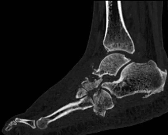
- İnce kesitli BT görüntülerinde kırık, fragmantasyon, dislokasyon ve yapısal deformiteler değerlendirilir; TMT eklemleri de sık tutulan bölgeler arasındadır.
Önerilen pencereler: Kemik (G 1800 / M 400), Yumuşak doku (G 400 / M 40).
BT bulguları
- Çok eklemli periartiküler dağılım — navikulokuneiform ve TMT eklemlerinin karşılıklı yüzlerinde birden fazla kemik etkilenir.
- Subkondral parçalanma — eklem yüzleri boyunca düzensiz çökme, parçalı korteks ve küçük kemik fragmanları görülür.
- TMT subluksasyonu — metatars tabanlarının kuneiform ve küboidlerle eklem ilişkisi bozulur; dorsal veya plantar yer değiştirme eşlik edebilir.
- Eklem içi kemik parçaları — Charcot nöro-osteoartropatisinde eklem çevresindeki kemik fragmantasyonu ve kemik debris’i görülebilir.
- Rocker-bottom konturu — orta ayak çökmesiyle ilişkili bu deformitede ayak tabanı konveks bir kontur kazanır.
- Kronik yeniden şekillenme — eklem çevresinde skleroz, kemik uçlarında yuvarlaklaşma, hipertrofik yeni kemik ve kısmi füzyon gelişebilir.
- Ülserle komşu kemik hasarı — rocker-bottom çıkıntısı üzerindeki plantar ülser doğrudan kemik yüzüne uzanıyorsa süperpoze osteomiyelit araştırılır.
- Aktif Charcot ayağında erken kemik iliği ödemi ve ince subkondral kırıklar BT’de güvenilir biçimde değerlendirilemeyebilir; klinik şüphe sürüyorsa erken kemik iliği ve yumuşak doku değişiklikleri için MR tercih edilir.
Ölçütler ve sınıflamalar
- Modifiye Eichenholtz evrelemesi
- Evre 0, klasik üç evreli Eichenholtz tanımına sonradan eklenmiştir: klinik sıcaklık/şişlik olabilir, radyografide belirgin kemik değişikliği yoktur; MR erken kemik iliği ve yumuşak doku değişikliklerini gösterebilir. Evre I (gelişim/parçalanma): fragmantasyon, subluksasyon ve eklem destrüksiyonu. Evre II (koalesans/birleşme): komşu kemik fragmanları birleşir ve yeni periosteal kemik oluşabilir. Evre III (rekonstrüksiyon/konsolidasyon): kemik yeniden şekillenir ve belirgin rezidüel deformite kalabilir.
Normalde
Görüntülemede çok kemik ve eklemi tutan periartiküler Charcot dağılımını, ülser veya sinüsle ilişkili tek kemik tutulumundan ayırın.
Normal BT ile kıyasla
Aynı bölgenin, kaynak veri setinde patoloji içermediği belirtilen bir BT incelemesini kesit kesit kaydırın; organ sınırlarını açarak anatomiyi hasta görüntüsüyle karşılaştırın. Küçük rastlantısal bulgular tümüyle dışlanmamıştır.
Kaynak: TotalSegmentator v2.01 veri seti (Wasserthal ve ark., Radiology: Artificial Intelligence 2023), CC BY 4.0, vaka s0685; organ sınırları veri setinin otomatik segmentasyonundan, birleştirilerek sadeleştirildi. Görüntüler 2,5 mm kesit aralığına yeniden örneklendi.
Ayırıcı tanı
- Diyabetik ayak osteomiyeliti sıklıkla ülser veya sinüsten komşu kemiğe doğrudan yayılır; Charcot ise çoğunlukla eklem merkezli, periartiküler ve çok kemiklidir. Bu dağılımlar ayırıcı tanıya yardımcı olur ancak kesin ayrım sağlamaz; iki süreç birlikte bulunabilir.
- Lisfranc travmatik kırıklı çıkığı
- belirgin travma öyküsüyle sınırlı kırık ve eklem ayrışması beklenir; yaygın periartiküler yeniden şekillenme ve çok eklemli parçalanma Charcot lehinedir.
- Gut ve diğer enflamatuar eklem hastalıkları klinik olarak Charcot ayağını taklit edebilir; görüntülemede Charcot nöro-osteoartropatisi kemik yıkımı, fragmantasyon ve subluksasyonlarla kendini gösterebilir.
- Nöropatik olmayan osteoartrit ile nöropatik artropati klinik olarak benzer olabilir; nöropatik artropati radyografide hızla ilerleyen osteoartrit benzeri görünümle birlikte kemik yıkımı, subluksasyon, dislokasyon ve kemik debris’i gösterebilir.
Tuzaklar
- Erken Charcot evresinde kemik korteksi henüz parçalanmamış olabilir; normal BT, nöropatik artropatiyi ve özellikle kemik iliği ödemini dışlamaz.
- Charcot ile osteomiyelit aynı ayakta birlikte bulunabilir; plantar ülser veya sinüsün ulaştığı kemiği, eklem merkezli genel dağılımdan ayrı izleyin.
- Yük verilmeyen BT, yük altında belirginleşen deformite ve eklem subluksasyonunu olduğundan az gösterebilir; bulguları yük vererek elde edilen görüntülerle birlikte değerlendirin.
Kendini dene
Nöropatik diyabetli bir hastada TMT eklemleri çevresinde birden fazla kemiği tutan periartiküler fragmantasyon ve subluksasyon görülüyor. En olası tanı hangisidir?
Cevabı göster
Charcot nöro-osteoartropatisi. Charcot nöro-osteoartropatisi eklem çevresinde birden fazla kemik ve eklemi tutabilir; osteomiyelit ise çoğunlukla ülser veya sinüs traktıyla ilişkili tek kemikte belirgindir.
Orta ayak tabanında ülserin hemen altındaki tek kemikte fokal kortikal yıkım ve sekestr var. Hangi tanı Charcot'tan daha olasıdır?
Cevabı göster
Diyabetik osteomiyelit. Ülserle doğrudan ilişkili tek kemik yıkımı ve sekestr kontinüite yoluyla gelişen osteomiyeliti destekler. Charcot tipik olarak eklem merkezli ve çok kemiklidir; osteoartrit osteofit/daralma yapar; bağ yaralanması kemik enfeksiyon bulgusu oluşturmaz.
İlgili konular
Kaynaklar
Bu sayfadaki 40 cümle ve 2 quiz sorusu aşağıdaki kaynaklardan alıntılarla otomatik olarak karşılaştırıldı; 12 cümle bu karşılaştırmada düzeltildi (2026-10-09).
- Burning Issues/Hot Topics: The Charcot Footpmc.ncbi.nlm.nih.gov
- Radiological Assessment of Charcot Neuro-Osteoarthropathy in Diabetic Foot: A Narrative Reviewpmc.ncbi.nlm.nih.gov
- To Assess the Variation in the Definitions Used to Describe the Stages of Acute Charcot Neuro‐Osteoarthropathy (Diagnosis, Remission and Relapse): A Systematic Reviewpmc.ncbi.nlm.nih.gov
- Charcot neuro-osteoarthropathy: a review of key concepts and an evidence-based surgical management algorithmpmc.ncbi.nlm.nih.gov
- The Role of Weight-Bearing Computed Tomography in the Assessment and Management of Charcot Foot Deformity: A Narrative Reviewpmc.ncbi.nlm.nih.gov
- Nonoperative Treatment of Charcot Neuro-osteoarthropathy.pubmed.ncbi.nlm.nih.gov
- Neuro-osteoarthropathy of the foot-radiologist: friend or foe?pubmed.ncbi.nlm.nih.gov
- Radiological Assessment of Charcot Neuro-Osteoarthropathy in Diabetic Foot: A Narrative Review.pubmed.ncbi.nlm.nih.gov
- Diagnostic imaging of the diabetic foot: an EANM evidence-based guidance.pubmed.ncbi.nlm.nih.gov
- The Role of Weight-Bearing Computed Tomography in the Assessment and Management of Charcot Foot Deformity: A Narrative Review.pubmed.ncbi.nlm.nih.gov
- Acute Charcot neuro-osteoarthropathy.pubmed.ncbi.nlm.nih.gov
- F-18 FDG PET/CT scanning in Charcot disease: a brief report.pubmed.ncbi.nlm.nih.gov
- Grading Fractures on Foot and Ankle X-rays and MRI Scans in the Active Charcot Foot in Diabetes: How Strong Is the Agreement Between Modalities?pubmed.ncbi.nlm.nih.gov
- Diagnostic imaging of the diabetic foot.pubmed.ncbi.nlm.nih.gov
- Osteomyelitis or Charcot neuro-osteoarthropathy? Differentiating these disorders in diabetic patients with a foot problem.pubmed.ncbi.nlm.nih.gov
- Charcot Neuro-osteoarthropathy: Critical Concepts and Practical Management Pearls for Dermatologists.pubmed.ncbi.nlm.nih.gov
- Sonographic features of active Charcot neuro-osteoarthropathy: A case series.pubmed.ncbi.nlm.nih.gov
- Denosumab in active Charcot neuro-osteoarthropathy of the foot.pubmed.ncbi.nlm.nih.gov
- A Rare Charcot Neuro-Osteoarthropathy of Hip with an Uncommon Cause.pubmed.ncbi.nlm.nih.gov
- Plantar Pressure and Radiographic Deformity at Diagnosis of Active Charcot Neuro-Osteoarthropathy as Potential Prognostic Markers for Time to Remission.pubmed.ncbi.nlm.nih.gov
- To Assess the Variation in the Definitions Used to Describe the Stages of Acute Charcot Neuro-Osteoarthropathy (Diagnosis, Remission and Relapse): A Systematic Review.pubmed.ncbi.nlm.nih.gov
- Charcot Neuro-Osteoarthropathy in Diabetes: Implications for Diabetic Foot Ulcers, Amputations, and Survival.pubmed.ncbi.nlm.nih.gov
- Charcot foot disease: A new approach.pubmed.ncbi.nlm.nih.gov
- Charcot foot in diabetes and an update on imaging.pubmed.ncbi.nlm.nih.gov
- Decoding Chronic Charcot Arthropathy: Molecular Mechanisms, Predictive Biomarkers, and Emerging Therapies.pubmed.ncbi.nlm.nih.gov
- Burning Issues/Hot Topics: The Charcot Foot.pubmed.ncbi.nlm.nih.gov
- Practical guidelines for the prevention and management of diabetic foot disease in China.pubmed.ncbi.nlm.nih.gov
- Charcot foot reconstruction with combined internal and external fixation: case report.pubmed.ncbi.nlm.nih.gov
- Asymptomatic Charcot foot in longstanding diabetes.pubmed.ncbi.nlm.nih.gov
- Imaging Insights into Midfoot and Forefoot Disorders.pubmed.ncbi.nlm.nih.gov
- Dufourmentel Rhomboid Flap for Plantar Charcot Midfoot Ulcer: A Novel Reconstructive Approach.pubmed.ncbi.nlm.nih.gov
- Imaging and Treatment of Posttraumatic Ankle and Hindfoot Osteoarthritis.pubmed.ncbi.nlm.nih.gov
- Supramalleolar osteotomies for degenerative joint disease of the ankle joint: indication, technique and results.pubmed.ncbi.nlm.nih.gov
- Clinical validity of radiographic parameters for evaluating pediatric foot deformities: correlation with pedobarographic analysis.pubmed.ncbi.nlm.nih.gov
- Subtalar Joint Statistical Shape Modeling Differentiates Cavus-to-Planus Foot Types From Weightbearing CT.pubmed.ncbi.nlm.nih.gov
- Surgical management of the diabetic foot: The current evidence.pubmed.ncbi.nlm.nih.gov
- Ischaemic Charcot Midfoot Reconstruction Combined with Lateral Tibial Cortex Transverse Transport: Case Report.pubmed.ncbi.nlm.nih.gov
- Explainable machine learning for differential diagnosis of diabetic foot infection and osteomyelitis: a two-center study and clinically applicable web calculator using routine blood biomarkers.pubmed.ncbi.nlm.nih.gov
- Magnetic Resonance Imaging and 99Tc WBC-SPECT/CT Scanning in Differential Diagnosis between Osteomyelitis and Charcot Neuroarthropathy: A Case Series.pubmed.ncbi.nlm.nih.gov
- Hallux valgus: Advances in weightbearing CT imaging and three-dimensional assessment of first ray deformity.pubmed.ncbi.nlm.nih.gov
- Three-Dimensional Morphologic Analysis of Foot Deformities Using Weightbearing CT and Semiautomated Segmentation Technique.pubmed.ncbi.nlm.nih.gov
- Imaging of osteoarthritis from the ankle through the midfoot.pubmed.ncbi.nlm.nih.gov
- Off-label use of a PHILOS plate in tibiotalocalcaneal arthrodesis with variable medial cannulated screw support: a retrospective evaluation of 32 ankles.pubmed.ncbi.nlm.nih.gov
- Moth-Eaten Osteolysis of Multiple Foot Bones After a Conservatively Treated "Stable" Ankle Fracture: A Case Report and Literature Review.pubmed.ncbi.nlm.nih.gov
- Vacuum Scaling Drainage in the Treatment of Infected Elbow Charcot Neuroarthropathy: A Case Report and Literature Review.pubmed.ncbi.nlm.nih.gov
- Case Report: Tibial plateau fracture in Charcot neuroarthropathy: a report of two cases with contrasting outcomes.pubmed.ncbi.nlm.nih.gov
- Case Report: Arthroscopic synovectomy and loose body removal for charcot knee in an adolescent with congenital insensitivity to pain with anhidrosis.pubmed.ncbi.nlm.nih.gov
- Hinged total knee arthroplasty in two female patients with type 1 diabetes-associated charcot neuroarthropathy: case reports and a review of the literature series.pubmed.ncbi.nlm.nih.gov
- Effectiveness of Pamidronate Infusion in the Treatment of Charcot Arthropathy.pubmed.ncbi.nlm.nih.gov
- PATHOGENESIS OF ACUTE DIABETIC CHARCOT ARTHROPATHY IN THE FOOT AND ANKLE: A COMPREHENSIVE LITERATURE REVIEW.pubmed.ncbi.nlm.nih.gov
- Musculoskeletal Infection Reporting and Data System (MSKI-RADS): reviewed and explained.pubmed.ncbi.nlm.nih.gov
- Stop Searching under the Streetlight! A Primer and Practical Guide to the Diagnosis of Joint Pain and Inflammation.pubmed.ncbi.nlm.nih.gov
- Imaging of lower extremity infections: predisposing conditions, atypical infections, mimics, and differentiating features.pubmed.ncbi.nlm.nih.gov
- RANKL-RANK-OPG Pathway in Charcot Diabetic Foot: Pathophysiology and Clinical-Therapeutic Implications.pubmed.ncbi.nlm.nih.gov
- HLA-B27-Positive Leprosy Masquerading as Peripheral Spondyloarthritis: A Case Report.pubmed.ncbi.nlm.nih.gov
- Management of hindfoot and ankle in Charcot arthropathy.pubmed.ncbi.nlm.nih.gov
- Congenital Insensitivity to Pain Syndrome With Hidrosis in a 17-Year-Old Female.pubmed.ncbi.nlm.nih.gov
- Non-Infectious Spinal Diseases Mimicking Infection: A Pictorial Essay.pubmed.ncbi.nlm.nih.gov
- When Glycaemic Control Comes Too Fast: Treatment-Induced Neuropathy of Diabetes and Charcot Neuro-Osteoarthropathy as Emerging Iatrogenic Complications of Rapid Metabolic Correction.pubmed.ncbi.nlm.nih.gov
- Pilot randomised controlled trial of probe to bone diabetic foot ulcer therapy (PROBE-DFU trial) - Study protocol.pubmed.ncbi.nlm.nih.gov
- Improving Diabetic Foot Care Through a Multidisciplinary Clinic: Experience From Pilgrim Hospital, UK.pubmed.ncbi.nlm.nih.gov
- Imaging modalities for non-acute pathologies of the foot and ankle.pubmed.ncbi.nlm.nih.gov
- Assessment of bone marrow edema on dual-energy CT scans in people with diabetes mellitus and suspected Charcot neuro-osteoarthropathy.pubmed.ncbi.nlm.nih.gov
- [Diabetic Charcot neuroarthropathy initially misdiagnosed as rheumatoid arthritis and gout: A case report].pubmed.ncbi.nlm.nih.gov
- Charcot arthropathy of the shoulder joint following Arnold-Chiari malformation: A case report.pubmed.ncbi.nlm.nih.gov
- MRI Distribution of Foot Bone Marrow Edema of Presumed Algodystrophic/BMES-Like Origin: A Small Retrospective Case Series with 3-Month Clinical Follow-Up.pubmed.ncbi.nlm.nih.gov
- Diagnostic imaging confusion in infectious spondylitis.pubmed.ncbi.nlm.nih.gov
- Charcot spinal arthropathy: a case series.pubmed.ncbi.nlm.nih.gov
- Application of patient-specific surgical guides in foot and ankle surgery.pubmed.ncbi.nlm.nih.gov
- Controversies in the management of active Charcot neuroarthropathy.pubmed.ncbi.nlm.nih.gov
- Utility of Quantitative and Semi-Quantitative SPECT/CT Metrics in Differentiating Mueller-Weiss Syndrome.pubmed.ncbi.nlm.nih.gov
- Imaging approach to deposition and neurogenic arthropathies.pubmed.ncbi.nlm.nih.gov
- Diabetic foot syndrome: Charcot arthropathy or osteomyelitis? Part I: Clinical picture and radiography.pubmed.ncbi.nlm.nih.gov
- Charcot Neuropathic Arthropathy of the Foot: A Literature Review and Single-Center Experience.pubmed.ncbi.nlm.nih.gov
- Radiographic analysis of diabetic midfoot charcot neuroarthropathy with and without midfoot ulceration.pubmed.ncbi.nlm.nih.gov
- Comparison of Arthrodesis with Total Contact Casting for Midfoot Ulcerations Associated with Charcot Neuroarthropathy.pubmed.ncbi.nlm.nih.gov
- Integrated mechanical and metabolic limb salvage for advanced diabetic Charcot hindfoot collapse with non-traumatic calcaneal fracture: an 8-year follow-up case report.pubmed.ncbi.nlm.nih.gov
- Brazilian Society of Angiology and Vascular Surgery 2023 guidelines on the diabetic foot.pubmed.ncbi.nlm.nih.gov
- The added value of ZTE MRI in the assessment of diabetic foot disease.pubmed.ncbi.nlm.nih.gov
- Guidelines on multidisciplinary approaches for the prevention and management of diabetic foot disease (2020 edition).pubmed.ncbi.nlm.nih.gov
- Conservative and surgical treatment of the chronic Charcot foot and ankle.pubmed.ncbi.nlm.nih.gov
- Postmortem computed tomography and histopathology of distal limb lesions in giraffe (Giraffa spp.) under managed care.pubmed.ncbi.nlm.nih.gov
- Osteotomies: Indications, Imaging Appearance, Surgical Techniques, and Complications.pubmed.ncbi.nlm.nih.gov
- Benign bone and soft tissue tumors of the foot.pubmed.ncbi.nlm.nih.gov
- Foot Osteochondroses.pubmed.ncbi.nlm.nih.gov
- Internal Brace Fixation Technique for Lisfranc Injury: A Retrospective Study.pubmed.ncbi.nlm.nih.gov
- The Missing Navicular: Bilateral Navicular Agenesis Presenting as Adult-acquired Flatfoot with Subtalar Arthritis: A Case Report.pubmed.ncbi.nlm.nih.gov
- 3D-printed navicular replacement for giant cell tumor: a case report on a feasible indication and surgical outcome.pubmed.ncbi.nlm.nih.gov
- Long-Term Outcomes of a Novel Surgical Approach for Early Charcot Neuroarthropathy.pubmed.ncbi.nlm.nih.gov
- Acute hot foot: Charcot neuroarthropathy or osteomyelitis? Untangling a diagnostic web.pubmed.ncbi.nlm.nih.gov
Eğitim amaçlıdır; hasta başında tanı ve tedavi kararının yerine geçmez, tıbbi tavsiye değildir. Metinler yapay zekâ desteğiyle yazıldı ve otomatik bir adımda kaynak alıntılarıyla karşılaştırıldı; bu bir tıbbi doğrulama değildir ve içerik uzman radyolog incelemesinden geçmedi. Klinik kararlarda güncel kılavuzları, kurum protokollerini ve radyoloji raporunu esas alın.