Özet
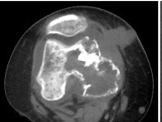
GCTB çoğunlukla 20–40 yaşlarında görülür; diz çevresinde distal femur ve proksimal tibianın metafiz-epifiz bölgesini tutabilir ve subkondral kemiğe uzanabilir. BT, tümör kenarı ile eklem yüzü arasındaki uzaklığı ve posterior kortikal kemik destrüksiyonunu değerlendirmede kullanılabilir. Kemik lezyonlarının ilk görüntüleme değerlendirmesinde radyografi temel yöntemdir; GCTB’nin lokal evrelemesinde radyografiye MR veya BT eklenebilir. Campanacci sınıflaması düz radyografi temellidir; GCTB’de subkondral kemik ilişkisi, kortikal destrüksiyon ve yumuşak doku uzanımı görüntülemede ayrıca değerlendirilir.
Faz ve pencere
- Kontrastsız tanısal
- Epimetafizer bölgede eksantrik, coğrafi litik alan subkondral kemiğe dayanabilir ve metafize uzanır; çoğunlukla belirgin sklerotik kenar veya mineralize matriks bulunmaz. Kortikal incelme, ekspansiyon, korteks dışına taşma ve kırık hattı değerlendirilir.
- Kontrastsız tanısal
- İnce kesitlerin kemik algoritmalı rekonstrüksiyonlarında subkondral plak, incelmiş/ekspansil korteks, kortikal perforasyon, eklem yüzü çökmesi ve patolojik kırık hattı gösterilir.
Önerilen pencereler G genişlik, M merkez; değerler Hounsfield birimi (HU)
- KemikG 1800 / M +400
- Yumuşak dokuG 400 / M +40
BT bulguları
- Eksantrik epimetafizer odak — lezyon distal femur kondili veya proksimal tibia platosunun eklem yüzüne yakın epifiz-metafiz bölgesinde görülür.
- Subkondral uzanım — lezyon eklem yüzüne yakın subkondral kemiğe ulaşabilir.
- Epifiz-metafiz tutulumu — lezyon metafiz ve epifiz bölgesini tutabilir; iskelet olgunluğunda fizis kapalıdır.
- Sklerotik kenarın bulunmayışı — agresif litik sınırın çevresinde belirgin reaktif sklerotik halka genellikle görülmez.
- Kortikal ekspansiyon — kemiğin çevresel konturu şişer, korteks incelir ve yer yer kesintiye uğrar.
- Kortikal aşılma — kemik dışı yumuşak doku komponenti korteks defektinden devam eder.
- Eklem yüzü veya kırık — subkondral çökme, eklem içi uzanım ve patolojik kırık ayrı ayrı aranır.
Ölçütler ve sınıflamalar
- Campanacci radyografik sınıflaması
- Derece 1: iyi sınırlı lezyon ve sağlam korteks. Derece 2: daha geniş intraosseöz lezyon, ince ancak kortikal sürekliliği korunmuş; sklerotik kenar bulunmayabilir. Derece 3: kortikal destrüksiyon ve ekstraosseöz yumuşak dokuya uzanım görülebilir. Sınıflama klasik olarak radyografik görünüme dayanır; BT korteks ve kırığı daha ayrıntılı gösterir.
- WHO tümör sınıflaması (6. baskı, çevrimiçi beta)
- GCTB osteoklastik dev hücreden zengin kemik tümörleri grubunda listelenir. Bu histopatolojik/taksonomik sınıflama, görüntüleme temelli Campanacci derecelendirmesinden ayrıdır.
Normalde
BT’de tümör kenarı ile eklem yüzü arasındaki uzaklığı ve posterior kortikal kemik destrüksiyonunu değerlendirin.
Normal BT ile kıyasla
Aynı bölgenin, kaynak veri setinde patoloji içermediği belirtilen bir BT incelemesini kesit kesit kaydırın; organ sınırlarını açarak anatomiyi hasta görüntüsüyle karşılaştırın. Küçük rastlantısal bulgular tümüyle dışlanmamıştır.
Kaynak: TotalSegmentator v2.01 veri seti (Wasserthal ve ark., Radiology: Artificial Intelligence 2023), CC BY 4.0, vaka s0685; organ sınırları veri setinin otomatik segmentasyonundan, birleştirilerek sadeleştirildi. Görüntüler 2,5 mm kesit aralığına yeniden örneklendi.
Ayırıcı tanı
- Kondroblastom
- açık fizise sahip daha genç hastada epifiz merkezli, ince sklerotik kenarlı ve bazen iç kalsifikasyonlu odak daha olasıdır.
- Anevrizmal kemik kisti
- ekspansil litik lezyon olarak GCTB’yi taklit edebilir; GCTB’de ikincil anevrizmal kemik kisti değişikliğine bağlı sıvı-sıvı düzeyleri görülebilir.
- Brown tümör
- hiperparatiroidi ile ilişkili litik kemik lezyonu GCTB ayırıcı tanısında yer alır.
- Berrak hücreli kondrosarkom
- özellikle 40 yaş üzerindeki hastada epimetafizer litik lezyon olarak GCTB’yi taklit edebilir; matriks mineralizasyonu varsa kıkırdak tümörünü destekler.
- İntraosseöz ganglion
- subkondral kistik lezyon olarak GCTB ayırıcı tanısında yer alır.
Tuzaklar
- Subkondral yerleşimi osteoartritik kist sanmayın; dev hücreli tümör genellikle daha geniş, eksantrik ve epifizden metafize uzanan litik alan oluşturur.
- Kortikal incelmeyi tek başına yumuşak doku invazyonu saymayın; gerçek kortikal perforasyon ve dışarıdaki kitleyi ayrı pencerelerde doğrulayın.
- Anevrizmal kemik kisti benzeri ikincil değişiklik tümörün temel epifiz yerleşimini örtebilir; sıvı seviyeleri için MR ile korelasyon kurun.
- Campanacci derecesini BT bulgusundan otomatik vermeyin; sistemin tanımı radyografik sınırlara dayanır ve görüntüleme modalitesini raporda belirtin.
Kendini dene
Diz çevresindeki dev hücreli kemik tümörü çoğunlukla hangi kemik bölgelerinde yerleşir?
Cevabı göster
Distal femur ve proksimal tibianın metafiz-epifiz bölgesinde. Diz çevresindeki GCTB çoğunlukla distal femur ve proksimal tibianın metafiz-epifiz bölgesinde yerleşir.
İyi sınırlı ancak sklerotik kenarı olmayan lezyonda korteks incelmiş ve orta derecede genişlemiş. Campanacci derecesi hangisidir?
Cevabı göster
Derece 2. Campanacci derece 2 tanımı, görece iyi sınırlı fakat opak kenarı olmayan lezyonda incelmiş ve orta derecede ekspansil kortekstir. Derece 1'de korteks sağlamdır; derece 3'te sınırlar belirsiz ve korteks destrükte olur.
İlgili konular
Kaynaklar
Bu sayfadaki 43 cümle ve 2 quiz sorusu aşağıdaki kaynaklardan alıntılarla otomatik olarak karşılaştırıldı; 20 cümle bu karşılaştırmada düzeltildi (2026-10-09).
- WHO Classification of Tumours: Soft Tissue and Bone Tumours, 6th edition online beta structurewhobluebooks.iarc.who.int
- WHO Classification of Tumours, 6th edition volume titles and online beta statuswhobluebooks.iarc.who.int
- Conventional radiography for the assessment of focal bone lesions of the appendicular skeleton: fundamental concepts in the modern imaging eralink.springer.com
- Giant Cell Tumor of Bone - StatPearlsncbi.nlm.nih.gov
- Giant Cell Tumor of Bone: Review, Mimics, and New Developments in Treatmentpubs.rsna.org
- The Clinical Approach Toward Giant Cell Tumor of Boneacademic.oup.com
- Giant cell tumor of bone revisitedpubmed.ncbi.nlm.nih.gov
- Deep learning for differentiation of osteolytic osteosarcoma and giant cell tumor around the knee joint on radiographs: a multicenter studypmc.ncbi.nlm.nih.gov
- A Tertiary Care Centre Experience of Recurrent Giant Cell Tumor Around the Knee Jointpmc.ncbi.nlm.nih.gov
- The epidemiological and clinical features of primary giant cell tumor around the knee: A report from the multicenter retrospective study in chinapmc.ncbi.nlm.nih.gov
- A novel subchondral bone-grafting procedure for the treatment of giant-cell tumor around the kneepmc.ncbi.nlm.nih.gov
- Bone combined cement grafting in giant cell tumor around the knee reduces mechanical failurepmc.ncbi.nlm.nih.gov
- Giant cell tumor of the rib with direct invasion into the thoracic spine.pubmed.ncbi.nlm.nih.gov
- Giant Cell Tumour of the Talus: A Rare Case Report.pubmed.ncbi.nlm.nih.gov
- [Treatment of giant cell tumor of bone around knee joint with three-dimensional printing personalized prosthesis].pubmed.ncbi.nlm.nih.gov
- Clinical characteristics and prognoses of six patients with multicentric giant cell tumor of the bone.pubmed.ncbi.nlm.nih.gov
- [Effectiveness of unicompartment allografts replacement for bone tumor around the knee].pubmed.ncbi.nlm.nih.gov
- Computerised tomography features of giant cell tumour of the knee are associated with local recurrence after extended curettage.pubmed.ncbi.nlm.nih.gov
- Implanted β -Tricalcium Phosphate Blocks Can Function as a Placeholder in Recurrent Giant Cell Tumor of Bone.pubmed.ncbi.nlm.nih.gov
- MRI and 18 F-FDG PET/CT findings of a giant cell tumor of the tendon sheath of the knee joint (pigmented villonodular synovitis): A case report and literature review.pubmed.ncbi.nlm.nih.gov
- Differentiation of giant cell tumours of bone, primary aneurysmal bone cysts, and aneurysmal bone cysts secondary to giant cell tumour of bone: using whole-tumour CT texture analysis parameters as quantitative biomarkers.pubmed.ncbi.nlm.nih.gov
- A prospective study on predicting local recurrence of giant cell tumour of bone by evaluating preoperative imaging features of the tumour around the knee joint.pubmed.ncbi.nlm.nih.gov
- Functional Outcome of Extended Curettage and Reconstruction using Sandwich Technique for Giant Cell Tumor around Knee.pubmed.ncbi.nlm.nih.gov
- Optimal management of giant cell tumor in the tibial plateau: A case report on curettage and cement reinforcement.pubmed.ncbi.nlm.nih.gov
- Giant Cell Tumor of Bone: Biology, Pathophysiology, and Histopathology in the Era of H3F3A.pubmed.ncbi.nlm.nih.gov
- Functional Outcome and Complications of Wide Excision of the Distal End of Radius and Reconstruction with Ulnar Transposition in Campanacci Grade III Giant Cell Tumor: A Longitudinal Study of 14 Cases in South India.pubmed.ncbi.nlm.nih.gov
- Anaplastic thyroid carcinoma: Clinicopathologic and immunohistochemical study of 144 cases with special emphasis on the spectrum of histologic features.pubmed.ncbi.nlm.nih.gov
- Multicentric metachronous giant cell tumor of the femur: a rare case report and review of the literature.pubmed.ncbi.nlm.nih.gov
- Unnoticed Giant Cell Tumor of the Left Distal Tibia in a 52-Year-Old Male: A Rare Case Report.pubmed.ncbi.nlm.nih.gov
- Diagnostic and Treatment Strategies for Adult Non-Ossifying Fibromas: A Case Report and Literature Review.pubmed.ncbi.nlm.nih.gov
- Proximal tibial tuberculous osteomyelitis with secondary knee joint arthritis: A case report.pubmed.ncbi.nlm.nih.gov
- Efficacy of microwave <i>in situ</i> ablation combined with intralesional curettage for locally aggressive benign bone tumors based on MSTS scores, bone healing time, and recurrence rate.pubmed.ncbi.nlm.nih.gov
- Preoperative denosumab combined with microwave ablation for joint preservation in advanced giant cell tumor of bone: a retrospective study.pubmed.ncbi.nlm.nih.gov
- Giant cell tumor of bone and secondary osteoarthritis.pubmed.ncbi.nlm.nih.gov
- Intraosseous tumours of the knee.pubmed.ncbi.nlm.nih.gov
- Unveiling Primary Bone Tumors of the Spine: A Review of Essential Imaging Clues.pubmed.ncbi.nlm.nih.gov
- Chondroblastoma of the femoral head: curettage without dislocation.pubmed.ncbi.nlm.nih.gov
- Patellofemoral Instability in the Adolescent Age Group.pubmed.ncbi.nlm.nih.gov
- Patellar Dislocation: Workup and Decision-Making.pubmed.ncbi.nlm.nih.gov
- Patellar Tracking: An Old Problem with New Insights.pubmed.ncbi.nlm.nih.gov
- Patient-specific osteoarticular reconstruction of the proximal tibia for giant cell tumor using a 3D-printed trabecular implant and hinged knee arthroplasty: a case report.pubmed.ncbi.nlm.nih.gov
- Case Report: Giant cell reparative granuloma of the humerus and femur-two rare cases with atypical skeletal involvement.pubmed.ncbi.nlm.nih.gov
- Case Report: Phosphaturic mesenchymal tumor of the scapula without calcification: a potential diagnostic pitfall.pubmed.ncbi.nlm.nih.gov
- <sup>18</sup>F-FDG PET/CT findings of intermediate bone tumors of the spine.pubmed.ncbi.nlm.nih.gov
- Trends in primary malignant bone cancer incidence and mortality in the United States, 2000-2017: A population-based study.pubmed.ncbi.nlm.nih.gov
- EACR 2025 Congress: Innovative Cancer Science, 16-19 June 2025.pubmed.ncbi.nlm.nih.gov
- The iliotibial band revisited: surgical anatomy and imaging-based guidelines for lateral tenodesis in ACL reconstruction.pubmed.ncbi.nlm.nih.gov
- Development of a dynamic anatomical model using 3D printing for the study of TPLO surgical technique in small-breed dogs.pubmed.ncbi.nlm.nih.gov
- Update from the 5th Edition of the World Health Organization Classification of Head and Neck Tumors: Odontogenic and Maxillofacial Bone Tumours.pubmed.ncbi.nlm.nih.gov
- Patterns of injury and violence in Yaoundé Cameroon: an analysis of hospital data.pubmed.ncbi.nlm.nih.gov
- Bone lesions of the tibia: Multimodal iconographic review and diagnostic algorithms, Part 2: Metaphyseal and epiphyseal lesions.pubmed.ncbi.nlm.nih.gov
- Glossary of terms for musculoskeletal radiology.pubmed.ncbi.nlm.nih.gov
- Osteoarthritis: Epidemiology, Diagnosis, and Treatment.pubmed.ncbi.nlm.nih.gov
- Hydrogel composite scaffold repairs knee cartilage defects: a systematic review.pubmed.ncbi.nlm.nih.gov
- The Osteoarthritis Natural Progress and Changes in Intraosseous Pressure of the Guinea Pig Model in Different Degeneration Stages.pubmed.ncbi.nlm.nih.gov
- Low-grade osteogenic tumor with secondary aneurysmal bone cyst: Distinguishing osteoblastoma from low-grade osteosarcoma in the setting of noncanonical molecular findings.pubmed.ncbi.nlm.nih.gov
- Excision of proximal fibular aggressive and malignant tumors: a new classification for surgical guidance.pubmed.ncbi.nlm.nih.gov
- Case Report: Direct anterior approach with surgical hip dislocation for management of juvenile femoral head chondroblastoma: a case series and systematic review of the literature.pubmed.ncbi.nlm.nih.gov
- Case Report: Adult proximal humeral aneurysmal bone cyst: radical resection and reconstruction with osteoconductive allograft & reverse arthroplasty-Ecuador's first reported case and functional outcomes.pubmed.ncbi.nlm.nih.gov
- Case Report: Arthroscopic management of a giant Baker cyst containing numerous rice body-like loose bodies in a patient with rheumatoid arthritis.pubmed.ncbi.nlm.nih.gov
- Proximal Tibial Giant Cell Tumor in a Skeletally Immature Child: A Diagnostic Dilemma and Review of the Literature.pubmed.ncbi.nlm.nih.gov
Eğitim amaçlıdır; hasta başında tanı ve tedavi kararının yerine geçmez, tıbbi tavsiye değildir. Metinler yapay zekâ desteğiyle yazıldı ve otomatik bir adımda kaynak alıntılarıyla karşılaştırıldı; bu bir tıbbi doğrulama değildir ve içerik uzman radyolog incelemesinden geçmedi. Klinik kararlarda güncel kılavuzları, kurum protokollerini ve radyoloji raporunu esas alın.