Özet
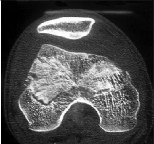
Hoffa kırığı, distal femur kondilinin koronal düzlemde ayrıldığı ve sıklıkla yüksek enerjili diz travması sonucu oluşan eklem içi kırık paternidir. Ön-arka grafilerde fragman diğer kondilin veya patellanın arkasında kalabildiğinden, kemik algoritmalı BT ve sagittal-koronal rekonstrüksiyonlar kırık hattının ve posterior fragmanın görünür olmasını sağlar. BT, medial, lateral ya da iki kondili tutan bileşeni ve eşlik eden metafizer veya interkondiler kırığı tanımlamaya yardım eder. Fragman deplasmanı ve eklem yüzü uyumsuzluğu gözden kaçarsa eklem yaralanmasının gerçek kapsamı eksik kalır; eşlik eden diz çıkığı veya damar bulgusu da ayrıca taranmalıdır.
Faz ve pencere
- Kontrastsız tanısal
- Kondil içindeki koronal kırık hattı, posterior fragmanın ayrılması, rotasyon/deplasman, eklem basamağı ve eşlik eden kırıklar BT ile detaylı olarak izlenir. Hoffa kırığının kemik değerlendirmesi bilgisayarlı tomografi ile yapılır; yumuşak doku veya damar yaralanması şüphesinde manyetik rezonans veya anjiyo ek değerlendirme gerekebilir.
- Kontrastsız tanısal
- Kemik algoritmalı sagittal ve koronal ince kesit rekonstrüksiyonları posterior kondil parçasının büyüklük ve konumunu, kondil yüzeyindeki basamağı ve medial, lateral veya bikondiler yayılımı gösterir.
- BTA tanısal
- Diz çıkığı, nabız/perfüzyon anormalliği veya popliteal arter yaralanması şüphesi varsa arter lümeninde kesinti, tromboz, daralma, psödoanevrizma ya da aktif ekstravazasyon araştırılır.
Önerilen pencereler: Kemik (G 1800 / M 400), Yumuşak doku (G 400 / M 40), Anjiyo (G 600 / M 150).
BT bulguları
- Koronal kondil yarığı — kırık düzlemi femoral kondili ön ve posterior parçaya ayırır; ön-arka ve lateral radyografilerde kemik örtüşmesi nedeniyle silik kalabilir.
- Posterior kondil fragmanı — femoral kondilin koronal düzlemde ayrışmış posterior parçasıdır; sagittal ve koronal MPR ile kırık hattı, deplasman ve komminüsyon net görülür.
- Eklem yüzü uyumsuzluğu — femoral kondilin yük taşıyan bölümünde seviye farkı, aralık veya çökme aranır.
- Kondil tarafı ve yayılımı — lateral, medial veya iki taraflı oluşu; karşı kondil ve interkondiler çentikle ilişkisi belirtilir.
- Parçalanma veya impaksiyon — posterior fragmanın içindeki küçük parçalar ve subkondral sıkışma eklem yüzü rekonstrüksiyonunu değiştirir.
- Eşlik eden kırık — suprakondiler metafiz, interkondiler çentik veya femur cismi boyunca devam eden hat ayrıca izlenir.
- Çevre hematomu ve damar bulguları — popliteal bölgede hematom veya popliteal arter diseksiyonu gibi damar yaralanmaları şüphesi varsa damar değerlendirmesi gereklidir.
Ölçütler ve sınıflamalar
- AO/OTA 2018 Compendium: distal femur 33B3.2 / 33B3.3
- 33B3, parsiyel eklem içi posterior koronal kondil (Hoffa) kırığıdır; 33B3.2 tek kondil, 33B3.3 bikondiler tutulumu gösterir.
- Letenneur
- Tip I tüm kondili tutar ve kırık hattı posterior femoral kortekse paraleldir; Tip II posteriorda daha ileridir ve tamamen eklem içi (ostekondral) olabilir; Tip III oblik hattıyla karakterizedir ve kırık hattı kapsül ve bağ yapışma yerleriyle ilişkilendirilir.
Normalde
Sağlam karşı kondil ile posterior kontur ve eklem yüzü kıyaslaması yapılabilir; ancak Hoffa kırığı değerlendirmesinde bilgisayarlı tomografi ile kırık hattı, fragman boyutu, deplasman ve komminüsyonun doğrudan ölçülmesi esastır.
Normal BT ile kıyasla
Aynı bölgenin, kaynak veri setinde patoloji içermediği belirtilen bir BT incelemesini kesit kesit kaydırın; organ sınırlarını açarak anatomiyi hasta görüntüsüyle karşılaştırın. Küçük rastlantısal bulgular tümüyle dışlanmamıştır.
Kaynak: TotalSegmentator v2.01 veri seti (Wasserthal ve ark., Radiology: Artificial Intelligence 2023), CC BY 4.0, vaka s0685; organ sınırları veri setinin otomatik segmentasyonundan, birleştirilerek sadeleştirildi. Görüntüler 2,5 mm kesit aralığına yeniden örneklendi.
Ayırıcı tanı
- Sagittal kondil kırığı
- ayrılma medial-lateral yöndedir ve koronal planda kondilin arka parçasını izole eden hat görülmez.
- Tibial plato posterior shear kırığı
- fragman femurda değil tibianın posterior eklem dudağındadır.
- Osteokondral fragmanlar genellikle sınırlı boyutta olup, koronal düzlemde kondili tüm uzunluğunda ikiye bölen karakteristik ayrılma hattı yerine, subkondral bölgede lokalize hasar olarak izlenir.
- İnterkondiler distal femur kırığı
- ayrılma çentik merkezinden ilerler; izole posterior kondil parçası olmayabilir.
- Epikondiler avulsiyon
- küçük kemik parçası ligament yapışma yerindedir; kondilin artiküler bölümünde koronal ayrılma görülmez.
Tuzaklar
- Ön-arka grafide görünmeyen kırığı yok saymayın; posterior fragman örtüşebilir ve kontrastsız BT'de sagittal rekonstrüksiyon gerekir.
- Kırık hattının devamlılığı tek bir düzlemde değil, koronal ve aksiyel rekonstrüksiyonlarla birlikte çoklu düzlemde doğrulanmalıdır; tek eksendeki görünüm yanıltıcı olabilir.
- Lateral kondile odaklanıp medial veya bikondiler uzanımı atlamayın; her iki posterior kondili ayrı ayrı takip edin.
- Posterior fragman komşuluğundaki hematomu otomatik olarak damar yaralanması kabul etmeyin; klinik şüphe varsa BTA'da arter lümenini inceleyin.
Kendini dene
Diz travmasında sagittal BT'de posterior lateral kondil, kondil gövdesinden koronal bir hatla ayrılmış. En olası tanı nedir?
Cevabı göster
Lateral Hoffa kırığı. Hoffa kırığı, distal femoral kondilin eklem yüzünü koronal düzlemde tutan bir kırık paternidir. Posterior koronal shear kırığı tibial platoda yer alırken, Hoffa kırığı distal femoral kondili tutar.
BT'de femoral kondil korteksi intakt; posterior tibial plato kenarında öne doğru yer değiştirmiş kemik parçası var. Hangi yaralanma düşünülür?
Cevabı göster
Posterior tibial shear kırığı. Fragmanın tibial plato posterior kenarından (posterior lip) kaynaklanması posterior tibial shear kırığını gösterir. Hoffa kırığı femoral kondil içinde koronal ayrılma oluşturur; femoral impaksiyonda kondil korteksi çöker ve patella kırığı patellada izlenir.
Kaynaklar
Bu sayfadaki 42 cümle ve 2 quiz sorusu aşağıdaki kaynaklardan alıntılarla tek tek karşılaştırıldı; 15 cümle bu karşılaştırmada düzeltildi (2026-10-08).
- Medial Hoffa Fracture: A Case Report and Literature Review of Approach and Managementpmc.ncbi.nlm.nih.gov
- Pediatric cartilaginous lateral femoral condyle Hoffa fracture: a case report and review of the literaturepmc.ncbi.nlm.nih.gov
- A case report of the Hoffa fracture and a review of literaturepmc.ncbi.nlm.nih.gov
- Surgical Treatment and Rehabilitation for Hoffa Fracture Nonunion: Two Case Reports and a Literature Reviewpmc.ncbi.nlm.nih.gov
- Busch-Hoffa fracture: A systematic reviewpmc.ncbi.nlm.nih.gov
- How to determine the surgical approach in Hoffa fractures?pubmed.ncbi.nlm.nih.gov
- Coronal plane partial articular fractures of the distal femoral condyle: current concepts in management.pubmed.ncbi.nlm.nih.gov
- Hoffa fracture of the femoral condyle: Injury mechanism, classification, diagnosis, and treatment.pubmed.ncbi.nlm.nih.gov
- Use of rapid prototyping and three-dimensional reconstruction modeling in the management of complex fractures.pubmed.ncbi.nlm.nih.gov
- Coronal fractures of the medial femoral condyle: a series of 6 cases and review of literature.pubmed.ncbi.nlm.nih.gov
- Non -Union of Lateral Hoffa Fracture - A Case Report.pubmed.ncbi.nlm.nih.gov
- Conjoint bicondylar Hoffa fracture in an adult.pubmed.ncbi.nlm.nih.gov
- [Research progress of Hoffa fracture].pubmed.ncbi.nlm.nih.gov
- Irreducible, incarcerated vertical dislocation of patella into a Hoffa fracture.pubmed.ncbi.nlm.nih.gov
- [Medial Hoffa fracture: a case report].pubmed.ncbi.nlm.nih.gov
- Hoffa's Fracture with Associated Injuries Around the Knee Joint: An Approach to a Rare Injury.pubmed.ncbi.nlm.nih.gov
- Surgical treatment of lateral Hoffa fracture with a locking plate through the lateral approach.pubmed.ncbi.nlm.nih.gov
- Hoffa fracture accompanied by dissection of the popliteal artery.pubmed.ncbi.nlm.nih.gov
- Hoffa Fracture of the Femoral Condyle.pubmed.ncbi.nlm.nih.gov
- Surgically treated Hoffa Fractures with poor long-term functional results.pubmed.ncbi.nlm.nih.gov
- A rare traumatic triad: Open bicondylar non-conjoint Hoffa fracture with partial tibial tubercle and medial epicondyle avulsions - A case report and biomechanical analysis.pubmed.ncbi.nlm.nih.gov
- Fixed Flexion Deformity due to Displaced Hoffa Fracture: A Case Report.pubmed.ncbi.nlm.nih.gov
- Medial Hoffa's Fracture Treated with Subvastus Approach - Case Series.pubmed.ncbi.nlm.nih.gov
- Multimodality imaging considerations for genicular artery embolization in knee osteoarthritis.pubmed.ncbi.nlm.nih.gov
- Hoffa fracture combined with rotational dislocation of the knee joint: A novel case report.pubmed.ncbi.nlm.nih.gov
- Irreducible Posterolateral Fracture Dislocation of the Knee in an Elderly Patient Treated with Rotating Hinge Total Knee Replacement.pubmed.ncbi.nlm.nih.gov
- Greater medial arterial supply revealed by 7-Tesla quantitative magnetic resonance imaging, histology and high-resolution computed tomography of the patellar tendon.pubmed.ncbi.nlm.nih.gov
- Secondary Displacement was Common in Healing Distal Femur Fractures in a Cohort of Elderly Patients.pubmed.ncbi.nlm.nih.gov
- The design of a sample rapid magnetic resonance imaging (MRI) acquisition protocol supporting assessment of multiple articular tissues and pathologies in knee osteoarthritis.pubmed.ncbi.nlm.nih.gov
- Fat Chance for Hidden Lesions: Pictorial Review of Hoffa's Fat Pad Lesions.pubmed.ncbi.nlm.nih.gov
- Calcified or ossified benign soft tissue lesions that may simulate malignancy.pubmed.ncbi.nlm.nih.gov
- Current Concepts in the Treatment of Distal Femur Fractures in Adults.pubmed.ncbi.nlm.nih.gov
- Surgical treatment of femoral medial condyle impaction fracture complicated with Hoffa fragment by contralateral medial distal tibia plate: a case report.pubmed.ncbi.nlm.nih.gov
- Case Report: Bicondylar conjoined Hoffa fracture with incarcerated patella.pubmed.ncbi.nlm.nih.gov
- Fracture mapping of coronal shear fractures of the distal humerus: morphological analysis and fixation efficacy.pubmed.ncbi.nlm.nih.gov
- Comparison of Gerdy's tubercle osteotomy versus conventional anterolateral approach for posterolateral tibial plateau fractures: a retrospective cohort study.pubmed.ncbi.nlm.nih.gov
- Preliminary retrospective analysis of combined retrograde tibial intramedullary nail and lateral less invasive stabilization system for distal femoral fractures.pubmed.ncbi.nlm.nih.gov
- Multiple fractures of the femur: Case report, literature review, and proposal for a shared algorithm of treatment.pubmed.ncbi.nlm.nih.gov
- Comparison of combined femoral nail and plate fixation versus dual plate fixation in the treatment of AO/OTA 33C distal femoral fractures.pubmed.ncbi.nlm.nih.gov
- Trochleoplasty for patellofemoral instability: current concepts.pubmed.ncbi.nlm.nih.gov
- Case Report: Three-dimensional planning for autologous osteoperiosteal transplantation in the treatment of a massive osteochondral defect of the medial femoral condyle.pubmed.ncbi.nlm.nih.gov
- Patellar Instability: A Narrative Review with an Integrated Treatment Algorithm.pubmed.ncbi.nlm.nih.gov
- Complexities in Hoffa Fracture Management: A Case Report on Diagnosis and Fixation Technique.pubmed.ncbi.nlm.nih.gov
- Treatment of displaced sagittal plane femoral condyle fractures (OTA 33 B1-2) with novel J-plate: technical trick and case series.pubmed.ncbi.nlm.nih.gov
- Bilateral Hoffa Fractures of the Medial Femoral Condyles: A Case Report and Review of the Literature.pubmed.ncbi.nlm.nih.gov
- Biomechanical study using the finite element analysis of a new method for fixation of coronal fracture of the femoral condyle.pubmed.ncbi.nlm.nih.gov
- Optimizing fixation in medial distal femur fractures: A biomechanical study.pubmed.ncbi.nlm.nih.gov
- The Role of Immediate Arthroplasty in Elderly Tibial Plateau Fractures.pubmed.ncbi.nlm.nih.gov
- High prevalence of generalized ligamentous laxity in patellar dislocation with posterior weight-bearing lateral femoral condyle osteochondral fractures: an observational study and treatment outcomes.pubmed.ncbi.nlm.nih.gov
- A Rare Hoffa Fracture with Sagittal Split and Intra-articular Comminution: A Case Report and Literature Review.pubmed.ncbi.nlm.nih.gov
- Three-dimensional fracture mapping and analysis of coronal fractures in AO/OTA types 33-B3 and C3.pubmed.ncbi.nlm.nih.gov
- The role of radiography and MRI for eligibility assessment in DMOAD trials of knee OA.pubmed.ncbi.nlm.nih.gov
- Pediatric lateral Busch-Hoffa fracture associated with lateral meniscus tear and incarcerated popliteus tendon: systematic review of literature and case report.pubmed.ncbi.nlm.nih.gov
- Novel CT-based classification of Hoffa fracture and optimal treatment strategies.pubmed.ncbi.nlm.nih.gov
- Introduction and An Analysis of Inter- and Intra-observer Validity to the Classification of Hoffa-Like Tibial Plateau Fractures.pubmed.ncbi.nlm.nih.gov
- The role of radiography and MRI as screening tools and outcome measures in clinical trials of knee osteoarthritis: a perspective on the current status and outlook.pubmed.ncbi.nlm.nih.gov
- Multifaceted imaging strategies for clinical trials of knee osteoarthritis-a tightly interlinked value and precision chain.pubmed.ncbi.nlm.nih.gov
- A structured evaluation of the symptomatic medial Oxford unicompartmental knee arthroplasty (UKA).pubmed.ncbi.nlm.nih.gov
- Outcomes of Surgical Management of Late Presenting Intra-Articular Distal Femoral Physeal Fracture: A Multicentric Retrospective Case Series.pubmed.ncbi.nlm.nih.gov
- Outcomes of Surgical Management of Late Presenting Intra-Articular Distal Femoral Physeal Fracture: A Multicentric Retrospective Case Series.pubmed.ncbi.nlm.nih.gov
- Megaprosthetic Total Knee Arthroplasty for Supracondylar Refracture in a Post-traumatic Osteoarthritic Knee: A Case Report.pubmed.ncbi.nlm.nih.gov
- Simulated gastrocnemius traction alters interfragmentary motion in Hoffa fracture fixation.pubmed.ncbi.nlm.nih.gov
- Robot-assisted versus conventional unicompartmental knee arthroplasty: a retrospective comparative study of postoperative complications.pubmed.ncbi.nlm.nih.gov
- Digging into the Cause of Abnormal Patellar Kinematics After Open-Wedge High Tibial Osteotomy via a Quantitative Study on In Vivo Soft Tissue Functional Changes.pubmed.ncbi.nlm.nih.gov
- Hoffa fractures are associated with concomitant soft tissue injures and a high postoperative complication rate.pubmed.ncbi.nlm.nih.gov
- Computer assisted orthopaedic surgery -- CAOS.pubmed.ncbi.nlm.nih.gov
- Progress of fracture mapping technology based on CT three-dimensional reconstruction.pubmed.ncbi.nlm.nih.gov
- A case report of the Hoffa fracture and a review of literature.pubmed.ncbi.nlm.nih.gov
- Management and Outcomes of Osteochondritis Dissecans of the Knee in the Pediatric and Adolescent Population.pubmed.ncbi.nlm.nih.gov
- A Multimodal Diagnostic Algorithm for Focal Knee Chondral Defects: Correlating Clinical Tests, Musculoskeletal Ultrasound, and MRI-Based ICRS Grading.pubmed.ncbi.nlm.nih.gov
- Osteochondritis Dissecans of the Elbow in Overhead Athletes: A Comprehensive Narrative Review.pubmed.ncbi.nlm.nih.gov
- Interpretation of Cartilage Damage at Routine Clinical MRI: How to Match Arthroscopic Findings.pubmed.ncbi.nlm.nih.gov
- The arrow and the target-description of the "Robin Hood" technique for fixation of tibial spine avulsion fractures: a technical note.pubmed.ncbi.nlm.nih.gov
- Tibial plateau depression and widening as predictors of meniscal and ligamentous injuries: a systematic review and meta-analysis.pubmed.ncbi.nlm.nih.gov
- Holding the line: predictors of coronal and sagittal loss of reduction after tibial plateau fracture fixation.pubmed.ncbi.nlm.nih.gov
- Multimodal positron emission tomography (PET) imaging in non-oncologic musculoskeletal radiology.pubmed.ncbi.nlm.nih.gov
- Fracture morphological characteristics and associated soft-tissue injuries in flexion varus tibial plateau fractures.pubmed.ncbi.nlm.nih.gov
- SPECT/CT and triple-phase bone scan: A valuable diagnostic approach for identifying indications for secondary patellar resurfacing in patients with unexplained anterior knee pain post-TKA.pubmed.ncbi.nlm.nih.gov
- Toward the Application of Dual-Energy Computed Tomography with Virtual Non-Hydroxyapatite Color-Coded Maps to Identify Traumatic Fractures in Daily Emergency Settings.pubmed.ncbi.nlm.nih.gov
- Isolated Double Hoffa Fracture of the Distal Femur without Intercondylar Extension: A Rare Case Report.pubmed.ncbi.nlm.nih.gov
- Rim plate fixation for a rare fracture pattern of the lateral femoral condyle: A case report.pubmed.ncbi.nlm.nih.gov
- Comparison of three fixation methods for posterolateral fragments in tibial plateau fractures: a biomechanical study.pubmed.ncbi.nlm.nih.gov
- Medial Tibial Plateau Fractures and Fracture-Dislocations: A Mechanism-Based Narrative Review of Instability Patterns, Imaging, and Contemporary Management.pubmed.ncbi.nlm.nih.gov
- Effects of axial malrotation on posterior tibial slope measurement: a digitally reconstructed radiograph study enabling automated quality assessment.pubmed.ncbi.nlm.nih.gov
- Plate-augmented fixation of comminuted Letenneur type II Hoffa fractures provides favorable stability compared to isolated posteroanterior screw fixation - a biomechanical study.pubmed.ncbi.nlm.nih.gov
- Osteotomies: Indications, Imaging Appearance, Surgical Techniques, and Complications.pubmed.ncbi.nlm.nih.gov
- Surgical Management of Complex Multiligament Knee Injury: Case Report.pubmed.ncbi.nlm.nih.gov
- Comparison of Ultrasonographic, Radiographic, Computed Tomographic, and Ex Vivo Measurements of Feline Femoral Trochlear Groove Depth.pubmed.ncbi.nlm.nih.gov
- Fascia Lata and Tensor Fasciae Latae in Lower-Limb Reconstruction.pubmed.ncbi.nlm.nih.gov
- Sex-Specific Adolescent Development of the Trochlea and Distal Femoral Physis: A 3D Shape Modeling Study.pubmed.ncbi.nlm.nih.gov
- The iliotibial band revisited: surgical anatomy and imaging-based guidelines for lateral tenodesis in ACL reconstruction.pubmed.ncbi.nlm.nih.gov
- Abnormal mechanical stress due to excessive femoral torsion is associated with dysplasia of the distal femoral epiphyseal plate and trochlea.pubmed.ncbi.nlm.nih.gov
- Comparison of the Frosch approach and the supra-fibular head approach in the treatment of posterolateral tibial plateau fractures combined with lateral column fractures.pubmed.ncbi.nlm.nih.gov
Eğitim amaçlıdır; hasta başında tanı ve tedavi kararının yerine geçmez. İçerik yapay zekâ destekli bir süreçle güncel literatüre göre cümle cümle kaynaklarla karşılaştırıldı, ancak uzman radyolog incelemesinden geçmedi. Klinik kararlarda güncel kılavuzları, kurum protokollerini ve radyoloji raporunu esas alın.