Özet
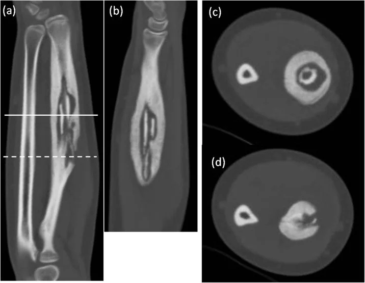
Kronik osteomiyelit, süregelen kemik enfeksiyonunun nekrotik kemik, reaktif yeni kemik ve zaman zaman cilde açılan sinüs yolu oluşturduğu tablodur. BT, kortikal mimariyi ve yoğun sekestrumu MR'dan daha ayrıntılı göstererek cerrahi değerlendirmeye ve hastalığın anatomik haritalanmasına katkı sağlar. Kronik osteomiyelitte BT sekestrumun gösterilmesinde önemlidir; MR erken osteomiyeliti, kemik iliği değişikliklerini ve yumuşak doku yayılımını değerlendirmede, radyografi ise kemik lezyonlarının başlangıç değerlendirmesinde tamamlayıcıdır. Sekestrum ve sinüs traktı varlığı, enfeksiyonun kemik yapısı içindeki kapsamını ve dışa yayılımını gösterir.
Faz ve pencere
- Kontrastsız tanısal
- BT'de kronik osteomiyelitte sekestrum ve kemik değişiklikleri değerlendirilir.
- İV kontrast (seçilmiş olguda) tanısal
- Kronik osteomiyelitte BT, sekestrumun gösterilmesinde önemlidir. Kemik iliği değişikliklerinin ve yumuşak doku yayılımının değerlendirilmesinde MR tamamlayıcıdır.
Önerilen pencereler G genişlik, M merkez; değerler Hounsfield birimi (HU)
- KemikG 1800 / M +400
- Yumuşak dokuG 400 / M +40
BT bulguları
- Sekestrum, kronik enfeksiyona bağlı nekrotik ve sklerotik kemik parçası olup, çevresinde inflamatuar reaksiyon veya sıvı nedeniyle düşük dansiteli alan izlenebilir.
- Kronik osteomiyelitte reaktif kemik trabekülleri izlenebilir.
- Sinüs traktı — enfekte kemik odaktan dış yumuşak doku veya cilt yüzeyine uzanan kanal.
- Sinüs yolu — kloakadan cilt yüzeyine uzanan yumuşak doku traktı; kontrastsız BT’de de görülebilir. İV kontrast çevresel inflamasyon/granülasyon dokusunu ve varsa koleksiyonu belirginleştirebilir; traktın uzanımını değerlendirmede MR daha duyarlıdır.
- Kortikal kalınlaşma ve skleroz — enfekte segment çevresinde gelişen reaktif yeni kemik oluşumu ve medüller alanda fibröz doku birikimi ile karakterize kronik yeniden yapılanma bulgularıdır.
- İntraosseöz kavite — sklerotik sınırla çevrili düzensiz lüsent alan, kronik enfeksiyon odağını veya sekestrum yatağını gösterebilir.
- Yumuşak doku apsesi — kontrastlanan duvarla çevrili sıvı dansitesi cloaca veya sinüs yoluyla kemik odağına bağlanabilir.
Normalde
Kronik osteomiyelit olgularda normal kemik yapısı bozulmuş, sklerotik değişiklikler ve periosteal reaksiyonlar mevcuttur. Enfekte bölgede sklerotik sekestrum, periosteal reaksiyon ve sinüs traktı varlığı araştırılmalıdır.
Normal BT ile kıyasla
Aynı bölgenin, kaynak veri setinde patoloji içermediği belirtilen bir BT incelemesini kesit kesit kaydırın; organ sınırlarını açarak anatomiyi hasta görüntüsüyle karşılaştırın. Küçük rastlantısal bulgular tümüyle dışlanmamıştır.
Kaynak: TotalSegmentator v2.01 veri seti (Wasserthal ve ark., Radiology: Artificial Intelligence 2023), CC BY 4.0, vaka s0685; organ sınırları veri setinin otomatik segmentasyonundan, birleştirilerek sadeleştirildi. Görüntüler 2,5 mm kesit aralığına yeniden örneklendi.
Ayırıcı tanı
- Brodie apsesi
- subakut osteomiyelit formudur; radyografide sınırlı radyolüsens lezyon ve çevresel skleroz ile kortikal hipertrofi görülür; içerde sekestrum bulunabilir.
- Osteoid osteoma
- küçük nidus çevresinde belirgin reaktif skleroz gelişir; merkezde ayrılmış nekrotik kortikal parça ve sinüs yolu beklenmez.
- Fibro-osseöz ve sklerozan lezyonlar
- genişleyici, non-destruktif kütleler veya yerel skleroz oluşturabilir; malignite şüphesinde kortikal yıkım ve yumuşak doku kitlesi ayırıcıdır.
- İyileşen kırık
- kallus oluşumu ve kemik birleşmesi ile karakterizedir; enfeksiyona özgü sekestrum veya sinüs traktı içermez.
- Nöropatik eklem hastalıkları
- eklem çevresinde parçalanma ve subluksasyon ile karakterizedir; osteomiyelitte görülen sekestrum ve medüller tutulum farklıdır.
Tuzaklar
- Sekestrumu normal kortikal ada veya kalın trabekül sanma; iki düzlemde ana kemikle bağlantısının kesildiğini ve çevresindeki lusent aralığı doğrula.
- Reaktif periosteal kemik oluşumunu basit bir reaksiyon olarak değillendirmek önemlidir; kronik enfeksiyolda kalın sklerotik kemik kılıfı ve sinüs traktı lezyonun kapsamını gösterir.
- Sinüs traktı ile travmatik kırık hattını ayırt etmek gerekir; sinüs traktı enfekte kemikten yumuşak doku yönüne uzanırken, kırık hattı travmatik kökenlidir ve kallusla ilişkilidir.
- Sinüs traktının varlığı, enfekte kemikten cilt yüzeyine uzanan yumuşak doku değişimi ve klinik drenaj bulguları ile desteklenmelidir.
Kendini dene
Kronik osteomiyelitte BT hangi bulgunun saptanmasında önemlidir?
Cevabı göster
Sekestrum. BT, kronik osteomiyelitte sekestrumun saptanmasında önemlidir.
BT’de sklerotik kenarlı, iyi sınırlı radyolüsent bir kemik lezyonu izleniyor. En olası taklitçi hangisi?
Cevabı göster
Osteoid osteoma. Sklerotik kenarlı, iyi sınırlı radyolüsent lezyon osteoid osteoma ile uyumlu olabilir. Kronik osteomiyelitte sekestrum ve sinüs traktları gelişebilir.
İlgili konular
Kaynaklar
Bu sayfadaki 37 cümle ve 2 quiz sorusu aşağıdaki kaynaklardan alıntılarla otomatik olarak karşılaştırıldı; 26 cümle bu karşılaştırmada düzeltildi (2026-10-09).
- Educational Case: Unraveling chronic recurrent multifocal osteomyelitis: An autoinflammatory bone disorderpmc.ncbi.nlm.nih.gov
- Role of three-phase bone scintigraphy and fluorodeoxyglucose positron emission tomography in monitoring hyperbaric oxygen therapy combined with conservative surgery for refractory osteomyelitispmc.ncbi.nlm.nih.gov
- A novel mouse calvarial defect model of osteomyelitis induced by Staphylococcus aureus (ATCC 6538)pmc.ncbi.nlm.nih.gov
- Primary tuberculous osteomyelitis of the maxilla mimicking a refractory post-endodontic infection: A diagnostic challengepmc.ncbi.nlm.nih.gov
- Combined application of antibiotic bone cement and bone transport technique in tibial osteomyelitis with bone defectpmc.ncbi.nlm.nih.gov
- [Osteomyelitis in adults].pubmed.ncbi.nlm.nih.gov
- Cone beam CT of the musculoskeletal system: clinical applications.pubmed.ncbi.nlm.nih.gov
- Floating labyrinth: A unique finding on CT scan.pubmed.ncbi.nlm.nih.gov
- Labyrinthine sequestrum: four case studies.pubmed.ncbi.nlm.nih.gov
- Chronic suppurative osteomyelitis of mandible: a case report.pubmed.ncbi.nlm.nih.gov
- Extensive Sequestration Chronic Maxillary Osteomyelitis in an Uncontrolled Diabetic Patient: Comprehensive Case Management of a Rare Entity.pubmed.ncbi.nlm.nih.gov
- [Comparative study of different concentrations of methicillin-resistant Staphylococcus aureus in the preparation of chronic femoral osteomyelitis models].pubmed.ncbi.nlm.nih.gov
- Mandible extensive intra osseous destructive lesion, a diagnostic challenge.pubmed.ncbi.nlm.nih.gov
- Treatment for Brodie's abscess of the radius in an adolescent: A case report.pubmed.ncbi.nlm.nih.gov
- A complication following tooth extraction: chronic suppurative osteomyelitis.pubmed.ncbi.nlm.nih.gov
- Foreign Body Induced Osteomyelitis in the Hand - Commonly Missed Clinical and Radiological Diagnosis.pubmed.ncbi.nlm.nih.gov
- Levofloxacin loaded mesoporous silica microspheres/nano-hydroxyapatite/polyurethane composite scaffold for the treatment of chronic osteomyelitis with bone defects.pubmed.ncbi.nlm.nih.gov
- Exfoliation and simultaneous formation of condylar process following chronic osteomyelitis of the mandible.pubmed.ncbi.nlm.nih.gov
- A Study of 29 Pediatric Patients With Osteomyelitis Resulting From Neuropathy and Pressure Effects.pubmed.ncbi.nlm.nih.gov
- A multiscale pathoanatomical atlas guides the design of functionally graded, anatomically intelligent implants for chronic osteomyelitis.pubmed.ncbi.nlm.nih.gov
- Effectiveness of Antibiotic Cement-Coated Rods in the Management of Chronic Post-traumatic Osteomyelitis.pubmed.ncbi.nlm.nih.gov
- How Clinical and Radiological Findings in Chronic Mandibular Osteomyelitis Do Not Always Correlate: Diagnostic Dilemmas in Dental-Related Bone Inflammations.pubmed.ncbi.nlm.nih.gov
- Advanced Maxillary Squamous Cell Carcinoma With Extension to the Infratemporal Fossa Masquerading as Extensive Osteomyelitis: A Case Report.pubmed.ncbi.nlm.nih.gov
- Anatomical and Systemic Risk Factors for Recurrence in Medication-Related Osteonecrosis of the Jaw (MRONJ): A Retrospective Study of 812 Patients.pubmed.ncbi.nlm.nih.gov
- Lower extremity infections: Essential anatomy and multimodality imaging findings.pubmed.ncbi.nlm.nih.gov
- Odontogenic Abscesses in Pet Rabbits: A Comprehensive Review of Pathogenesis, Diagnosis, and Treatment Advances.pubmed.ncbi.nlm.nih.gov
- Malignant Transformation of Musculoskeletal Lesions with Imaging-Pathology Correlation-Part 1: Bone Lesions.pubmed.ncbi.nlm.nih.gov
- Musculoskeletal infections through direct inoculation.pubmed.ncbi.nlm.nih.gov
- MRI nomenclature for musculoskeletal infection.pubmed.ncbi.nlm.nih.gov
- Staged antibiotic-loaded cement-based reconstruction versus flap-based reconstruction for complex diabetic foot defects in patients with osteoporosis: a retrospective cohort study.pubmed.ncbi.nlm.nih.gov
- Study of the extent of surgical osteotomy in Cierny-Mader stages III and IV chronic osteomyelitis.pubmed.ncbi.nlm.nih.gov
- Osteomyelitis of the Jaw Bones and Its Mimics: Resolving the Diagnostic Enigma.pubmed.ncbi.nlm.nih.gov
- Musculoskeletal Infection Reporting and Data System (MSKI-RADS): reviewed and explained.pubmed.ncbi.nlm.nih.gov
- The imaging of osteomyelitis.pubmed.ncbi.nlm.nih.gov
- When Ear Pain Signals More: Otomastoiditis-Related Bilateral Cranial Neuropathies in an Immunocompromised Patient.pubmed.ncbi.nlm.nih.gov
- Imaging of acute musculoskeletal infections in children and their differential diagnoses.pubmed.ncbi.nlm.nih.gov
- Unveiling Primary Bone Tumors of the Spine: A Review of Essential Imaging Clues.pubmed.ncbi.nlm.nih.gov
- Review of imaging modalities and radiological findings of calvarial lesions.pubmed.ncbi.nlm.nih.gov
- Diagnostic imaging confusion in infectious spondylitis.pubmed.ncbi.nlm.nih.gov
- Intraosseous tumours of the knee.pubmed.ncbi.nlm.nih.gov
- Spinal lesions: a comprehensive radiologic overview.pubmed.ncbi.nlm.nih.gov
- Musculoskeletal Infection: The Great Mimickers on Imaging.pubmed.ncbi.nlm.nih.gov
- Fibrous dysplasia of the jaws: four cases with conservative and resective surgical management.pubmed.ncbi.nlm.nih.gov
- Osteohistological analysis of metatarsals reveals new information on pathology and life history of troodontids from the Campanian Dinosaur Park Formation, Alberta, Canada.pubmed.ncbi.nlm.nih.gov
- Bizarre Parosteal Osteochondromatous Proliferation (Nora Lesion): A Narrative Review.pubmed.ncbi.nlm.nih.gov
- Current updates in MSK infection imaging: A narrative review.pubmed.ncbi.nlm.nih.gov
- Osteoid Osteoma of Talus - A Rare Occurrence Mimicking Ankle Arthritis: A Case Report.pubmed.ncbi.nlm.nih.gov
- Subacute knee pain and swelling in a healthy male: a case of Brodie's abscess.pubmed.ncbi.nlm.nih.gov
- Glossary of terms for musculoskeletal radiology.pubmed.ncbi.nlm.nih.gov
- The quiet flame: whole-body MRI and the underestimated burden of pediatric chronic non-bacterial osteomyelitis (CNO).pubmed.ncbi.nlm.nih.gov
- Benign and malignant lesions of the mandible and maxilla: imaging findings and systematic approach.pubmed.ncbi.nlm.nih.gov
- Magnetic resonance imaging of musculoskeletal infections.pubmed.ncbi.nlm.nih.gov
- Melorheostosis of The Leg: A Case Report.pubmed.ncbi.nlm.nih.gov
- Spontaneous bacteremia and spontaneous bacterial peritonitis share similar prognosis in patients with cirrhosis: a cohort study.pubmed.ncbi.nlm.nih.gov
- An integrated clinical and imaging model for predicting post-traumatic nonunion.pubmed.ncbi.nlm.nih.gov
- Advancements in biomaterials and bioactive solutions for lumbar spine fusion cages: Current trends and future perspectives.pubmed.ncbi.nlm.nih.gov
- Long-term safety and effectiveness of the "OptEase" vena cava filter.pubmed.ncbi.nlm.nih.gov
- Multimodal imaging in neuro-ophthalmology.pubmed.ncbi.nlm.nih.gov
- Ultrasound in Women's Health: Mechanisms, Applications, and Emerging Opportunities.pubmed.ncbi.nlm.nih.gov
- MRI of total hip arthroplasty: technical aspects and imaging findings.pubmed.ncbi.nlm.nih.gov
- Candida Femoral Osteomyelitis in a Young Adult Without Known Risk Factors for Immunosuppression: A Case Report.pubmed.ncbi.nlm.nih.gov
- The epidemiology and management of chronic osteomyelitis in pediatrics - A systematic review.pubmed.ncbi.nlm.nih.gov
- Madura foot: Atypical MRI presentation beyond the classic dot-in-circle sign.pubmed.ncbi.nlm.nih.gov
- Osteoid osteoma of the triquetrum: A rare cause of ulnar-sided wrist pain in an adolescent female.pubmed.ncbi.nlm.nih.gov
- Beyond Chordoma: A Comprehensive Review of Sacral Lesions.pubmed.ncbi.nlm.nih.gov
- Osteoid osteoma of the thumb: A case report.pubmed.ncbi.nlm.nih.gov
- Role of three-phase bone scintigraphy and fluorodeoxyglucose positron emission tomography in monitoring hyperbaric oxygen therapy combined with conservative surgery for refractory osteomyelitis.pubmed.ncbi.nlm.nih.gov
- Salmonella Typhi Osteomyelitis of the Ulnar Diaphysis in an Immunocompetent Adult: A Case Report.pubmed.ncbi.nlm.nih.gov
- Purely osteolytic bone lesions in the early osteoarticular phase of synovitis, acne, pustulosis, hyperostosis and osteitis syndrome.pubmed.ncbi.nlm.nih.gov
- Acute sacral osteomyelitis mimicking hip pathology in a child: A diagnostic challenge.pubmed.ncbi.nlm.nih.gov
- MRI Findings of Acute on Chronic Osteomyelitis of Tibia in a 12-Year-Old Child.pubmed.ncbi.nlm.nih.gov
- Primary Tuberculous Osteomyelitis of the Proximal Tibia Presenting as Chronic Osteomyelitis: A Diagnostic Challenge in Musculoskeletal Tuberculosis.pubmed.ncbi.nlm.nih.gov
- Chronic Osteomyelitis or Squamous Cell Carcinoma of the Mandible: A Case Report With Diagnostic Challenges.pubmed.ncbi.nlm.nih.gov
- Skeletal coccidioidomycosis: an atypical pediatric presentation.pubmed.ncbi.nlm.nih.gov
- Extensive Temporal Bone Necrosis in Petrous Bone Cholesteatoma Complicated by Actinomycosis: A Case Report.pubmed.ncbi.nlm.nih.gov
- Brodie's Abscess in a Pediatric Patient: A Diagnostic Pitfall.pubmed.ncbi.nlm.nih.gov
- Conventional radiography for the assessment of focal bone lesions of the appendicular skeleton: fundamental concepts in the modern imaging era.pubmed.ncbi.nlm.nih.gov
- Antibiotic-Loaded PMMA Beads for Recurrent Sternocutaneous Fistula: Expanding the Surgical Armamentarium in Post-Sternotomy Osteomyelitis: Case Report and Literature Review.pubmed.ncbi.nlm.nih.gov
- Managing Bone Infections Beyond Systemic Antibiotics: A Scoping Review.pubmed.ncbi.nlm.nih.gov
- Spatiotemporal mapping of osteomyelitis pathogenesis guides the design of functionally graded bone regeneration scaffolds.pubmed.ncbi.nlm.nih.gov
Eğitim amaçlıdır; hasta başında tanı ve tedavi kararının yerine geçmez, tıbbi tavsiye değildir. Metinler yapay zekâ desteğiyle yazıldı ve otomatik bir adımda kaynak alıntılarıyla karşılaştırıldı; bu bir tıbbi doğrulama değildir ve içerik uzman radyolog incelemesinden geçmedi. Klinik kararlarda güncel kılavuzları, kurum protokollerini ve radyoloji raporunu esas alın.