Özet
Stres kırığı, normal kemiğe tekrarlayan aşırı yük binmesiyle veya zayıflamış kemiğin olağan yük altında yetmezliğe uğramasıyla gelişir; sporcu, osteoporozlu yaşlı ve pelvis radyoterapisi öyküsü olan hastada farklı dağılımlar görülebilir. Kontrastsız ince kesit BT kortikal kırık çizgisini, sklerozu, kallusu ve sakral kırığın foramene uzanımını ve spinopelvik devamlılığa etkisini gösterir; erken kemik iliği stres yanıtında MR daha duyarlıdır. Sakrumda iki alar çizgiye bağlanan transvers bileşen, koronal ve sagittal rekonstrüksiyonlarda aranmalıdır. Hattın gözden kaçması tamamlanmamış femur boyun kırığının ilerlemesine veya sakral deplasmanın ve sinir foramenine uzanımın eksik bildirilmesine neden olabilir.
Faz ve pencere
- Kontrastsız tanısal
- Kontrast madde gerekmez; hedef kemikte ince kortikal radyolusen (lusent) çizgi, kortikal kalınlaşma, çevresel skleroz ve iyileşen kırık kallusu değerlendirilir. Sakrumda alar skleroz, kırık çizgisi seçilmese de eşlik edebilir. Aynı kontrastsız serinin aksiyel, koronal ve sagittal kemik algoritmalı rekonstrüksiyonlarında sakral alar çizgiler, olası transvers bileşen, femur boynu hattı ve kallus izlenir.
Önerilen pencereler: Kemik (G 1800 / M 400).
BT bulguları
- Kortikal kırık çizgisi — yük taşıyan kortekse ulaşan ince çizgisel kesinti; yönünü uzun kemik eksenine ve eklem yüzüne göre tanımlayın.
- Periostal kallus — özellikle tibia veya metatars korteksinde çizgiye komşu düzgün kortikal kalınlaşma, tekrarlayan yüklenmeye bağlı iyileşmeyi destekler.
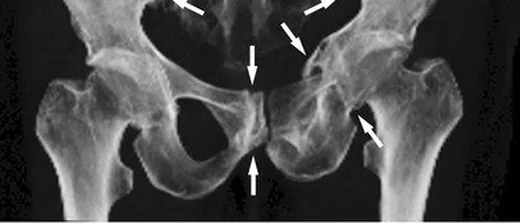
- Sakral alar skleroz — sakroiliak ekleme paralel, yamalı yoğunluk artışı; sakrum yetmezlik kırığında çizgiye eşlik edebilir veya tek görünür bulgu olabilir.
- Sakral vertikal kırık — alar kemikte sagittal yönelimli çizgi, aksiyel kesitlerde izlenir; iki tarafı ve foraminal uzanımı ayrı ayrı kontrol edin.
- Transvers sakral bileşen — koronal veya sagittal planda vertikal alar çizgileri birleştiren yatay kırık; H/U biçimli konfigürasyon oluşturabilir.
- Eklem yüzüne uzanım — femur boynu, tibia platosu veya metatars kırığında çizginin eklem yüzüne ulaşıp ulaşmadığını gösterin.
- Deplasman ve kemik yapısı — kortikal basamak veya açılanma; sakrumda deplasman, açılanma ve eşlik eden kemik yapısal değişiklikler aranır.
Normalde
Normal sakrumda alar kemik trabekülleri iki tarafta benzer yoğunluktadır; sakroiliak ekleme paralel sklerotik bant, kortikal çizgi veya transvers bağlantı bulunmaz. Normal kemikte korteks bütündür; ancak normal kemiğe tekrarlayan (döngüsel) anormal yüklenme lokal kortikal rezorpsiyona ve kırığa yol açabilir.
Normal BT ile kıyasla
Aynı bölgenin, kaynak veri setinde patoloji içermediği belirtilen bir BT incelemesini kesit kesit kaydırın; organ sınırlarını açarak anatomiyi hasta görüntüsüyle karşılaştırın. Küçük rastlantısal bulgular tümüyle dışlanmamıştır.
Kaynak: TotalSegmentator v2.01 veri seti (Wasserthal ve ark., Radiology: Artificial Intelligence 2023), CC BY 4.0, vaka s1256; organ sınırları veri setinin otomatik segmentasyonundan, birleştirilerek sadeleştirildi. Görüntüler 2,5 mm kesit aralığına yeniden örneklendi.
Ayırıcı tanı
- Sakral metastaz
- fokal medüller litik/sklerotik destrüksiyon, kortikal yıkım veya yumuşak doku kitlesi oluşturabilir; kırıkta çizgisel hat ve çevresel skleroz daha belirgindir.
- Enfeksiyon ve primer kemik tümörü gibi diğer durumlardan ayırt edilmelidir.
- Osteomiyelit
- kortikal erozyon, düzensiz periost reaksiyonu ve yumuşak doku koleksiyonu varlığı enfeksiyonu destekler; mekanik yük eksenine uyan düzenli kırık çizgisi beklenmez.
- Osteonekroz
- eklem altı segmentte serpijinöz sınır ve olası subkondral çökme yapar; uzun kemik diafizinde kortekse dik stres hattı gibi görünmez.
- Sakral yetmezlik kırığında alar skleroz, foraminel uzanım ve transvers bileşen değerlendirilir; malign metastazdan ayırt edilir.
Tuzaklar
- Sakrumdaki transvers bağlantı yalnız koronal veya sagittal planda belirginleşebilir; iki vertikal alar hat görüldüğünde diğer düzlemlerde bağlantıyı özellikle arayın.
- BT'de kırık çizgisinin görülmemesi yetmezlik kırığını dışlamaz; yalnız yamalı alar skleroz varsa ve klinik şüphe sürüyorsa MR ile erken kemik iliği yanıtını araştırın.
- Sakral alar sklerozu metastaz kabul etmeden önce kortikal destrüksiyon, yumuşak doku kitlesi ve çizgisel kırık hattını karşılıklı değerlendirin.
- Femur boynu stres kırığında çizginin eklem yüzü ve boyun boyunca uzanımı önemlidir; tek aksiyel görüntüyle sınırlayıp tamamlanmamış hattı atlamayın.
Kendini dene
Osteoporozlu hastanın pelvis BT'sinde iki sakral alada vertikal çizgi ve S2 düzeyinde bunları birleştiren hat var. En olası patern nedir?
Cevabı göster
H tipi sakral yetmezlik kırığı. Bilateral alar kırıkların transvers bileşenle birleşmesi H/U biçimli sakral yetmezlik paternini oluşturur. Metastazda fokal destrüksiyon veya yumuşak doku kitlesi beklenir; sakroiliit eklem yüzlerini etkiler; koksigeal çıkık sakrum içindeki alar çizgileri açıklamaz.
Koşucu tibia BT'sinde kortikal kalınlaşma içinde uzun eksene dik ince çizgi görülüyor; yumuşak doku kitlesi yok. En olası tanı nedir?
Cevabı göster
Stres kırığı. Tekrarlayan yüklenme, kortikal çizgi ve eşlik eden düzgün periostal kalınlaşma stres kırığıyla uyumludur. Stres kırığı tanısı koyarken, enfeksiyon, neoplazma ve patolojik kırıklar klinik ve görüntüleme özellikleriyle birlikte değerlendirilerek dışlanmalıdır.
İlgili konular
Kaynaklar
Bu sayfadaki 38 cümle ve 2 quiz sorusu aşağıdaki kaynaklardan alıntılarla tek tek karşılaştırıldı; 10 cümle bu karşılaştırmada düzeltildi (2026-10-08).
- Sequential stress fractures of the ulna and radius in a patient with neurofibromatosis type 1: A case reportpmc.ncbi.nlm.nih.gov
- Risk Factors for Sacral Insufficiency Fracture After Pelvic Irradiation for Cervical Cancerpmc.ncbi.nlm.nih.gov
- Concurrent Acute Fracture and Dysplasia‐Related Fragmentation of the Medial Coronoid Process of the Ulna in a Dog—A Case Reportpmc.ncbi.nlm.nih.gov
- Complex posterolateral rotatory instability due to malunited radius fracture: diagnosis with four-dimensional computed tomography scan and corrective osteotomypmc.ncbi.nlm.nih.gov
- Bilateral L3-L4 pedicular insufficiency fractures in a patient with breast cancer and treated spinal metastasis: A diagnostic pitfallpmc.ncbi.nlm.nih.gov
- [Stress fractures].pubmed.ncbi.nlm.nih.gov
- Nonspinal Fragility Fractures.pubmed.ncbi.nlm.nih.gov
- Stress fractures: pathophysiology, clinical presentation, imaging features, and treatment options.pubmed.ncbi.nlm.nih.gov
- Stress fractures of the foot - current evidence on management.pubmed.ncbi.nlm.nih.gov
- [Cartilage repair of the knee joint].pubmed.ncbi.nlm.nih.gov
- Sacral Insufficiency Fractures.pubmed.ncbi.nlm.nih.gov
- Chronic Anterior Pelvic Instability: Diagnosis and Management.pubmed.ncbi.nlm.nih.gov
- [Special aspects of stress fractures].pubmed.ncbi.nlm.nih.gov
- Tumour-induced osteomalacia: a literature review and a case report.pubmed.ncbi.nlm.nih.gov
- [Radiologic diagnosis of pathologic fractures].pubmed.ncbi.nlm.nih.gov
- Imaging of insufficiency fractures.pubmed.ncbi.nlm.nih.gov
- Red flags to screen for vertebral fracture in people presenting with low back pain.pubmed.ncbi.nlm.nih.gov
- The pathophysiology, diagnosis, and management of stress fractures in postmenopausal women.pubmed.ncbi.nlm.nih.gov
- Sacral and Pelvic Insufficiency Fractures Following Adult Spinal Deformity Surgery: A Case Report and Systematic Literature Review.pubmed.ncbi.nlm.nih.gov
- Atypical Femoral Fracture Secondary to Long-Term Bisphosphonate Use: A Case Report.pubmed.ncbi.nlm.nih.gov
- Chronic pelvic insufficiency fractures and their treatment.pubmed.ncbi.nlm.nih.gov
- Physical Factors Associated With Stage Progression of Lumbar Spondylolysis: A Prospective Cohort Study in Male Adolescent Soccer Players.pubmed.ncbi.nlm.nih.gov
- Beyond Chordoma: A Comprehensive Review of Sacral Lesions.pubmed.ncbi.nlm.nih.gov
- Pelvic insufficiency fractures in cervical cancer patients treated with definitive chemoradiation: impact of dosimetric and clinical factors.pubmed.ncbi.nlm.nih.gov
- CT of Sacral Fractures: Classification Systems and Management.pubmed.ncbi.nlm.nih.gov
- Lumbar spine MRI versus non-lumbar imaging modalities in the diagnosis of sacral insufficiency fracture: a retrospective observational study.pubmed.ncbi.nlm.nih.gov
- Neural Basis of Etiopathogenesis and Treatment of Cervicogenic Orofacial Pain.pubmed.ncbi.nlm.nih.gov
- Fragility Fractures of the Pelvis-Current Understanding and Open Questions.pubmed.ncbi.nlm.nih.gov
- A Guide to Improving the Care of Patients with Fragility Fractures, Edition 2.pubmed.ncbi.nlm.nih.gov
- International Society of Paediatric Surgical Oncology (IPSO) Surgical Practice Guidelines.pubmed.ncbi.nlm.nih.gov
- Glossary of terms for musculoskeletal radiology.pubmed.ncbi.nlm.nih.gov
- Risk Factors for Sacral Insufficiency Fracture After Pelvic Irradiation for Cervical Cancer.pubmed.ncbi.nlm.nih.gov
- Clinical and 18F-FDG PET/CT Imaging Characteristics of Post-radiotherapy Sacral Insufficiency Fractures in Cervical Cancer Patients.pubmed.ncbi.nlm.nih.gov
- Imaging of Sacroiliac Pain: The Current State-of-the-Art.pubmed.ncbi.nlm.nih.gov
- Atraumatic anterior hip dislocation after cephalomedullary nailing for an intertrochanteric femoral fracture: a case report highlighting the importance of initial reduction and prevention of excessive telescoping.pubmed.ncbi.nlm.nih.gov
- Musculoskeletal Ultrasound and Protein-Based Hydrogels: Novel Approaches for the Diagnosis and Treatment of Sports Injuries.pubmed.ncbi.nlm.nih.gov
- Structural characteristics, biomechanics and clinical significance of calcar femorale: A review.pubmed.ncbi.nlm.nih.gov
- Incidence, Anatomic Distribution, and Return to Training After Stress Fractures in Okinawan Athletes.pubmed.ncbi.nlm.nih.gov
- A Plea for a Paradigm Shift from X-Ray to Ultrasound in Adults: An Update for Emergency Physicians, General Practitioners, Orthopedists and Sports Medicine Physicians.pubmed.ncbi.nlm.nih.gov
- Republication of "Stress Fractures of the Foot and Ankle in Athletes".pubmed.ncbi.nlm.nih.gov
- Musculoskeletal pitfalls and pseudotumours in the pelvis: a pictorial review for body imagers.pubmed.ncbi.nlm.nih.gov
- High-affinity alphavbeta3 integrin targeted optical probe as a new imaging biomarker for early atherosclerosis: initial studies in Watanabe rabbits.pubmed.ncbi.nlm.nih.gov
- L5-S1 Anatomic Features Relevant to Minimally Invasive Decompression and Fusion: A Cadaveric and Imaging-Based Study.pubmed.ncbi.nlm.nih.gov
- Essential knowledge and technical tips for total mesorectal excision and related procedures for rectal cancer.pubmed.ncbi.nlm.nih.gov
- Consensus Guidelines for the Use of Peripheral Nerve Stimulation in the Treatment of Chronic Pain and Neurological Diseases: A Neuron Project from the American Society of Pain and Neuroscience.pubmed.ncbi.nlm.nih.gov
- A Systematic Approach to the Evaluation of Acute Musculoskeletal Injuries in the Emergency Department.pubmed.ncbi.nlm.nih.gov
- A novel approach to distal femur: a minimally invasive technique for supracondylar and intercondylar fracture.pubmed.ncbi.nlm.nih.gov
- Ultrasound and bone: a pictorial review.pubmed.ncbi.nlm.nih.gov
- Anatomy of the intraosseous arterial microvasculature of the adult lumbar vertebral body and its role in fracture healing: a literature review and historical perspective.pubmed.ncbi.nlm.nih.gov
- Vertebral Augmentation Involving Vertebroplasty or Kyphoplasty for Cancer-Related Vertebral Compression Fractures: A Systematic Review.pubmed.ncbi.nlm.nih.gov
- Long-term safety and effectiveness of the "OptEase" vena cava filter.pubmed.ncbi.nlm.nih.gov
- Stress fracture of the pelvis and lower limbs including atypical femoral fractures-a review.pubmed.ncbi.nlm.nih.gov
- Diagnostic Musculoskeletal Ultrasound in the Evaluation of the Plantar Fascia.pubmed.ncbi.nlm.nih.gov
- Innovative photobiomodulation-enhanced T-PRF in posterior mandibular fractures: a randomized pilot study.pubmed.ncbi.nlm.nih.gov
- Research progress and clinical application of all-inside reconstruction techniques: narrative review.pubmed.ncbi.nlm.nih.gov
- Expert consensus on a multidisciplinary approach for the management of multiple myeloma-related bone disease.pubmed.ncbi.nlm.nih.gov
- Minimally Invasive Interventional Procedures for Metastatic Bone Disease: A Comprehensive Review.pubmed.ncbi.nlm.nih.gov
- Zero Echo Time MRI for Osseous Assessment of Sports-Related Pathology in Athletes: A Pictorial Essay.pubmed.ncbi.nlm.nih.gov
- Intramedullary osteosclerosis of the tibia with inflammatory features: Multimodality imaging findings.pubmed.ncbi.nlm.nih.gov
- Preclinical evaluation of patient-specific 3D-printed PDLLA/SrHAp implants in a minipig segmental mandibular defect model.pubmed.ncbi.nlm.nih.gov
- Axial Spondyloarthritis: Mimics and Pitfalls of Imaging Assessment.pubmed.ncbi.nlm.nih.gov
- Sacral Insufficiency Fractures : How to Classify?pubmed.ncbi.nlm.nih.gov
- Optimizing diagnosis and management of ONFH, FNSF, and SIF.pubmed.ncbi.nlm.nih.gov
- Multiple Occult Fractures at Hemodialysis Initiation in a Chronic Kidney Disease Patient: Diagnostic Value of MRI and Potential Role of Zinc Deficiency.pubmed.ncbi.nlm.nih.gov
- Deep learning and hybrid architectures for atypical and complex bone fracture diagnosis: a systematic review of performance and clinical validity.pubmed.ncbi.nlm.nih.gov
- A case of pelvic Ewing sarcoma misinterpreted as osteomyelitis: diagnostic and clinical implications.pubmed.ncbi.nlm.nih.gov
- Research Progress on the Structure and Properties of Medical Bone Implants Based on Additive Manufacturing.pubmed.ncbi.nlm.nih.gov
- Fascia Lata and Tensor Fasciae Latae in Lower-Limb Reconstruction.pubmed.ncbi.nlm.nih.gov
- Time-Dependent Anchor Hole Expansion May Associate with Meniscal Extrusion After Open-Wedge High Tibial Osteotomy Combined with Medial Meniscus Posterior Root Tear Repair and Meniscal Centralization.pubmed.ncbi.nlm.nih.gov
- Chronic Low Back Pain: History, Symptoms, Pain Mechanisms, and Treatment.pubmed.ncbi.nlm.nih.gov
- Guidelines on multidisciplinary approaches for the prevention and management of diabetic foot disease (2020 edition).pubmed.ncbi.nlm.nih.gov
- Five-year clinical outcomes with endoscopic transforaminal outside-in foraminoplasty techniques for symptomatic degenerative conditions of the lumbar spine.pubmed.ncbi.nlm.nih.gov
- Bilateral abducens nerve palsy following ligamentous C1-C2 distraction.pubmed.ncbi.nlm.nih.gov
- Knowledge gaps in high-energy sacral fracture treatment and management.pubmed.ncbi.nlm.nih.gov
- Sacral and Pelvic Fractures: Historical Systems and Advancements with the AO Spine Classification.pubmed.ncbi.nlm.nih.gov
- Top 15 musculoskeletal lesions in the aging recreational sporter: a pictorial review.pubmed.ncbi.nlm.nih.gov
- Ultrasound-Diagnosed Tibia Stress Fracture: A Case Report.pubmed.ncbi.nlm.nih.gov
- Intraosseous tumours of the knee.pubmed.ncbi.nlm.nih.gov
- Conventional radiography for the assessment of focal bone lesions of the appendicular skeleton: fundamental concepts in the modern imaging era.pubmed.ncbi.nlm.nih.gov
- Sequential stress fractures of the ulna and radius in a patient with neurofibromatosis type 1: A case report.pubmed.ncbi.nlm.nih.gov
- Bilateral L3-L4 pedicular insufficiency fractures in a patient with breast cancer and treated spinal metastasis: A diagnostic pitfall.pubmed.ncbi.nlm.nih.gov
- When it is not sacroiliitis.pubmed.ncbi.nlm.nih.gov
- Spinal lesions: a comprehensive radiologic overview.pubmed.ncbi.nlm.nih.gov
- Retinal biological age correlates with bone mineral density and fracture risk score and predicts incident osteoporosis.pubmed.ncbi.nlm.nih.gov
- Fracture Risk in Chronic Kidney Disease: Addressing an Overlooked Complication.pubmed.ncbi.nlm.nih.gov
- Fragility Fracture: 10 Commandments.pubmed.ncbi.nlm.nih.gov
- Abusive spinal injury: imaging and updates.pubmed.ncbi.nlm.nih.gov
- Analysis of Fracture Patterns and Characteristics in Sacral Insufficiency Fracture: Do Sacral Fractures Occur in Patients Who Had Previous Lumbosacral Fusion Insufficiency Fractures or Stress Fractures?pubmed.ncbi.nlm.nih.gov
- Unraveling stress fractures: from cellular and molecular mechanisms to therapeutic approaches.pubmed.ncbi.nlm.nih.gov
- Mechano-immune interactions in musculoskeletal aging: Mechanisms and translational perspectives.pubmed.ncbi.nlm.nih.gov
- From nerve to bone: a tale of autonomic nervous system, bone cells, and beyond.pubmed.ncbi.nlm.nih.gov
- Fracture healing: from molecular and cellular mechanisms to therapeutic strategies.pubmed.ncbi.nlm.nih.gov
- Effects of hypochlorous acid, povidone-iodine, and Carrel-Dakin solution irrigation on open fracture healing: a rat femur model.pubmed.ncbi.nlm.nih.gov
- Osteomyelitis of the Jaw Bones and Its Mimics: Resolving the Diagnostic Enigma.pubmed.ncbi.nlm.nih.gov
- Maxillofacial tubercular osteomyelitis: diagnostic and therapeutic perspectives from a case series.pubmed.ncbi.nlm.nih.gov
- Cone-Beam Computed Tomography Imaging of Primary Non-Hodgkin Lymphoma of the Jaws: A 6-Case Series.pubmed.ncbi.nlm.nih.gov
- The added value of ZTE MRI in the assessment of diabetic foot disease.pubmed.ncbi.nlm.nih.gov
Eğitim amaçlıdır; hasta başında tanı ve tedavi kararının yerine geçmez. İçerik yapay zekâ destekli bir süreçle güncel literatüre göre cümle cümle kaynaklarla karşılaştırıldı, ancak uzman radyolog incelemesinden geçmedi. Klinik kararlarda güncel kılavuzları, kurum protokollerini ve radyoloji raporunu esas alın.