Özet
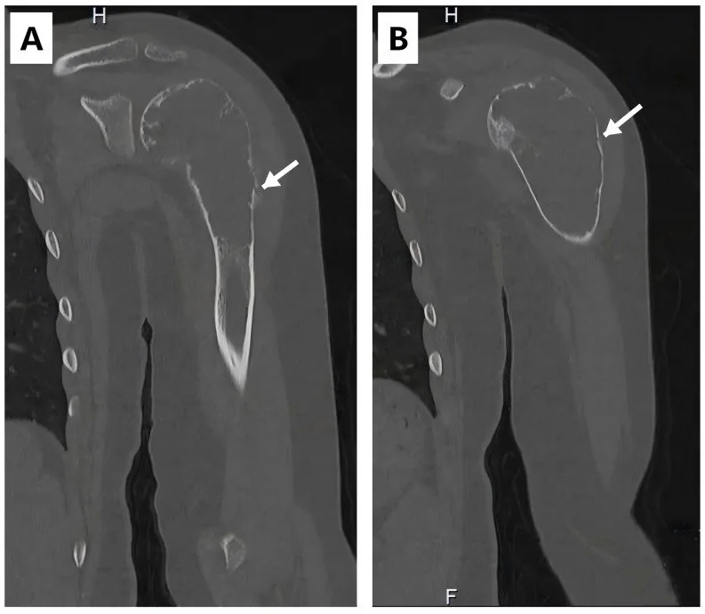
Patolojik kırık, yeterli bir travma olmaksızın fokal benign veya malign bir kemik lezyonu nedeniyle oluşan kırıktır; düşük enerjili ya da belirgin travma olmadan gelişmesi altta yatan lezyon olasılığını düşündürür. BT kemik mimarisini, matriks özelliklerini ve korteksi değerlendirmeye yardımcı olur; yumuşak doku uzanımı ve kompartman ilişkisi için MR kullanılabilir. BT kemik anatomisini, MR ise komşu kompartmanlara yumuşak doku uzanımını değerlendirmeye yardımcı olur. Yeterli travma olmadan gelişen kırıkta miyelom ve kemik metastazları gibi altta yatan lezyonları ayırıcı tanıda değerlendirin; BT korteksi ayrıntılı gösterir ve biyopsi kılavuzluğunda kullanılabilir.
Faz ve pencere
- Kontrastsız tanısal
- BT, kemik mimarisini ve matriks özelliklerini değerlendirmeye yardımcı olur; görüntülemede periostal reaksiyonun tipi ve matriks mineralizasyonu ayırıcı tanıyı daraltabilir. BT kortikal tabakayı ayrıntılı gösterir ve kemik lezyonunun değerlendirilmesine katkı sağlar.
- Kemik algoritması tanısal
- İnce aksiyel-koronal-sagittal kemik rekonstrüksiyonlarında kortikal kopuş, parçalanma, lezyonun kemik eksenindeki uzunluğu ve eklem yüzüne uzanım izlenir; yumuşak doku kitlesi kemik penceresiyle sınırlı kalmadan ayrıca değerlendirilir.
Önerilen pencereler: Kemik (G 1800 / M 400), Yumuşak doku (G 400 / M 40).
BT bulguları
- Kırık hattının lezyon içinden geçmesi — çizgiyi çok düzlemli izleyin; kırık, medüller odağın veya korteksin zayıfladığı noktada başlar.
- Litik kemik odağı — medüller trabekül kaybı ve coğrafi ya da permeatif destrüksiyon; geçiş zonu ve kortikal penetrasyon lezyonun saldırganlığını gösterir.
- Kortikal incelme veya yıkım — kırık hattının bulunduğu segmentte tek korteksin balonlaşması, incelmesi ya da kesintiye uğraması mekanik zayıflama odağını açıklar.
- Matriks mineralizasyonu — kondroid halka-yay, osteoid bulutlanması veya kaba fibröz görünüm lezyon tipini daraltabilir.
- Periostal reaksiyonun tipi, kemik lezyonlarının ayırıcı tanısını daraltmaya yardımcı olabilir.
- Ekstraosseöz yumuşak doku kitlesi — kortikal defektten dışarı uzanan solid komponent özellikle primer malign kemik tümöründe önemlidir.
- Eklem ve kompartman uzanımı — kırığın subkondral kemiğe/eklem yüzüne, komşu ekleme veya nörovasküler yapıya yaklaşımını gösterin.
- Çoklu kemik lezyonları — diğer kesitlerde benzer litik veya sklerotik odaklar metastaz ya da plazma hücreli hastalık olasılığını artırır.
Normalde
Normal aynı segmentte medüller trabeküller süreklidir, korteks düzgün kalınlıkta ve kesintisizdir; periost altında kitle ya da fokal kemik yıkımı bulunmaz. Kırık tarafını karşı tarafın aynı kemiği ve aynı anatomik seviyesiyle kıyaslayarak kortikal kalınlık, medüller yoğunluk, kemik konturu ve eklem yüzü sürekliliğine bakın.
Normal BT ile kıyasla
Aynı bölgenin, kaynak veri setinde patoloji içermediği belirtilen bir BT incelemesini kesit kesit kaydırın; organ sınırlarını açarak anatomiyi hasta görüntüsüyle karşılaştırın. Küçük rastlantısal bulgular tümüyle dışlanmamıştır.
Kaynak: TotalSegmentator v2.01 veri seti (Wasserthal ve ark., Radiology: Artificial Intelligence 2023), CC BY 4.0, vaka s0685; organ sınırları veri setinin otomatik segmentasyonundan, birleştirilerek sadeleştirildi. Görüntüler 2,5 mm kesit aralığına yeniden örneklendi.
Ayırıcı tanı
- Stres/yetmezlik kırığı
- yük eksenine uyan ince çizgi ve düzgün periostal kallus vardır; kırığı çevreleyen medüller tümör odağı veya ekstraosseöz kitle bulunmaz.
- Osteoporotik travmatik kırık
- yaygın trabeküler azalma zemininde kırık oluşur, fakat kırık dışında fokal litik/sklerotik lezyon ve kortikal destrüksiyon görülmez.
- Primer kemik sarkomu
- geniş geçiş zonu, kortikal yıkım, tümör matriksi ve ekstraosseöz solid kitle birlikte bulunabilir; kırık bu lezyonun içinden geçer.
- Kemik metastazı
- çoklu, değişken litik veya sklerotik odaklar ve bilinen primer tümör öyküsü destekler; kırık çevresinde yumuşak doku uzanımı olabilir.
- Multipl miyelom
- birden fazla keskin sınırlı litik odak veya yaygın medüller destrüksiyon görülebilir; sklerotik matriks ve mineralize tümör kitlesi tipik değildir.
Tuzaklar
- Ekstraosseöz yumuşak doku komponenti varsa MR, komşu kompartmanlara uzanımın değerlendirilmesine; BT ise kortikal tabakanın ayrıntılı incelenmesine yardımcı olur.
- Kırık parçalanması asıl lezyonun sınırlarını gizleyebilir; kırık hattının iki ucundan sağlam kemiğe doğru takip ederek kırık öncesi litik veya sklerotik odağı yeniden kurun.
- Kallus ve periostal yeni kemik primer tümör reaksiyonuyla karışabilir; düzgün, kırık merkezli iyileşme ile lezyon boyunca uzanan agresif periost cevabını ayırın.
- Yetersiz travma sonrasında gelişen kırıkta, fokal benign veya malign bir kemik lezyonu olasılığını değerlendirin.
Kendini dene
Yeterli travma olmadan, fokal benign veya malign bir kemik lezyonu nedeniyle oluşan kırık nasıl adlandırılır?
Cevabı göster
Patolojik kırık. Fokal benign veya malign iskelet lezyonuna bağlı ve yeterli travma olmadan oluşan kırık patolojik kırık olarak tanımlanır.
Kemik lezyonunda kortikal tabakayı daha ayrıntılı gösteren görüntüleme yöntemi hangisidir?
Cevabı göster
Bilgisayarlı tomografi. Kaynak, bilgisayarlı tomografinin kortikal tabakayı daha ayrıntılı gösterdiğini belirtir.
İlgili konular
Kaynaklar
Bu sayfadaki 35 cümle ve 2 quiz sorusu aşağıdaki kaynaklardan alıntılarla otomatik olarak karşılaştırıldı; 3 cümle bu karşılaştırmada düzeltildi (2026-10-09).
- Combined Ultrasound- and Fluoroscopy-guided Percutaneous Cementoplasty and Ablation for a C2 Pathologic Fracture: A Case Reportpmc.ncbi.nlm.nih.gov
- Sequential stress fractures of the ulna and radius in a patient with neurofibromatosis type 1: A case reportpmc.ncbi.nlm.nih.gov
- ABM/P-15-enhanced bone graft in oncologic cervical spine reconstruction: two case reports and narrative discussionpmc.ncbi.nlm.nih.gov
- Surgical Treatment of Pathologic Spinal Fractures in Osteoporosis: Two Clinical Casespmc.ncbi.nlm.nih.gov
- Primary gastric plasmacytoma evolving to non-secretory multiple myelomapmc.ncbi.nlm.nih.gov
- Gorham Disease.pubmed.ncbi.nlm.nih.gov
- IMAGING DIAGNOSIS-RADIOGRAPHY AND COMPUTED TOMOGRAPHY OF RADIOULNAR ISCHEMIC NECROSIS IN A JACK RUSSELL TERRIER.pubmed.ncbi.nlm.nih.gov
- [Radiologic diagnosis of pathologic fractures].pubmed.ncbi.nlm.nih.gov
- Musculoskeletal tumours throughout history and beyond: Clinical features, imaging, staging and biopsy.pubmed.ncbi.nlm.nih.gov
- [Marble bone disease: The role of SPECT/CT hybrid imaging].pubmed.ncbi.nlm.nih.gov
- Primary bone lymphoma: pictorial essay.pubmed.ncbi.nlm.nih.gov
- Single-center outcomes for percutaneous pedicle screw fixation in metastatic spinal lesions: can spontaneous facet fusion occur?pubmed.ncbi.nlm.nih.gov
- Pseudomalignant osteoblastoma of the odontoid process.pubmed.ncbi.nlm.nih.gov
- Finite Element Assessment of Bone Fragility from Clinical Images.pubmed.ncbi.nlm.nih.gov
- Quantification of the effect of osteolytic metastases on bone strain within whole vertebrae using image registration.pubmed.ncbi.nlm.nih.gov
- A rare presentation of cervical osteochondroma arising in a spinous process.pubmed.ncbi.nlm.nih.gov
- Right iliac bone pathological fracture mimicking acute appendicitis.pubmed.ncbi.nlm.nih.gov
- Sequential stress fractures of the ulna and radius in a patient with neurofibromatosis type 1: A case report.pubmed.ncbi.nlm.nih.gov
- Management of a Severe L4/5 Andersson Lesion with Three-Column Instability in Ankylosing Spondylitis Using Single-Stage Circumferential Reconstruction: A Case Report.pubmed.ncbi.nlm.nih.gov
- Management of Cancer-Induced Bone Pain in Hospice and Palliative Care.pubmed.ncbi.nlm.nih.gov
- A Primer on Anatomy, Biophysics, Pathology, Imaging and Treatment of the Intervertebral Disc and the Anterior Spinal Column: The Discogenic, Intervertebral, Spinal Column (DISC) ASPN Workgroup.pubmed.ncbi.nlm.nih.gov
- A Pattern-based Imaging Approach to Pediatric Jaw Lesions.pubmed.ncbi.nlm.nih.gov
- Review of the Imaging Features of Benign Osteoporotic and Malignant Vertebral Compression Fractures.pubmed.ncbi.nlm.nih.gov
- ESR Essentials: bone marrow MRI in oncology-practice recommendations by the European Society of Musculoskeletal Radiology.pubmed.ncbi.nlm.nih.gov
- Sphenoid sinus hyperpneumatization: anatomical variants, molecular blueprints, and AI-augmented roadmaps for skull base surgery.pubmed.ncbi.nlm.nih.gov
- Novelties for increased safety in cranio-vertebral surgery: a review.pubmed.ncbi.nlm.nih.gov
- Musculoskeletal Manifestations of T-Cell Lymphomas: Clinical Presentation, Imaging Features, Pathobiology, and Orthopedic Considerations-A Narrative Review.pubmed.ncbi.nlm.nih.gov
- Radiomics in spinal research: a narrative review.pubmed.ncbi.nlm.nih.gov
- Malignant Transformation of Musculoskeletal Lesions with Imaging-Pathology Correlation-Part 1: Bone Lesions.pubmed.ncbi.nlm.nih.gov
- Unveiling Primary Bone Tumors of the Spine: A Review of Essential Imaging Clues.pubmed.ncbi.nlm.nih.gov
- Beyond Chordoma: A Comprehensive Review of Sacral Lesions.pubmed.ncbi.nlm.nih.gov
- Delayed Diagnosis of an Occult Femoral Neck Fracture Undetected by MRI and Dual-Energy CT: A Case Report.pubmed.ncbi.nlm.nih.gov
- Osteoid osteoma of the femoral neck as a rare cause of hip pain in a 50-year-old male: A case report.pubmed.ncbi.nlm.nih.gov
- AI-Based Approaches for the Identification and Quantification of Traumatic Brain Injury in Computed Tomography Imaging: Systematic Review.pubmed.ncbi.nlm.nih.gov
- Transphyseal Proximal Humeral Aneurysmal Bone Cyst with Pathologic Fracture in a Child: A Case Report.pubmed.ncbi.nlm.nih.gov
- Imaging Features of Intraosseous Schwannoma: A Case Series and Review of the Literature.pubmed.ncbi.nlm.nih.gov
- Images of giant cell tumor and chondroblastoma around the knee: retrospective analysis of 99 cases.pubmed.ncbi.nlm.nih.gov
- Review of imaging modalities and radiological findings of calvarial lesions.pubmed.ncbi.nlm.nih.gov
- Clinical Scenario and Imaging with Illustrations of Giant Cell Tumor of Bone: A Retrospective Analysis.pubmed.ncbi.nlm.nih.gov
- Chondrosarcoma: A Clinical Review.pubmed.ncbi.nlm.nih.gov
- Management of aneurysmal bone cysts of the spine and sacrum: A narrative review.pubmed.ncbi.nlm.nih.gov
- Pseudodislocation of the Shoulder After a Pathologic Scapula Fracture: A Rare Case Report.pubmed.ncbi.nlm.nih.gov
- SHIELD: An AI Framework for Skeletal Health Intelligence and Early Lesion Detection to Improve Orthopedic Referrals.pubmed.ncbi.nlm.nih.gov
- Calcified Chondroid Mesenchymal Neoplasm of the Temporomandibular Joint: A Rare and Recently Described Benign Tumor.pubmed.ncbi.nlm.nih.gov
- Xanthogranulomatous Osteomyelitis as a Bone Tumor Mimic: A Systematic Review of Pre-biopsy Diagnostic Clues and Management Decision Points.pubmed.ncbi.nlm.nih.gov
- Paget's Disease of the Spine as a Catalyst for Osteosarcoma: A Narrative Review.pubmed.ncbi.nlm.nih.gov
- Low-grade osteogenic tumor with secondary aneurysmal bone cyst: Distinguishing osteoblastoma from low-grade osteosarcoma in the setting of noncanonical molecular findings.pubmed.ncbi.nlm.nih.gov
- Osteosarcoma in the Setting of Genetically Confirmed <i>IFITM5-</i>Related Osteogenesis Imperfecta.pubmed.ncbi.nlm.nih.gov
- Proximal Tibial Resection for Bone Tumor and Prosthetic Reconstruction Combined with Medial Gastrocnemius Flap.pubmed.ncbi.nlm.nih.gov
- Assessment and management of periacetabular aneurysmal bone cysts-a series of four cases.pubmed.ncbi.nlm.nih.gov
- Benign bone and soft tissue tumors of the foot.pubmed.ncbi.nlm.nih.gov
- Dialysis-related amyloidosis mimicking metastatic bone disease: a scintigraphic pitfall of non-MDP-avid osteolytic lesions on bone SPECT/CT.pubmed.ncbi.nlm.nih.gov
- Primary gastric plasmacytoma evolving to non-secretory multiple myeloma: A case report.pubmed.ncbi.nlm.nih.gov
- Giant expansile humeral lesion with soap-bubble appearance mimicking a primary bone tumor: Multiple myeloma as the final diagnosis.pubmed.ncbi.nlm.nih.gov
- Diagnostic Imaging Analysis of Humerus Bone Structure in Long-Legged Buzzard (Buteo rufinus): Radiology and CT-Scan Insights.pubmed.ncbi.nlm.nih.gov
- Comparative Diagnostic Performance of Cone Beam Computed Tomography and Dual-Energy X-Ray Absorptiometry for Low Bone Density Assessment. A Systematic Review and Meta-Analysis.pubmed.ncbi.nlm.nih.gov
- Intraskeletal Variation in Cortical Bone Quantity in a Medieval Italian Sample: A Multivariate Exploratory Approach.pubmed.ncbi.nlm.nih.gov
- Ultrasound of the distal tibiofibular syndesmosis.pubmed.ncbi.nlm.nih.gov
- Case Report: Total hip arthroplasty via the direct anterior approach using a cementless revision stem without corrective osteotomy for fibrous dysplasia.pubmed.ncbi.nlm.nih.gov
- A Systematic Approach to the Evaluation of Acute Musculoskeletal Injuries in the Emergency Department.pubmed.ncbi.nlm.nih.gov
- Orthopedic Mechanophenotype: A Structured Narrative Review of Multiscale Biomechanics, Mechanotransduction, and Clinical Decision-Making.pubmed.ncbi.nlm.nih.gov
- Rugby Injury Imaging.pubmed.ncbi.nlm.nih.gov
- Human biomarker navigator.pubmed.ncbi.nlm.nih.gov
- Anatomy of the intraosseous arterial microvasculature of the adult lumbar vertebral body and its role in fracture healing: a literature review and historical perspective.pubmed.ncbi.nlm.nih.gov
- A practical guide to the diagnosis and management of osteoporosis in childhood and adolescence.pubmed.ncbi.nlm.nih.gov
- Geriatric spine fractures - Demography, changing trends, challenges and special considerations: A narrative review.pubmed.ncbi.nlm.nih.gov
- Fibrosarcoma of bone masquerading as fibrous dysplasia: diagnostic pitfall and molecular correlates of aggressive behavior.pubmed.ncbi.nlm.nih.gov
- Metastatic Retroperitoneal Rhabdomyosarcoma in a Child: A Case Report Highlighting the Role of Imaging in Diagnosis, Staging, and Follow-Up.pubmed.ncbi.nlm.nih.gov
- Risk Factors for Pathologic Fracture in Dogs With Confirmed or Suspected Appendicular Osteosarcoma Treated With Low- or Standard-Dose-Rate Palliative External Beam Radiation Therapy.pubmed.ncbi.nlm.nih.gov
- Clinicopathologic and Molecular Analysis of Primary Angiosarcoma of Bone-A Single Institution Experience.pubmed.ncbi.nlm.nih.gov
- Conventional radiography for the assessment of focal bone lesions of the appendicular skeleton: fundamental concepts in the modern imaging era.pubmed.ncbi.nlm.nih.gov
- Collaborative Diagnostic Pathways for Unilateral Orbital Disease: A Clinicopathological Series.pubmed.ncbi.nlm.nih.gov
- Imaging of acute musculoskeletal infections in children and their differential diagnoses.pubmed.ncbi.nlm.nih.gov
- Current State-of-the-Art 3D MRI Sequences for Assessing Bone Morphology with Emphasis on Cranial and Spinal Imaging: A Narrative Review.pubmed.ncbi.nlm.nih.gov
- Post-COVID-19 Jaw Osteonecrosis: A Narrative Review.pubmed.ncbi.nlm.nih.gov
- Mechanisms of aortic dissection: From pathological changes to experimental and <i>in silico</i> models.pubmed.ncbi.nlm.nih.gov
- High-affinity alphavbeta3 integrin targeted optical probe as a new imaging biomarker for early atherosclerosis: initial studies in Watanabe rabbits.pubmed.ncbi.nlm.nih.gov
- The most common bone tumors of the upper extremity in childhood and adolescence and their treatment: a review of the current literature.pubmed.ncbi.nlm.nih.gov
- Fifty Shades of PSMA-Avid Rib Lesions: A Comprehensive Review.pubmed.ncbi.nlm.nih.gov
- Osteosarcoma of the appendicular skeleton in dogs: consensus and guidelines.pubmed.ncbi.nlm.nih.gov
- A large-scale vision foundation model for musculoskeletal radiographs.pubmed.ncbi.nlm.nih.gov
- Health system learning enables generalist neuroimaging models.pubmed.ncbi.nlm.nih.gov
- Bone Marrow Edema and Tyrosine Kinase Inhibitors Treatment in Chronic Myeloid Leukemia.pubmed.ncbi.nlm.nih.gov
- Combined Microwave Ablation and Osteosynthesis for Long Bone Metastases.pubmed.ncbi.nlm.nih.gov
- Avulsion injuries: an update on radiologic findings.pubmed.ncbi.nlm.nih.gov
- Imaging in Hip Arthroplasty Management Part 2: Postoperative Diagnostic Imaging Strategy.pubmed.ncbi.nlm.nih.gov
- Decoding Osteoradionecrosis of the Jaw: Radiological Progression and a Novel CT-Based Grading System.pubmed.ncbi.nlm.nih.gov
- A clinically applicable energy-based approach for evaluating bone strength.pubmed.ncbi.nlm.nih.gov
- Ewing Sarcoma-Diagnosis, Treatment, Clinical Challenges and Future Perspectives.pubmed.ncbi.nlm.nih.gov
- Rare Diseases of the Orbit.pubmed.ncbi.nlm.nih.gov
- Commentary: an eye on PET quantification.pubmed.ncbi.nlm.nih.gov
- Isolated Lesser Trochanter Fractures in Adults: A Systematic Review of Malignant, Infectious, and Benign Etiologies.pubmed.ncbi.nlm.nih.gov
- The Unique Features of Pediatric Sports Injuries: The Hip.pubmed.ncbi.nlm.nih.gov
- Comparing the Outcomes of Intramedullary Nail Fixation for Subtrochanteric Femur Fractures Performed by Trauma Fellowship-Trained and Non-Trauma-Trained Surgeons.pubmed.ncbi.nlm.nih.gov
- The solitary skull mass: An atypical presentation of anaplastic large cell lymphoma.pubmed.ncbi.nlm.nih.gov
- Imaging of ewing sarcoma: an updated analysis including presenting features, prognostic imaging biomarkers, and treatment response assessment.pubmed.ncbi.nlm.nih.gov
- Venous Thromboembolism in Pediatric Bone Sarcoma Patients: A 10-Year, Single-Institution Experience Encompassing the COVID-19 Pandemic.pubmed.ncbi.nlm.nih.gov
- Melorheostosis of the Right Lower Limb Presenting as Progressive Painful Swelling in a Young Adult: A Case Report.pubmed.ncbi.nlm.nih.gov
- In the shadow of bone: calvarial metastasis unmasking the undiagnosed prostate adenocarcinoma: a case report.pubmed.ncbi.nlm.nih.gov
- Lumbosacral aggressive osteoblastoma with extensive flare phenomenon: A diagnostic pitfall.pubmed.ncbi.nlm.nih.gov
- Acetabular Giant Cell Tumor Presenting with Protrusio Acetabuli: A Rare Case Report.pubmed.ncbi.nlm.nih.gov
- Pathological fractures in pediatric extremities: a comparative analysis of patient characteristics, trauma mechanisms, and lesion types.pubmed.ncbi.nlm.nih.gov
- Use of the IlluminOss Photodynamic Bone Stabilisation System Following Stereotactic Body Radiation Therapy for Limb-Sparing in Six Dogs With Presumptive Appendicular Osteosarcoma.pubmed.ncbi.nlm.nih.gov
- The Importance of Closed Reduction and Internal Fixation done with Intramedullary Implants in Pathological Fractures - A Case Series.pubmed.ncbi.nlm.nih.gov
- Outcomes of Percutaneous Stabilization for Pelvic Metastatic Bone Disease: A Systematic Review and Meta-Analysis.pubmed.ncbi.nlm.nih.gov
- Liposomal multimodal theranostics for bone disorders: from rational responsive delivery and biomimetic strategies to clinical translation.pubmed.ncbi.nlm.nih.gov
- Diffusion-Weighted Imaging in the Musculoskeletal System: Evolving Role in Modern Imaging Practice.pubmed.ncbi.nlm.nih.gov
- Lymphoma masquerading as Paget's disease of bone: a rare diagnostic challenge.pubmed.ncbi.nlm.nih.gov
- Bone metastases in thyroid cancer.pubmed.ncbi.nlm.nih.gov
- NCCN Task Force Report: Bone Health in Cancer Care.pubmed.ncbi.nlm.nih.gov
- Paget's disease of bone with superimposed osseous metastasis from lung carcinoma: A case report.pubmed.ncbi.nlm.nih.gov
- Comparison of FRACTURE MRI, Conventional Radiography, and CT in the Evaluation of Bone Tumors.pubmed.ncbi.nlm.nih.gov
- Imaging the brain: diagnosis aided by structural features on neuroimaging studies.pubmed.ncbi.nlm.nih.gov
- Deep Learning-Assisted Radiomics for Predicting Thoracolumbar Osteoporotic Vertebral Fractures.pubmed.ncbi.nlm.nih.gov
- Imaging-Guided Surgical Decision-Making and Bone Healing in Mandibular Cystic and Tumor-like Lesions: A Case-Based Radiologic Observation.pubmed.ncbi.nlm.nih.gov
Eğitim amaçlıdır; hasta başında tanı ve tedavi kararının yerine geçmez, tıbbi tavsiye değildir. Metinler yapay zekâ desteğiyle yazıldı ve otomatik bir adımda kaynak alıntılarıyla karşılaştırıldı; bu bir tıbbi doğrulama değildir ve içerik uzman radyolog incelemesinden geçmedi. Klinik kararlarda güncel kılavuzları, kurum protokollerini ve radyoloji raporunu esas alın.