Özet
Transvers sakral kırıklar, düşük enerjili travma sonrası görülen yetersizlik kırıklarından yüksek enerjili travmayla oluşan bilateral dikey ve yatay bileşenli spinopelvik ayrışmaya kadar farklı örüntülerde görülebilir. Kontrastsız BT, kırığın sakral segmentini, fragmantasyonu, kifotik angulasyonu, translasyonu ve kanal lümenine etkisini gösterir. Sagittal MPR yatay hattın ve deformitenin değerlendirilmesinde, koronal MPR ise eşlik eden dikey bileşenleri aramada önemlidir. Eşlik eden U/H tipi bağlantı kopmasını atlamak instabiliteyi ve olası kanal/foramen yaralanmasını eksik tanımlar.
Faz ve pencere
- Kontrastsız tanısal
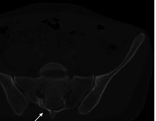
- Kemik penceresinde sakral korpus veya posterior elemanları çaprazlayan yatay lusent çizgi, kortikal basamak ve olası angulasyon/deplasman görülür; retropulse fragman kanal boşluğunu daraltabilir. Kırık morfolojisi için kontrast fazı gerekmez.
Önerilen pencereler G genişlik, M merkez; değerler Hounsfield birimi (HU)
- KemikG 1800 / M +400
- Yumuşak dokuG 400 / M +40
BT bulguları
- Transvers kırık hattı — korpus boyunca sağ-sol yönde uzanır; sagittal planda kesintili korteks ve yatay lusensi izlenir.
- Kırık seviyesi — S1-S2 bileşkesi, sakral korpus veya daha kaudal segment ilişkisini açıkça belirt; seviye deformite yorumunu değiştirir.
- Kifotik angulasyon — üst ve alt sakral parçaların eksenindeki açısal kırılma sagittal MPR'de görülür.
- Posterior translasyon — kraniyal parçanın kaudal parçaya göre geriye kayması kanal konturunu bozabilir.
- Kanal içi fragman — posterior korpus duvarından kopan parça sakral kanala taşabilir; aksiyel ve sagittal planda takip et.
- Eşlik eden dikey çizgiler — koronal MPR'de bir ya da iki foramen/ala boyunca uzanıp U, H, T veya λ biçimi oluşturabilir.
- Kraniyal ve kaudal fragmanların deplasman yönünü ve sakral kanaldaki kemik daralmasını BT'de tarif edin; BT sinir basısı veya gerilmesine ilişkin dolaylı bulgular gösterebilir, ancak nörolojik yaralanmayı tek başına kesinleştirmez.
Ölçütler ve sınıflamalar
- Roy-Camille / Strange-Vognsen-Lebech transvers kırık sınıflaması
- Roy-Camille tip I örneğinde kifotik deformite görülebilir; tip II üst sakral fragmanın distal segmente göre posterior, tip III ise anterior translasyonuyla tanımlanır; Strange-Vognsen–Lebech'in eklediği tip IV, S1 korpusunda segmental parçalanma ile karakterizedir. Deformiteyi sagittal MPR’de, parçalanmayı aksiyel kesitlerde doğrula.
- Denis zonları
- Sakral kanala uzanan transvers kırık, Denis zon III örüntüsüyle uyumludur. Foramen düzeyindeki sakral kırık bileşenleri Denis zon II veya III kapsamında yer alabilir.
Normalde
Normal sakral korpuslar kaynaşmış yapı oluşturur; posterior kortikal kontur ve sakral kanalın kemik sınırı kesintisizdir. Kırık seviyesini komşu segmentlerle kıyaslayarak yatay kortikal çizgiyi, açı değişimini, translasyonu ve kanal yönünde kemik çıkıntısını araştır.
Normal BT ile kıyasla
Aynı bölgenin, kaynak veri setinde patoloji içermediği belirtilen bir BT incelemesini kesit kesit kaydırın; organ sınırlarını açarak anatomiyi hasta görüntüsüyle karşılaştırın. Küçük rastlantısal bulgular tümüyle dışlanmamıştır.
Kaynak: TotalSegmentator v2.01 veri seti (Wasserthal ve ark., Radiology: Artificial Intelligence 2023), CC BY 4.0, vaka s0119; organ sınırları veri setinin otomatik segmentasyonundan, birleştirilerek sadeleştirildi. Görüntüler 2,5 mm kesit aralığına yeniden örneklendi.
Ayırıcı tanı
- Sakral yetersizlikte yatay bileşen
- çoğu kez bilateral dikey alar çizgilerle birlikte ve belirgin travmatik deplasman olmadan H biçimi verir.
- Sakrokoksigeal eklem konturu
- eklem aralığı ve düzgün kortike yüzeyler kırık hattındaki kortikal kesinti/angulasyondan ayrılır.
- Doğumsal segmentasyon varyantı
- düzgün kortike sınırlar ve uyumlu disk/eklem aralığı keskin, düzensiz travmatik hattın aksinedir.
- Denis zon 2 dikey kırığı
- foramenden kraniokaudal uzanır; transvers korpus hattı oluşturmaz.
Tuzaklar
- Yatay çizgi yalnız tek düzlemde görünüyorsa hacim etkisi veya kısmi füzyon çizgisi olabilir; sagittal ve koronal devamlılığı ile kortikal basamağı doğrula.
- Alt sakrumdaki küçük transvers kırıklar kemik penceresinde gözden kaçabilir; S2-S5 korpuslarını ve sakrokoksigeal bileşkeyi ardışık sagittal görüntülerde tara.
- Yalnız transvers bileşeni adlandırmak dikey kanat kırıklarını saklayabilir; koronal reformatta iki sakral ala boyunca tam tarama yap.
Kendini dene
Roy-Camille sınıflamasında, üst sakral fragmanın distal segmente karşı posterior translasyonu ile karakterize olan sakral kırık tipi hangisidir?
Cevabı göster
Tip II. Kaynaklar, Tip II sakral kırıkları üst fragmanın alt segmana göre posterior kayması ile tanımlamaktadır.
Sagittal BT'de transvers sakral çizgiye ek olarak iki dikey alar hat seçiliyor. Hangi ek değerlendirme gerekir?
Cevabı göster
Spinopelvik devamlılık. İki dikey ve bir transvers bileşen U/H biçimli spinopelvik ayrışma oluşturabilir; koronal MPR ve pelvik bağlantılar kontrol edilmelidir. Diğer seçenekler sakral kırık örüntüsüne ilişkin değildir.
İlgili konular
- Sakral kanal kırığı (Denis zon 3)
- Sakral U/H tipi kırık ve spinopelvik ayrışma
- Transforaminal sakral kırık (Denis zon 2)
Kaynaklar
Bu sayfadaki 39 cümle ve 2 quiz sorusu aşağıdaki kaynaklardan alıntılarla otomatik olarak karşılaştırıldı; 4 cümle bu karşılaştırmada düzeltildi (2026-10-09).
- A novel approach to Roy-Camille type 2 sacral fracture: Percutaneous lumbopelvic fixation following manual hyperextension-assisted closed reductionpmc.ncbi.nlm.nih.gov
- Retrograde gluteal cleft sacral screw fixation for a transverse sacral fracture (AO A3) with cauda equina: A technical notepmc.ncbi.nlm.nih.gov
- Roy-Camille Type 3 suicidal jumper's fractures: Case series and review of the literaturepmc.ncbi.nlm.nih.gov
- Usefulness of Sacral Sublaminar Wire for Low Transverse Sacral Fractures: Two Cases' Reportpmc.ncbi.nlm.nih.gov
- A novel technique to repair a transverse sacral fracture in a previously fused lumbosacral spondylolisthesispmc.ncbi.nlm.nih.gov
- Spondylopelvic dissociation.pubmed.ncbi.nlm.nih.gov
- Traumatic spinopelvic dissociation or U-shaped sacral fracture: a review of the literature.pubmed.ncbi.nlm.nih.gov
- Isolated transverse sacral fractures.pubmed.ncbi.nlm.nih.gov
- A minimally invasive surgical technique for the management of U-shape sacral fractures.pubmed.ncbi.nlm.nih.gov
- Risk factors for lumbosacral plexus palsy related to pelvic fracture.pubmed.ncbi.nlm.nih.gov
- Open reduction and posterior instrumentation of type 3 high transverse sacral fracture-dislocation: technical note.pubmed.ncbi.nlm.nih.gov
- Stepladder Pattern Transverse Sacral Fracture: A Previously Unreported Morphology With Neurological Recovery.pubmed.ncbi.nlm.nih.gov
- A Rare Case of Isolated Lower Segment Transverse Sacral Fracture in a 12-Year-Old Girl and Its Management by Fixation with K-Wire.pubmed.ncbi.nlm.nih.gov
- A novel approach to Roy-Camille type 2 sacral fracture: Percutaneous lumbopelvic fixation following manual hyperextension-assisted closed reduction.pubmed.ncbi.nlm.nih.gov
- Variations in sacral morphology and implications for iliosacral screw fixation.pubmed.ncbi.nlm.nih.gov
- Morphometric measurement and applicable feature analysis of sacral alar-iliac screw fixation using forward engineering.pubmed.ncbi.nlm.nih.gov
- Anatomical conditions and patient-specific locked navigation templates for transverse sacroiliac screw placement: a retrospective study.pubmed.ncbi.nlm.nih.gov
- Computational analysis on the feasibility of transverse iliosacral screw fixation for different sacral segments.pubmed.ncbi.nlm.nih.gov
- Current concepts in spondylopelvic dissociation.pubmed.ncbi.nlm.nih.gov
- Turning the backbone into an ankylosed concrete-like structure: Case report.pubmed.ncbi.nlm.nih.gov
- Minimally Invasive Lumbopelvic and Transiliac-Transsacral Fixation for a Pathological Sacral Fracture Due to Metastatic Breast Cancer: A Case Report.pubmed.ncbi.nlm.nih.gov
- Open and Percutaneous Fixation of Traumatic Sacral Fracture-Dislocation with Spinopelvic Dissociation: Two Adolescent Cases and a Systematic Literature Review.pubmed.ncbi.nlm.nih.gov
- S1-pedicle-based triangle fixation for U-type sacral fracture: a technical note.pubmed.ncbi.nlm.nih.gov
- Emergency radiology of severe acute injury in extreme alpine summer sports: a pictorial essay.pubmed.ncbi.nlm.nih.gov
- Traumatic lumbosacral instability: part 2-indications and techniques for surgical management.pubmed.ncbi.nlm.nih.gov
- Modified spinopelvic crab-shaped fixation using offset connectors for a H-shaped sacral fracture with a floating Roy-Camille type 3 transverse component: a case report.pubmed.ncbi.nlm.nih.gov
- Knowledge gaps in high-energy sacral fracture treatment and management.pubmed.ncbi.nlm.nih.gov
- Sacral U-Type Fractures: A Comparative Study of Treatment Approaches.pubmed.ncbi.nlm.nih.gov
- Vertebral Augmentation Involving Vertebroplasty or Kyphoplasty for Cancer-Related Vertebral Compression Fractures: A Systematic Review.pubmed.ncbi.nlm.nih.gov
- A Guide to Improving the Care of Patients with Fragility Fractures, Edition 2.pubmed.ncbi.nlm.nih.gov
- Neurolymphomatosis: A Comprehensive Review of Clinical and Imaging Features.pubmed.ncbi.nlm.nih.gov
- A case of pelvic Ewing sarcoma misinterpreted as osteomyelitis: diagnostic and clinical implications.pubmed.ncbi.nlm.nih.gov
- Iconographies of rare causes of medullary compression due to primary vertebro-medullary and epidural hydatidosis: Contribution of imaging.pubmed.ncbi.nlm.nih.gov
- Comparative analysis of morphologival parameters in isolated and fused L5 spondylolysis patients on the basis of CT features.pubmed.ncbi.nlm.nih.gov
- Navigation-Guided Thoracolumbar Transpedicular Instrumentation Using the 7D Flash™ Navigation System (SeaSpine) in a Patient With L1 Vertebral Fracture and Spinal Deformity Secondary to Prior Instrumentation for Idiopathic Scoliosis: A Case Report.pubmed.ncbi.nlm.nih.gov
- Anterior Sacroiliac Fracture Dislocation: A Comparative Radiologic analysis of Crescent Fractures in Pelvic Ring Injuries: A Retrospective Study.pubmed.ncbi.nlm.nih.gov
- CT-based feasibility of transsacral osseous corridors in older adults: the impact of lumbosacral transitional anatomy and sacral dysmorphism.pubmed.ncbi.nlm.nih.gov
- Recognizing Sacral Insufficiency Fractures Hidden in Plain Sight: An Illustrative Case Report.pubmed.ncbi.nlm.nih.gov
- Proximal junctional kyphosis in distraction-based growth-friendly treatment: Definition, risk factors, and timeline.pubmed.ncbi.nlm.nih.gov
- Comparison of short-segment vs. long-segment fixation in posterior osteotomy for kyphotic deformity: effects on postoperative alignment and complications.pubmed.ncbi.nlm.nih.gov
- Traumatic Bilateral Lumbosacral Jumped Facet Without Fracture in Childhood: Case Report and Systematic Review.pubmed.ncbi.nlm.nih.gov
- Traumatic lumbosacral instability: part 1 - proposing a definition and identifying underlying injury patterns.pubmed.ncbi.nlm.nih.gov
- Failed back surgery syndrome successfully ameliorated with Chiropractic Biophysics ® structural rehabilitation improving pain, disability as well as sagittal and coronal balance: a Chiropractic Biophysics ® case report with a 6 year follow-up.pubmed.ncbi.nlm.nih.gov
- Added Value of Three-Plane Multiecho Fast Field Echo MRI Sequence in the Evaluation of Acute Spinal Trauma Using Sensitivity: A Prospective Study.pubmed.ncbi.nlm.nih.gov
- The American Society of Pain and Neuroscience (ASPN) Evidence-Based Clinical Guideline of Interventional Treatments for Low Back Pain.pubmed.ncbi.nlm.nih.gov
- Novel 3D-navigated screw corridors for posterior pelvic ring stabilization: a finite element analysis.pubmed.ncbi.nlm.nih.gov
- Sacral and Pelvic Fractures: Historical Systems and Advancements with the AO Spine Classification.pubmed.ncbi.nlm.nih.gov
- Chronic pelvic insufficiency fractures and their treatment.pubmed.ncbi.nlm.nih.gov
- Effectiveness of Hybrid Surgery Using Polyetheretherketone Rods for Preventing Proximal Junctional Kyphosis and Failure After Adult Spinal Deformity Surgery: Biomechanical Evaluation.pubmed.ncbi.nlm.nih.gov
- Magnetic resonance imaging (MRI) vs. computed tomography (CT) in the diagnosis and classification of spondylolysis and spondylolisthesis-a narrative review.pubmed.ncbi.nlm.nih.gov
- Preserving Posterior Complex Can Prevent Adjacent Segment Disease following Posterior Lumbar Interbody Fusion Surgeries: A Finite Element Analysis.pubmed.ncbi.nlm.nih.gov
- Agreement rate of surgically treated sacral fractures with the recommendations proposed by the sacral AOSpine Injury Score.pubmed.ncbi.nlm.nih.gov
- CT of Sacral Fractures: Classification Systems and Management.pubmed.ncbi.nlm.nih.gov
- Denis pattern-dependent biomechanical behavior of posterior trans-iliac plate fixation compared with bilateral triangular osteosynthesis in unilateral sacral fractures: a finite element study.pubmed.ncbi.nlm.nih.gov
- Neuronavigated placement of iliosacral screws in sacral fractures: an additional tool in the spine surgeon's arsenal.pubmed.ncbi.nlm.nih.gov
- A positioning method for the transiliac-transsacral screw bone channel based on a manual tracing of a transparent overlay of preoperative computed tomography images in patients with pelvic fracture and a unilateral trans-sacroiliac corridor.pubmed.ncbi.nlm.nih.gov
- Preferential sacral fracture sites in fragility fractures of the pelvis type IVb and comparison of internal fixation methods: CT-based morphological mapping and finite element analysis.pubmed.ncbi.nlm.nih.gov
- Congenital and Breed-Associated Vertebral Anomalies in Dogs and Cats: Developmental Anatomy, Genetic Determinants, and Biological Significance.pubmed.ncbi.nlm.nih.gov
- Filamin B (<i>FLNB</i>)-Related Spondylocarpotarsal Synostosis Syndrome: Systematic Literature Review and Novel Case Report.pubmed.ncbi.nlm.nih.gov
- Deciphering the Genetic Basis of Congenital Vertebral Malformations Through a Stepwise Diagnostic Approach.pubmed.ncbi.nlm.nih.gov
- Axial Spondyloarthritis: Mimics and Pitfalls of Imaging Assessment.pubmed.ncbi.nlm.nih.gov
- Multidisciplinary management of spinal metastases: what the radiologist needs to know.pubmed.ncbi.nlm.nih.gov
- Clinical manifestations, classification, and surgical management of sacral tumors and the need for personalized approach to sacrectomy.pubmed.ncbi.nlm.nih.gov
- Sacral Fractures and Associated Injuries.pubmed.ncbi.nlm.nih.gov
- Patterns of injury and violence in Yaoundé Cameroon: an analysis of hospital data.pubmed.ncbi.nlm.nih.gov
- Long-term safety and effectiveness of the "OptEase" vena cava filter.pubmed.ncbi.nlm.nih.gov
- Iliosacral Screw Fixation for Treating Sacral Fractures: A Modified Pedicle-Guided Fixation Technique in Prone Position.pubmed.ncbi.nlm.nih.gov
- Specifying the Entry Point for a Transiliac-Transsacral Screw in Posterior Pelvic Ring Fixation.pubmed.ncbi.nlm.nih.gov
- Lower extremity and spinopelvic alignment changes after total hip arthroplasty in unilateral Crowe III-IV dysplasia: clinical and radiological outcomes.pubmed.ncbi.nlm.nih.gov
- Dural tears from traumatic spinal fractures: an illustrative case and systematic review of its incidence, diagnosis, risk factors, and management.pubmed.ncbi.nlm.nih.gov
- Complications of vertebral augmentation in metastatic spinal fractures: a systematic review of multiple study designs.pubmed.ncbi.nlm.nih.gov
- Staged management of a Tile C3 pelvic ring injury with bilateral sacroiliac disruption and T-shaped sacral fracture.pubmed.ncbi.nlm.nih.gov
- Clinical application study of E3D-assisted design combined with personalized 3D printed guide system in the treatment of pelvic fractures.pubmed.ncbi.nlm.nih.gov
- Sacral U-type Fractures in Patients Older Than 65 years.pubmed.ncbi.nlm.nih.gov
- Surgical Management of Unstable U-Shaped Sacral Fractures and Tile C Pelvic Ring Disruptions: Institutional Experience in Light of a Narrative Literature Review.pubmed.ncbi.nlm.nih.gov
- Sacral Dysmorphism as an Independent Risk Factor for Cortical Breach During Percutaneous S1 Iliosacral Screw Fixation.pubmed.ncbi.nlm.nih.gov
- Preliminary Retrospective Analysis of X-Ray, CT, and MRI Imaging Features of Brucellar Spondylodiscitis Diagnosis.pubmed.ncbi.nlm.nih.gov
- Effect of lumbosacral transitional vertebra on developmental alterations of the hip: a quantitative investigation of the lumbo-pelvic-hip complex via whole-body computed tomography.pubmed.ncbi.nlm.nih.gov
- Factors associated with U-type sacral fractures in geriatric patients: A retrospective cohort study.pubmed.ncbi.nlm.nih.gov
- A novel internal fixation system for sacral fractures: a finite element biomechanical study.pubmed.ncbi.nlm.nih.gov
- Preoperative 3D-Planned S1 Corridors Transferred into 2D Fluoroscopy Allow for Safe Intraoperative Large-Diameter Implant Placement: Description of a Novel Sacroiliac Fixation Technique and Proof of Concept in 137 Implantations.pubmed.ncbi.nlm.nih.gov
- Anatomy-driven automated subsegmentation of pelvic and proximal femoral CT into clinically relevant subregions and landmarks.pubmed.ncbi.nlm.nih.gov
- New Minimally Invasive Method for Treating Posterior Pelvic Ring Fractures: Biomechanical Validation and Clinical Application of Sacroiliac Joint Locking Plate.pubmed.ncbi.nlm.nih.gov
- Minimally invasive percutaneous cannulated polyaxial screw and rod fixation thoracolumbar and lumbosacral vertebrae: cadaveric evaluation of accuracy and safety of a fluoroscopic-guided technique, in large breed dogs.pubmed.ncbi.nlm.nih.gov
- Paraspinal muscle CT measurements in predicting subsequent fractures after percutaneous kyphoplasty in elderly women.pubmed.ncbi.nlm.nih.gov
- Sacral Fractures: A Review.pubmed.ncbi.nlm.nih.gov
- Spinopelvic Dissociation Case Report and Literature Review.pubmed.ncbi.nlm.nih.gov
- Acute management of spinal fractures: What you need to know.pubmed.ncbi.nlm.nih.gov
Eğitim amaçlıdır; hasta başında tanı ve tedavi kararının yerine geçmez, tıbbi tavsiye değildir. Metinler yapay zekâ desteğiyle yazıldı ve otomatik bir adımda kaynak alıntılarıyla karşılaştırıldı; bu bir tıbbi doğrulama değildir ve içerik uzman radyolog incelemesinden geçmedi. Klinik kararlarda güncel kılavuzları, kurum protokollerini ve radyoloji raporunu esas alın.