Özet
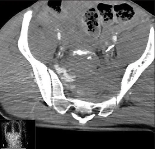
U/H tipi sakral kırıkta iki longitudinal sakral hat, bir transvers bileşenle birleşir ve omurgaya bağlı üst sakral segmentin pelvisle kemik devamlılığını keser. Yüksek enerjili düşme ve araç çarpışmalarında görülebilir; yetersizlik kırığında da benzer geometrik hatlar oluşabileceği için travma mekanizması ve deplasman birlikte yorumlanır. Kontrastsız BT'nin koronal, sagittal ve aksiyel MPR'leri kırık geometrisini, ayrışan segmentleri, kanal/foramen basısını ve eşlik eden pelvik halka hasarını ortaya koyar. Örüntüyü kaçırmak ciddi mekanik instabiliteyi ve kanal içi fragmanla ilişkili nörolojik riski gözden kaçırabilir.
Faz ve pencere
- Kontrastsız tanısal
- Kemik algoritmalı BT'de iki dikey sakral ala/foramen çizgisi ve bunları birleştiren yatay kırık U veya H konturu oluşturur; sagittal planda posterior translasyon, kifoz ve retropulse fragman görülebilir. Akut yüksek enerjili yaralanmada sakral kırığın değerlendirilmesinde BT tercih edilen yöntemdir.
Önerilen pencereler G genişlik, M merkez; değerler Hounsfield birimi (HU)
- KemikG 1800 / M +400
- Yumuşak dokuG 400 / M +40
BT bulguları
- İki dikey sakral hat — koronal reformatta sağ ve sol taraftaki çizgiler aladan, foramenden veya kanal komşuluğundan geçebilir.
- Birleştirici transvers hat — aksiyel ve sagittal MPR'de seviyesini, tamlığını ve dikey bileşenlerle bağlantısını belirle.
- U/H geometrisi — kesintisiz üst sakral blok ile alt sakrum arasındaki kırık konfigürasyonu pelvisin omurgaya tutunmasını bozar.
- Spinopelvik translasyon — kraniyal sakral parçanın posterior veya anterior kayması ile sakral kifoz sagittal planda gösterilir.
- Kanal ve foraminal fragman — posteriora itilmiş kemik parçası merkezi kanalı ya da nöral çıkış deliklerini daraltabilir.
- SI eklem/pelvik halka — iliak kanat, asetabulum ve anterior halka yaralanmaları ile presakral hematom eşlik edebilir; pelvis BT'sinin tamamını tara.
- U tipi sakral kırık spinopelvik ayrışmadır; sagittal BT rekonstrüksiyonu transvers bileşeni ve kırık deplasmanını değerlendirmeye yardımcı olur.
- AO Spine'da C0 deplase olmayan U tipi kırık, C2 transvers bileşeni olmayan bilateral tam B tipi kırıklar, C3 deplase U tipi kırıktır.
Ölçütler ve sınıflamalar
- Deskriptif şekil sınıflaması
- U veya H biçimi iki longitudinal bileşen ile transvers bağlantının görünümünü adlandırır. AO Spine sınıflamasında C1, arka pelvik stabilitesi korunmuş U tipi sakral kırık varyantı olarak tanımlanır.
- Roy-Camille sınıflaması
- Roy-Camille tip 2 U tipi kırık posteriora, tip 3 ise anteriora deplase olarak tanımlanır.
- AO Spine sakral sınıflaması
- C tipi yaralanma spinopelvik ayrışmayı ifade eder; transvers bileşenle tamamlanan dikey kırık düzeni C grubu içinde sınıflandırılır. Nörolojik durum ve hasta-özel değiştiriciler morfoloji kodundan ayrı kaydedilir.
Normalde
Normalde lomber omurga, sakrum ve iki iliak kemik arasında kesintisiz yük aktarım hattı vardır; sakral korteks ve SI eklem yüzleri düzgün konturludur. Koronal planda iki ala, sagittal planda sakral segment dizilimi ve aksiyel planda kanal/foramen açıklıkları karşılaştırılarak kemik köprü devamlılığındaki kopma ve segment kayması gösterilir.
Normal BT ile kıyasla
Aynı bölgenin, kaynak veri setinde patoloji içermediği belirtilen bir BT incelemesini kesit kesit kaydırın; organ sınırlarını açarak anatomiyi hasta görüntüsüyle karşılaştırın. Küçük rastlantısal bulgular tümüyle dışlanmamıştır.
Kaynak: TotalSegmentator v2.01 veri seti (Wasserthal ve ark., Radiology: Artificial Intelligence 2023), CC BY 4.0, vaka s0119; organ sınırları veri setinin otomatik segmentasyonundan, birleştirilerek sadeleştirildi. Görüntüler 2,5 mm kesit aralığına yeniden örneklendi.
Ayırıcı tanı
- Bilateral sakral yetersizlik kırığı
- H biçimi oluşturabilir; eşlik eden osteoporoz, ala boyunca sklerotik bant ve genellikle az deplasman destekler.
- AO Spine'da yatay bileşeni olmayan longitudinal sakral kırıklar B tipi posterior pelvik yaralanma olarak sınıflanır; spinopelvik ayrışma ise omurga ile pelvik halka bağlantısının bozulduğu C tipi yaralanmadır.
- AO Spine'da sakroiliak eklemin altında kalan deplase olmayan transvers kırık A2, deplase olan A3'tür; bilateral longitudinal ve transvers bileşenli U tipi kırıklar C grubunda değerlendirilir.
- Sakrum tümörleri ile tümör dışı lezyonların görüntüleme özellikleri örtüşebilir; belirli örüntüler etiyolojiyi düşündürse de kesin tanı için biyopsi gerekebilir.
Tuzaklar
- Osteoporotik hastalarda bilateral dikey sakral ala kırıkları saptandığında, eşlik eden transvers bileşeni aramak için sagittal BT rekonstrüksiyonları da incelenmelidir.
- Yetersizlik kırığı U/H benzeri görünüm oluşturabilir; alar skleroz, kemiğin genel osteopenisi ve belirgin segment translasyonu olup olmadığını birlikte değerlendir.
- Sagittal BT rekonstrüksiyonu, deplase olmayan veya minimal deplase sakral kırıkların tanınmasında en duyarlı görüntüleme yöntemidir.
Kendini dene
BT'de iki dikey sakral çizgi, bunları birleştiren S2 transvers kırık ve posteriora kaymış kraniyal blok var. En uygun tanım?
Cevabı göster
U tipi ayrışma. İki dikey kol ve birleştirici transvers bileşenle üst sakral blok pelvik bağlantısını kaybeder; bu U tipi spinopelvik ayrışmadır. Diğer seçenekler bu üç kollu geometriyi açıklamaz.
Bilateral alar dikey çizgiler ve yatay S2 bileşeni var; yaygın osteopeni, bant sklerozu ve belirgin translasyon yok. En olası eşlik eden mekanizma?
Cevabı göster
Yetersizlik kırığı. Kaynaklar, osteopeni ve belirlenmiş alar bantlarla oluşan H/U tipi kırıkların yetersizlik kırığı ile uyumlu olduğunu belirtir.
İlgili konular
- Sakral kanal kırığı (Denis zon 3)
- Transvers sakral kırık
- Transforaminal sakral kırık (Denis zon 2)
- Sakral ala kırığı (Denis zon 1)
Kaynaklar
Bu sayfadaki 43 cümle ve 2 quiz sorusu aşağıdaki kaynaklardan alıntılarla otomatik olarak karşılaştırıldı; 7 cümle bu karşılaştırmada düzeltildi (2026-10-09).
- Management of C0 Sacral Fractures Based on the AO Spine Sacral Injury Classificationpmc.ncbi.nlm.nih.gov
- Surgical management strategies for unstable sacral fractures: Case series and surgical guideline.pubmed.ncbi.nlm.nih.gov
- A Case of U-shaped Sacral Fracture After Longstanding Spinopelvic Fixation Treated With Percutaneous Sacroiliac Joint Fusion and Iliosacral Osteosynthesis.pubmed.ncbi.nlm.nih.gov
- Surgical management of lumbosacral and sacral fractures: roles of the pelvic and spinal surgeons.pubmed.ncbi.nlm.nih.gov
- Sacral Fractures: A Review.pubmed.ncbi.nlm.nih.gov
- Long-term safety and effectiveness of the "OptEase" vena cava filter.pubmed.ncbi.nlm.nih.gov
- High-affinity alphavbeta3 integrin targeted optical probe as a new imaging biomarker for early atherosclerosis: initial studies in Watanabe rabbits.pubmed.ncbi.nlm.nih.gov
- Radiographic and Clinical Adjacent Segment Disease After Posterior Instrumented Fusion for Traumatic Thoracolumbar and Lumbar Vertebral Fractures.pubmed.ncbi.nlm.nih.gov
- Traumatic lumbosacral instability: part 1 - proposing a definition and identifying underlying injury patterns.pubmed.ncbi.nlm.nih.gov
- Emergency treatment of pelvic ring injuries: state of the art.pubmed.ncbi.nlm.nih.gov
- CT of Sacral Fractures: Classification Systems and Management.pubmed.ncbi.nlm.nih.gov
- Emergency radiology of severe acute injury in extreme alpine summer sports: a pictorial essay.pubmed.ncbi.nlm.nih.gov
- Towards Automated Spine Fracture Detection on Whole-Body CT of Polytraumatized Patients.pubmed.ncbi.nlm.nih.gov
- Radiomics in spinal research: a narrative review.pubmed.ncbi.nlm.nih.gov
- Reliability of Fossae Lumbales Laterales and Pelvic Incidence for Estimating Transsacral Corridors Assessed Using Reconstruction Computed Tomography.pubmed.ncbi.nlm.nih.gov
- Imaging and treatment of sacral insufficiency fractures.pubmed.ncbi.nlm.nih.gov
- Imaging Review of Pelvic Ring Fractures and Its Complications in High-Energy Trauma.pubmed.ncbi.nlm.nih.gov
- S1-pedicle-based triangle fixation for U-type sacral fracture: a technical note.pubmed.ncbi.nlm.nih.gov
- Percutaneous spinopelvic fixation technique using external fixation for focal kyphosis reduction in U-type sacral fractures: a case report.pubmed.ncbi.nlm.nih.gov
- Traumatic lumbosacral instability: part 2-indications and techniques for surgical management.pubmed.ncbi.nlm.nih.gov
- The American Society of Pain and Neuroscience (ASPN) Evidence-Based Clinical Guideline of Interventional Treatments for Low Back Pain.pubmed.ncbi.nlm.nih.gov
- Artificial intelligence for abdominopelvic trauma imaging: trends, gaps, and future directions.pubmed.ncbi.nlm.nih.gov
- Orthopaedic fracture surgery in polytraumatized patients while on extracorporeal membrane oxygenation (ECMO): A report of two cases.pubmed.ncbi.nlm.nih.gov
- Traumatic hemorrhage and chain of survival.pubmed.ncbi.nlm.nih.gov
- Congenital and Breed-Associated Vertebral Anomalies in Dogs and Cats: Developmental Anatomy, Genetic Determinants, and Biological Significance.pubmed.ncbi.nlm.nih.gov
- Partial sacrectomy with en bloc tumor resection without instrumentation. What level is safe?pubmed.ncbi.nlm.nih.gov
- Pelvic-girdle reconstruction with three-dimensional-printed endoprostheses after limb-salvage surgery for pelvic sarcomas: current landscape.pubmed.ncbi.nlm.nih.gov
- Sacroiliac Joint Anatomical Variants and Their Association with Spinopelvic Alignment: A Whole-Spine CT Study.pubmed.ncbi.nlm.nih.gov
- Zero Echo Time MRI for Osseous Assessment of Sports-Related Pathology in Athletes: A Pictorial Essay.pubmed.ncbi.nlm.nih.gov
- Sacral Dysmorphism as an Independent Risk Factor for Cortical Breach During Percutaneous S1 Iliosacral Screw Fixation.pubmed.ncbi.nlm.nih.gov
- Comparative Radiologic and Morphologic Analysis of Posterolateral Fusion and Percutaneous Pedicle Screw Fixation for Thoracolumbar Junction Burst Fractures.pubmed.ncbi.nlm.nih.gov
- Integrating radiomics with clinical data for enhanced prediction of vertebral fracture risk.pubmed.ncbi.nlm.nih.gov
- Altered distribution of intervertebral disc degeneration in degenerative thoracolumbar kyphosis: A retrospective clinical study and finite element analysis.pubmed.ncbi.nlm.nih.gov
- Padua Days on Muscle and Mobility Medicine, March 25-29, 2025, Hotel Petrarca, Euganean Thermae, Italy: Program and Abstracts.pubmed.ncbi.nlm.nih.gov
- Spinopelvic Dissociation Case Report and Literature Review.pubmed.ncbi.nlm.nih.gov
- Minimally Invasive Inline Spinopelvic Fixation Without Offset Connectors for Unstable Sacral Fractures: Technical Description and Preliminary Clinical Experience.pubmed.ncbi.nlm.nih.gov
- Knowledge gaps in high-energy sacral fracture treatment and management.pubmed.ncbi.nlm.nih.gov
- Open and Percutaneous Fixation of Traumatic Sacral Fracture-Dislocation with Spinopelvic Dissociation: Two Adolescent Cases and a Systematic Literature Review.pubmed.ncbi.nlm.nih.gov
- Imaging of Sacroiliac Pain: The Current State-of-the-Art.pubmed.ncbi.nlm.nih.gov
- Axial Spondyloarthritis: Mimics and Pitfalls of Imaging Assessment.pubmed.ncbi.nlm.nih.gov
- Microdamage accumulation in the femoral head and its association with subchondral insufficiency fractures: A retrospective study.pubmed.ncbi.nlm.nih.gov
- Comparative analysis of morphologival parameters in isolated and fused L5 spondylolysis patients on the basis of CT features.pubmed.ncbi.nlm.nih.gov
- Deep Learning-Assisted Radiomics for Predicting Thoracolumbar Osteoporotic Vertebral Fractures.pubmed.ncbi.nlm.nih.gov
- Fluid transport in the brain.pubmed.ncbi.nlm.nih.gov
- Beyond Chordoma: A Comprehensive Review of Sacral Lesions.pubmed.ncbi.nlm.nih.gov
- Augmented-reality-assisted minimally invasive quad-rod spinopelvic fixation for spinopelvic dissociation caused by pathological fracture due to extramedullary plasmacytoma: an evaluation of technique and its benefits.pubmed.ncbi.nlm.nih.gov
- Clinical applications of MRI-based artificial intelligence in spinal metastases: A systematic review.pubmed.ncbi.nlm.nih.gov
- Total laparoscopic radical hysterectomy for cervical cancer in prolapsed uterus.pubmed.ncbi.nlm.nih.gov
- Management strategies of pediatric coccydynia: a narrative review.pubmed.ncbi.nlm.nih.gov
- Spinal lesions: a comprehensive radiologic overview.pubmed.ncbi.nlm.nih.gov
- Three-Dimensional Printing of Medical Devices Used Directly to Treat Patients: A Systematic Review.pubmed.ncbi.nlm.nih.gov
- Magnetic resonance imaging of the spine.pubmed.ncbi.nlm.nih.gov
- Minimally Invasive Lumbopelvic and Transiliac-Transsacral Fixation for a Pathological Sacral Fracture Due to Metastatic Breast Cancer: A Case Report.pubmed.ncbi.nlm.nih.gov
- Post-traumatic Exacerbation of Grade II L5-S1 Spondylolisthesis Requiring Surgical Stabilization.pubmed.ncbi.nlm.nih.gov
- Application of Three-Dimensionally Printed Surgical Guides in Precise Sacral Tumor Excision and Defect Reconstruction.pubmed.ncbi.nlm.nih.gov
- Ramadanov-Zabler Safe Zone for Sacroiliac Screw Placement: A CT-Based Computational Pilot Study.pubmed.ncbi.nlm.nih.gov
- Consensus practice guidelines on sacroiliac joint complex pain from a multispecialty, international working group.pubmed.ncbi.nlm.nih.gov
- Iliosacral Screw Fixation for Treating Sacral Fractures: A Modified Pedicle-Guided Fixation Technique in Prone Position.pubmed.ncbi.nlm.nih.gov
- Explainable AI for classifying vertebral fracture histology in digital spine pathology.pubmed.ncbi.nlm.nih.gov
- Traumatic bilateral acetabular fracture secondary to high-energy trauma in healthy adults.pubmed.ncbi.nlm.nih.gov
- Outcomes of Lumbosacral Fixation in Patients with Suicidal Jumper's Fractures: A Retrospective Study from a Single Center in Turkey.pubmed.ncbi.nlm.nih.gov
- Preferential sacral fracture sites in fragility fractures of the pelvis type IVb and comparison of internal fixation methods: CT-based morphological mapping and finite element analysis.pubmed.ncbi.nlm.nih.gov
- Fatal Injuries Caused by Falling Trees: Correlation Between Postmortem Computed Tomography and Forensic Autopsy Findings in Eight Cases.pubmed.ncbi.nlm.nih.gov
- Clinical application study of E3D-assisted design combined with personalized 3D printed guide system in the treatment of pelvic fractures.pubmed.ncbi.nlm.nih.gov
- Multilevel Lumbar CT Attenuation Beyond L1: Comparison with QCT-Derived Volumetric Bone Mineral Density and Prevalent Fragility Fracture Status in a Diagnostic Referral Cohort.pubmed.ncbi.nlm.nih.gov
- Posterior Extradural Spinal Tumor Extending From T3 to T6 Presenting With Paraplegia.pubmed.ncbi.nlm.nih.gov
- Clinical Nononcologic Applications of PET/CT and PET/MRI in Musculoskeletal, Orthopedic, and Rheumatologic Imaging.pubmed.ncbi.nlm.nih.gov
- Anatomy-driven automated subsegmentation of pelvic and proximal femoral CT into clinically relevant subregions and landmarks.pubmed.ncbi.nlm.nih.gov
- Causes of Increased Use of Closed Reduction and Internal Fixation for High-Energy-Related Traumatic Sacral Fractures.pubmed.ncbi.nlm.nih.gov
- Surgical Management of Unstable U-Shaped Sacral Fractures and Tile C Pelvic Ring Disruptions: Institutional Experience in Light of a Narrative Literature Review.pubmed.ncbi.nlm.nih.gov
- Clinical and radiological outcomes of lumbopelvic fixation in unstable Isler type II and III sacral fractures.pubmed.ncbi.nlm.nih.gov
- Recent Progress in the Classification and Operation of Sacral Fractures.pubmed.ncbi.nlm.nih.gov
- Agreement rate of surgically treated sacral fractures with the recommendations proposed by the sacral AOSpine Injury Score.pubmed.ncbi.nlm.nih.gov
- Sacral and Pelvic Fractures: Historical Systems and Advancements with the AO Spine Classification.pubmed.ncbi.nlm.nih.gov
- Open reduction and internal fixation versus percutaneous multiple-trajectory screw fixation for unstable pelvic fractures with severe polytrauma: a prospective study.pubmed.ncbi.nlm.nih.gov
- Machine learning, decision tree and nomogram for predicting screw loosening after PLIF in osteoporotic patients: a retrospective multicenter study.pubmed.ncbi.nlm.nih.gov
- Management of aneurysmal bone cysts of the spine and sacrum: A narrative review.pubmed.ncbi.nlm.nih.gov
- Patterns of injury and violence in Yaoundé Cameroon: an analysis of hospital data.pubmed.ncbi.nlm.nih.gov
- Minimally invasive percutaneous cannulated polyaxial screw and rod fixation thoracolumbar and lumbosacral vertebrae: cadaveric evaluation of accuracy and safety of a fluoroscopic-guided technique, in large breed dogs.pubmed.ncbi.nlm.nih.gov
- Paraspinal muscle CT measurements in predicting subsequent fractures after percutaneous kyphoplasty in elderly women.pubmed.ncbi.nlm.nih.gov
- Fixation of unstable sacral fractures by transpedicular system: a prospective study.pubmed.ncbi.nlm.nih.gov
- Spinopelvic dissociation: extended definition, physical examination, classification, and therapy.pubmed.ncbi.nlm.nih.gov
- Explainable Multimodal Machine Learning Predicts 90-Day Treatment Failure in Older Patients with Fragility Fractures of the Pelvis.pubmed.ncbi.nlm.nih.gov
- Surgical stabilization of rib fractures (SSRF): the WSES and CWIS position paper.pubmed.ncbi.nlm.nih.gov
- Glossary of terms for musculoskeletal radiology.pubmed.ncbi.nlm.nih.gov
- Staged management of a Tile C3 pelvic ring injury with bilateral sacroiliac disruption and T-shaped sacral fracture.pubmed.ncbi.nlm.nih.gov
- Progress of fracture mapping technology based on CT three-dimensional reconstruction.pubmed.ncbi.nlm.nih.gov
- International Society of Paediatric Surgical Oncology (IPSO) Surgical Practice Guidelines.pubmed.ncbi.nlm.nih.gov
- L4-sparing versus L4-L5 spinopelvic fixation in traumatic spinopelvic dissociation: A randomized controlled trial.pubmed.ncbi.nlm.nih.gov
- Fragility Fractures of the Pelvis-Current Understanding and Open Questions.pubmed.ncbi.nlm.nih.gov
- Sacral U-type Fractures in Patients Older Than 65 years.pubmed.ncbi.nlm.nih.gov
- Sacral Fractures and Associated Injuries.pubmed.ncbi.nlm.nih.gov
- Lumbo-sacral Junction Instability by Traumatic Sacral Fractures: Isler's Classification Revisited - A Narrative Review.pubmed.ncbi.nlm.nih.gov
- Sacral fractures: issues, challenges, solutions.pubmed.ncbi.nlm.nih.gov
- A case of nontraumatic acetabular fracture during epileptic seizure in an elderly patient: Imaging diagnosis and endovascular treatment.pubmed.ncbi.nlm.nih.gov
- Iconographies of rare causes of medullary compression due to primary vertebro-medullary and epidural hydatidosis: Contribution of imaging.pubmed.ncbi.nlm.nih.gov
- A rare incidental finding of splenogonadal fusion in a trauma patient: A case report and review of the literature.pubmed.ncbi.nlm.nih.gov
- An investigation to the clinical application of personalized 3D printed guides combined with preoperative virtual iliosacral screws for the management of posterior pelvic ring injury.pubmed.ncbi.nlm.nih.gov
- Demystifying MR Neurography of the Lumbosacral Plexus: From Protocols to Pathologies.pubmed.ncbi.nlm.nih.gov
- Delayed sacroiliac joint infection after robot-assisted percutaneous iliac screw fixation for tile type C2 pelvic fractures: A case report and literature review.pubmed.ncbi.nlm.nih.gov
- Modified spinopelvic crab-shaped fixation using offset connectors for a H-shaped sacral fracture with a floating Roy-Camille type 3 transverse component: a case report.pubmed.ncbi.nlm.nih.gov
- A positioning method for the transiliac-transsacral screw bone channel based on a manual tracing of a transparent overlay of preoperative computed tomography images in patients with pelvic fracture and a unilateral trans-sacroiliac corridor.pubmed.ncbi.nlm.nih.gov
- Sacral U-Type Fractures: A Comparative Study of Treatment Approaches.pubmed.ncbi.nlm.nih.gov
- Recognizing Sacral Insufficiency Fractures Hidden in Plain Sight: An Illustrative Case Report.pubmed.ncbi.nlm.nih.gov
- Comparison of the AO/OTA 1996/2007 and 2018 pelvic ring fracture classifications.pubmed.ncbi.nlm.nih.gov
- Preoperative 3D-Planned S1 Corridors Transferred into 2D Fluoroscopy Allow for Safe Intraoperative Large-Diameter Implant Placement: Description of a Novel Sacroiliac Fixation Technique and Proof of Concept in 137 Implantations.pubmed.ncbi.nlm.nih.gov
Eğitim amaçlıdır; hasta başında tanı ve tedavi kararının yerine geçmez, tıbbi tavsiye değildir. Metinler yapay zekâ desteğiyle yazıldı ve otomatik bir adımda kaynak alıntılarıyla karşılaştırıldı; bu bir tıbbi doğrulama değildir ve içerik uzman radyolog incelemesinden geçmedi. Klinik kararlarda güncel kılavuzları, kurum protokollerini ve radyoloji raporunu esas alın.